Autocrine/paracrine TGF-β1 inhibits Langerhans cell migration (original) (raw)
Abstract
Langerhans cells (LCs) are skin-resident dendritic cells (DC) located in the epidermis that migrate to skin-draining lymph nodes during the steady state and in response to inflammatory stimuli. TGF-β1 is a critical immune regulator that is highly expressed by LCs. The ability to test the functional importance of LC-derived TGF-β1 is complicated by the requirement of TGF-β1 for LC development and by the absence of LCs in mice with an LC-specific ablation of TGF-β1 or its receptor. To overcome these problems, we have engineered transgenic huLangerin-CreERT2 mice that allow for inducible LC-specific excision. Highly efficient and LC-specific expression was confirmed in mice bred onto a YFP Cre reporter strain. We next generated huLangerin-CreERT2 × TGF-βRIIfl and huLangerin-CreERT2 × TGF-β1fl mice. Excision of the TGFβRII or TGFβ1 genes induced mass migration of LCs to the regional lymph node. Expression of costimulatory markers and inflammatory cytokines was unaffected, consistent with homeostatic migration. In addition, levels of p-SMAD2/3 were decreased in LCs from wild-type mice before inflammation-induced migration. We conclude that TGF-β1 acts directly on LCs in an autocrine/paracrine manner to inhibit steady-state and inflammation-induced migration. This is a readily targetable pathway with potential therapeutic implications for skin disease.
Keywords: skin immunology, pSMAD2/3, tolerance
Langerhans cells (LCs) are a subset of skin-resident dendritic cells (DCs) that forms a dense network in the epidermis, where they actively acquire antigen (1). In response to maturation stimuli, LCs become activated and increase surface expression of costimulatory markers, such as CD86, as they migrate out of the epidermis, through the lymphatics, and into the regional draining lymph node (LN). Once in the LN, they present antigen acquired in the periphery to naïve T cells and thereby modulate adaptive immune responses. LCs also migrate during the steady state and may participate with the maintenance of peripheral self-tolerance to epidermal self-antigen (2).
Unlike other DC subsets, LCs have a unique ontogeny (3). LC precursors seed the epidermis shortly before birth. During the first postnatal week of life, LCs in mice undergo a proliferative burst until they occupy the entire epidermis (4). After the first week the LC network is established, and LCs assume a low level of proliferation to replace those LCs that have migrated under steady-state, homeostatic conditions (5). In response to strong inflammatory stimuli, bone marrow-derived precursors are recruited to repopulate the epidermis (6).
LC ontogeny requires TGF-β1. Bone marrow or monocyte precursors cultured in GM-CSF and TGF-β1 generate LC-like cells (7, 8). In addition, LCs are absent from TGF-β1−/− mice and from mice with a genetic ablation of id2 or Runx3, which are both part of the TGF-β1 pathway (9–11). There are three isoforms of TGF-β, but TGF-β1 is the dominant isoform within the immune system. TGF-β binds to the TGF-β receptor II (TGF-βRII) and ALK5 (TGF-βRI) to activate the intracellular signaling proteins SMAD2,3 and -4 (12).
Using mice engineered to have LC-specific expression of Cre (Langerin-Cre) that were combined with floxed TGF-β1fl and TGF-βRIIfl mice, we have previously shown that autocrine/paracrine TGF-β1 is required for formation of the LC network (13). A small number of LCs can be transiently observed in CD11c-Cre TGF-βRIfl mice during the first few postnatal days, suggesting that in addition to the formation of LC precursors, TGF-β may also participate in the early proliferative burst that establishes the LC network and/or LC homeostasis (14). The requirement for TGF-β1 during LC ontogeny prevents the examination of this pathway in fully developed LCs using models of constitutive gene ablation.
To determine whether TGF-β1 is involved with LC homeostasis in vivo, we generated Langerin-CreERT2 mice. CreERT2 is a fusion protein of Cre with a modified form of the estrogen receptor that remains nonfunctional in cytoplasm until a synthetic estrogen (i.e., tamoxifen) is administered, thereby allowing entry of CreERT2 into the nucleus, where it becomes functional (15). Thus, Langerin-CreERT2 mice allow for the inducible ablation of genes selectively in LCs. We have combined these mice with TGF-βRIIfl or TGF-β1fl mice to examine the consequence of ablation of sensitivity to TGF-β1 or the cytokine itself in fully differentiated LCs.
Results
Generation and Validation of huLangerin CreERT2 Mice.
The sequence encoding CreERT2 was inserted in the 3′ UTR of the human langerin gene in the bacterial artificial chromosome RP11-504O1 by homologous recombination (15, 16). Successful recombination was confirmed by restriction digestion (Fig. S1). A 61-kb linear Not-I fragment containing the langerin gene was used to generate transgenic founders. Founders were bred to the Rosa26.STOPfl.YFP Cre reporter strain (henceforth referred to as YFP mice) to evaluate the specificity and efficiency of Cre-mediated excision.
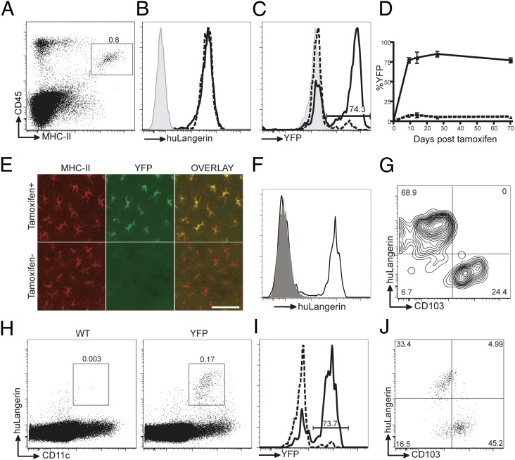
Because the insertion of CreERT2 into the 3′ UTR did not disrupt the human langerin gene, transgene expression could be assessed according to the expression of human Langerin. Flow cytometry of single-cell suspensions of epidermal cells revealed that all LCs (MHC-II+, CD45+) from transgenic mice expressed huLangerin (Fig. 1 A and B). To test CreERT2 efficiency, mice were treated with 1.0 mg tamoxifen i.p. for 5 consecutive days. On day +9, ∼75% of epidermal LCs expressed YFP (Fig. 1_C_, solid line). Cells expressing YFP were absent in littermate control (shaded) and very infrequent in untreated huLangerin-CreERT2 YFP (broken line) mice (Fig. 1_C_). The percentage of YFP-positive LCs in epidermis from tamoxifen-treated (solid line) and untreated (broken line) mice remained constant up to 70 d after injection (Fig. 1_D_). Similarly, immunofluorescent imaging of whole-mounted epidermal sheets showed YFP expression in virtually all LCs in tamoxifen-treated but not sham-injected mice (Fig. 1_E_).
Fig. 1.
Specificity and efficiency of huLangerin-CreERT2. (A) Flow cytometry of epidermal single-cell suspension from huLangerin-CreERT2 YFP mice stained with MHC II and CD45 to identify LCs. (B and C) Expression of (B) huLangerin and (C) YFP in LCs from tamoxifen-treated wild-type (shaded), untreated YFP (dashed line), and tamoxifen-treated YFP mice (solid line). (D) Percent of LCs expressing YFP over time in treated (solid line) and untreated (dashed line) YFP mice. (E) Immunofluorescence of epidermal whole mounts stained for YFP (green) and MHC-II (red) from untreated and tamoxifen-treated YFP mice. (Scale bar, 30 μm.) (F) Expression of huLangerin on singlets gated on MHC-II and muLangerin from the dermis of YFP (solid line) or littermate control (shaded) mice. (G) Expression of huLangerin and CD103 from YFP mice gated as in F. (H) Singlets gated cells from skin-draining LNs from wild-type (Left) and YFP (Right) mice stained for CD11c and huLangerin. (I) Expression of YFP in huLangerin+ cells as gated in H from untreated YFP (dashed line) and tamoxifen-treated YFP mice (solid line). (J) CD103 and huLangerin expression on CD11c+ muLangerin+ cells isolated from skin-draining LNs of YFP mice. All data are representative of at least three independent experiments.
As we have seen with other huLangerin transgenic mice, transgene expression was specific for LCs (13, 16, 17). In the dermis of YFP mice, only migratory LCs as defined by expression of muLangerin in the absence of CD103 expressed huLangerin (Fig. 1 F and G). In skin draining LNs, LCs expressing huLangerin were readily detectable in YFP mice (Fig. 1_H_). As expected, ∼75% of these cells isolated from tamoxifen-treated mice expressed YFP (Fig. 1_I_). HuLangerin expression was found only in DCs that coexpressed murine Langerin and were CD103dim (Fig. 1_J_), confirming selective expression in LCs. Thus, huLangerin-CreERT2 mice have a highly selective expression of CreERT2 within LCs that efficiently excises floxed genes in response to tamoxifen administration.
Interruption of Autocrine/Paracrine TGF-β Induces LC Migration.
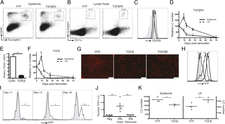
We next bred huLangerin-CreERT2 YFP mice to TGF-β receptor II floxed mice and generated huLangerin-CreERT2 YFP/TGF-βRIIfl (henceforth called TGF-βRII mice) (18). In response to tamoxifen-induced activation of CreERT2, LCs in these mice will begin to express YFP and become insensitive to TGF-β. We confirmed deletion of TGF-βRII by flow cytometry of LCs isolated 9 d after administration of tamoxifen (Fig. 2_C_). We were surprised to find that on day +9 after tamoxifen treatment the percentage of LCs in the epidermis of TGF-βRII mice was significantly lower than in YFP mice (Fig. 2_A_). In addition, the percentage and number of LCs in skin-draining LNs of TGF-βRII mice was almost double that of YFP mice (Fig. 2_B_). A time course revealed that the number of YFP+ LCs in the epidermis declined rapidly over ∼2 wk (Fig. 2_D_). This is accompanied by an increased number of LCs in skin-draining LNs that peaks on day +9 after tamoxifen administration and gradually diminishes over time. Thus, the absence of sensitivity to TGF-β results in spontaneous migration of LCs from the epidermis into the skin-draining LNs.
Fig. 2.
LC-specific inducible excision of TGF-βRII or TGF-β1 results in LCs migration. Flow cytometry of (A) epidermis and (B) LNs from YFP (Left) and TGF-βRII (Right) mice on day +9 after tamoxifen injection. (C) Expression of TGF-βRII on LCs gated as in B, isolated from LNs of tamoxifen-treated (solid line) or untreated (dashed line) TGF-βRII mice. The shaded line represents isotype control. (D) Relative number of LCs in the epidermis (solid line) or skin-draining LNs (dashed line) of TGF-βRII mice at the indicated number of days after tamoxifen treatment, as a percentage compared with control YFP mice. (E) Relative expression of TGF-β1 mRNA in LCs isolated from untreated (Cntl) or tamoxifen-treated TGF-β mice. (F) Relative number of LCs in the epidermis (solid line) or skin-draining LNs (dashed line) from tamoxifen-treated TGF-β1 mice, as in D. (G) Immunofluorescence of epidermal whole mounts stained for MHC-II (red) isolated from YFP, TGF-β, and TGF-βRII mice on day +17 after tamoxifen treatment. (Original magnification: 100×; scale bar, 100 μm.) (H) YFP expression in LCs isolated from the epidermis of YFP mice after a single topical application of tamoxifen. Numbers indicate the number of days after tamoxifen application. (I) Percentage of YFP+ LCs isolated from the cervical LNs of YFP mice at the indicated day after a single application of topical tamoxifen. (J) Ears of YFP mice were painted with vehicle (Neg) or tamoxifen and either left intact (Site intact) or surgically excised after 18 h (Site removed). The number of YFP+ LCs isolated from cervical LNs on day +6 is shown. Each symbol represents an individual animal. (K) Number of epidermal YFP+ LCs per ear (Left) and ipsilateral cervical LNs (Right) from YFP and TGF-β mice on day 9 after topical tamoxifen application. Each symbol represents an individual animal. All data are representative of at least three independent experiments with cohorts of three to six mice except for C, E, and H, which used three individual mice. *P < 0.05; **P < 0.01.
To test whether LC-derived TGF-β1 participates in migration, we bred huLangerin-CreERT2 YFP mice to TGF-β1fl mice, thereby generating huLangerin-CreERT2 YFP/TGF-β1fl mice (henceforth called TGF-β mice) (19). We observed an approximate 10-fold decrease in TGF-β1 mRNA on day +9 after tamoxifen injection, which confirmed efficient excision of TGFβ1 (Fig. 2_E_). As with TGF-βRII mice, we noted a gradual decrease in the total number of YFP+ LCs in the epidermis (Fig. 2_F_). Decreased numbers of LCs were also visualized in epidermal whole mounts isolated on day +17 after tamoxifen treatment (Fig. 2_G_). The remaining LCs have an altered morphology that is similar to that seen with activation. Dendritic epidermal T cells were unaffected in all three strains of mice (Fig. S2_C_). By day +26, less than 2% of the total number of LCs remained in the epidermis. Interestingly, the excision efficiency of TGF-β1fl (∼98%) seems to be much greater than the STOPfl cassette in YFP mice (Fig. 1_D_). The efficiency of floxed target excision is known to vary between genes. Other genes, such as I-Aβfl, have 100% tamoxifen-induced excision in huLangerin-CreERT2 mice (Fig. S2 A and B). The total number of LCs in the skin-draining LNs of TGF-β mice increased almost twofold, with a peak on day +9, and then gradually declined (Fig. 2_F_). These data are consistent with a synchronized egress of LCs from the epidermis in response to ablation of TGF-β1 or TGF-βRII. LCs that migrate from the skin accumulate in the LNs but are relatively short-lived. As the pool of LCs in the epidermis available to migrate declines, so does the number of LCs present in the LNs.
To confirm that the reduced number of LCs in the epidermis was in fact due to LC migration, we first excluded the possibility of LC apoptosis. Expression of activated caspase 3 was unaffected in tamoxifen-treated TGF-β mice (Fig. S3). We next applied tamoxifen epicutaneously to ears rather than by i.p. injection. In YFP mice treated with a single epicutaneous application of tamoxifen, we observed the gradual accumulation of YFP, so that by day +6 virtually all LCs in the epidermis expressed easily detectable levels of YFP (Fig. 2_H_). YFP+ LCs could also be seen accumulating over time in the cervical LNs (Fig. 2_I_). To confirm that YFP+ LCs found in the LNs had migrated from the skin and not become YFP+ because of lymphatic draining of tamoxifen, ears of mice were painted with vehicle (Neg) or tamoxifen and either left intact (Site intact) or surgically excised after 18 h (Site removed) (Fig. 2_J_). Six days later YFP+ LCs were present in the cervical LNs of intact mice but not in surgically treated or control animals, indicating that expression of YFP+ accurately identifies LCs that were present in the epidermis when tamoxifen was applied. When we compared numbers of YFP+ LCs between YFP and TGF-β mice, we observed that the number of LCs in the epidermis of TGF-β mice 9 d after epicutaneous application of tamoxifen was reduced by ∼25%. LC numbers in LNs were increased by ∼50% (Fig. 2_K_). These data are consistent with mice treated with tamoxifen i.p. and indicate that the observed phenotype results from increased LC migration. Thus, we conclude that autocrine/paracrine TGF-β1 is required for the maintenance of LCs in the epidermis.
LC Migration Induced by TGF-β1 Deficiency Is MyD88 Independent.
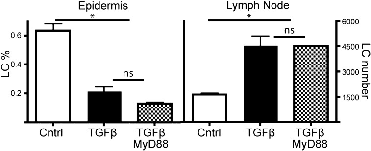
One of the properties of TGF-β1 is its ability to inhibit cell-intrinsic recognition of many toll like receptor (TLR) agonists through ubiquitination and degradation of the adapter molecule MyD88 (20). Because LCs are chronically exposed to skin commensal microbiota, we hypothesized that the absence of TGF-β signaling could release suppression of MyD88 and render LCs more sensitive to TLR agonists, thus promoting increased LC migration (21, 22). To test this, we bred huLangerin-CreERT2 TGF-β1fl mice to MyD88fl, generating huLangerin-CreERT2 TGF-β1fl/MyD88fl (henceforth called TGF-β/MyD88 mice) (23). We compared the number of LCs in the epidermis and LNs in YFP, TGF-β, and TGF-β/MyD88 mice on day +9 after i.p. tamoxifen injection. TGF-β/MyD88 mice showed reduced numbers of LCs in the epidermis and increased numbers in the LNs that were identical to those in TGF-β mice (Fig. 3). Thus, LC migration in response to the interruption of the autocrine/paracrine TGF-β1 circuit occurs independently of MyD88.
Fig. 3.
LC migration in response to loss of TGF-β1 is MyD88 independent. Percentage of LCs found in the epidermis (Left) and number of LCs found in skin-draining LNs (Right) of YFP (Cntl), TGF-β, and TGF-β/MyD88 mice on day +9 after i.p. administration of tamoxifen. Data are representative of three independent experiments with cohorts of three to five mice. ns, not significant. *P < 0.05.
TGF-β–Deficient LCs Acquire a Homeostatic Migratory Phenotype Before Migration.
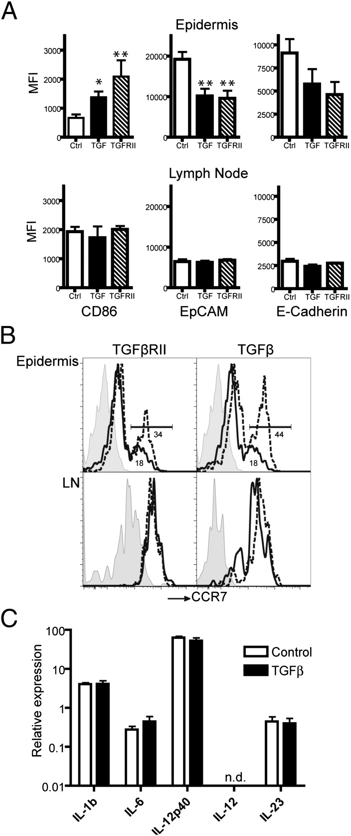
Migration of LCs is associated with increased expression of the costimulatory molecule CD86 (B7-2) and the chemokine receptor CCR7, along with decreased expression of the adhesion molecules EpCAM and E-cadherin. We evaluated the expression of these markers of activation in LCs isolated from the epidermis of YFP, TGF-β, and TGF-βRII mice by flow cytometry. As expected, we found increased expression of CD86 and CCR7 and decreased expression of EpCAM and E-cadherin in TGF-β and TGF-βRII mice, which suggested that LCs from these mice assumed a mature phenotype (Fig. 4 A and B, Top, and Fig. S4_A_). A similar phenotype was reported in LCs isolated from the epidermis of neonatal CD11c-Cre TGF-βRIfl mice (14). We were thus surprised to find that in the LNs, expression of these markers of activation was no longer different between the groups (Fig. 4 A and B, Lower, and Fig. S4_B_). CD86 expression by LCs in the LNs of TGF-β and TGF-βRII mice was similar to cells in the epidermis. Rather, the expression of CD86 on LCs from YFP mice had increased after migration. Similarly, expression of EpCAM and E-cadherin decreased after migration in YFP mice but was relatively unaltered in TGF-β and TGF-βRII mice. Thus, LCs in the epidermis of TGF-β and TGF-βRII mice prematurely adopt the phenotype of homeostatically migrated LCs while still in the epidermis. Once they have migrated to the LNs, they become phenotypically similar to wild-type, steady-state LCs.
Fig. 4.
TGF-β1–deficient LCs maintain steady-state phenotype. (A) Mean fluorescence intensity of CD86, EpCAM, and E-cadherin staining for LCs isolated from the epidermis and gated as CD45+ MHC-II+ (Upper) or skin-draining lymph node gates as CD11c+, huLangerin+ (Lower) of TGF-β, TGF-βRII and control mice on day +9 after tamoxifen treatment. Histograms from representative mice are shown in Fig. S4. Data are representative of at least three independent experiments with groups of four to five mice each. (B) Expression of CCR7 on LCs isolated from epidermis (Upper) or LNs (Lower) on day +9 after tamoxifen treatment. Cells from YFP mice are represented by solid lines, whereas cells from TGF-βRII (Left) or TGF-β (Right) are shown as broken lines. Percentage of CCR7 cells is indicated. Shaded lines are isotype staining. (C) Relative expression of IL-1β, IL-6, IL-12β (IL-12p40), IL-12α (IL-12p35), and IL-23 is shown as assessed by quantitative PCR of mRNA isolated from LCs in the skin-draining LNs of control and TGF-β mice. Data are from two independent experiments. n.d., not detected. *P < 0.05, **P < 0.01.
The similarity between TGF-β–deficient LC migration and steady-state LC migration was further supported by examination of cytokine expression. LCs were sorted from skin-draining LNs of YFP and TGF-β mice on day +9 after tamoxifen treatment. Expression of mRNA for the inflammatory cytokines IL-1β, IL-6, IL-12α (IL-12p35), IL-12β (IL-12p40), and IL-23 was equivalent in LCs from both mouse strains (Fig. 4_C_). The levels of expression were similar to what we have previously seen in steady-state LCs but well below the levels that occur during acute infection with Candida albicans (24).
Disruption of TGF-β Signaling Occurs During Inflammation-Induced LC Migration.
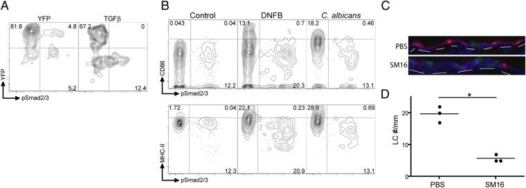
To generalize our finding that interruption of TGF-β1 signaling results in LC migration, we next sought to determine whether TGF-β signaling is disrupted during inflammation-induced migration. Phosphorylation of the proteins SMAD2 and SMAD3 is a major pathway for TGF-β intracellular signaling (25). In the steady state, we found that ∼10–20% of LCs in YFP mice contain detectable levels of pSMAD2/3 at any given time (Fig. 5_A_). The relatively low percentage of LCs containing pSMAD2/3 despite the functional requirement of TGF-β signaling for epidermal residence likely represents transient feedback inhibition of TGF-β signaling by SMAD7 (25). As a control, we also examined LCs from TGF-β mice and found that only LCs that had not excised YFP contained pSMAD2/3.
Fig. 5.
TGF-β signaling is interrupted during inflammation. (A) Expression of pSMAD2/3 and YFP expression in LCs (CD45+, MHC-II+), in cells isolated from unmanipulated YFP and TGF-β mice on day +9 after tamoxifen treatment. (B) Expression of CD86 (Upper) and MHC II (Lower) vs. pSMAD2/3 is shown in LCs isolated from the epidermis of wild-type mice 18 h after application of 0.5% DNFB or infection with C. albicans (24, 26). Data represent two independent experiments with groups of three mice each. (C) Wild-type mice were treated with the TGF-βRI kinase inhibitor SM16 for 47 d. Immunofluorescence of transverse sections of ear skin from PBS- and SM16-treated mice is shown. (D) Number of LCs per millimeter of epidermis. Each symbol represents an individual animal. *P < 0.05.
We next examined expression of pSMAD2/3 in LCs from wild-type mice. LCs isolated from the epidermis of control mice were compared with LCs isolated from mice painted with 2,4-dinitrofluorobenzene (DNFB) or infected with C. albicans 24 h earlier. Both epicutaneous application of DNFB and infection with C. albicans are strong inducers of LC migration (26). We found that pSMAD2/3 expression was limited to those LCs that had not become activated, as determined by lower expression of CD86 or MHC-II (Fig. 5_B_). Thus, activated LCs in the epidermis lack evidence of TGF-β signaling. This suggests that interruption of autocrine/paracrine TGF-β1 participates in inflammation-induced LC migration.
Recently, a specific in vivo inhibitor of TGF-β receptor I kinase activity (SM16) has been described (27). To determine whether pharmacological inhibition of TGF-β signaling with SM16 would affect LCs, we supplemented the diet of wild-type mice with 0.45 g/kg SM16 for 47 d. Mice treated with SM16 had dramatically fewer epidermal LCs compared with control mice (Fig. 5 C and D). Thus, pharmacologic blockade of TGF-β signaling recapitulated our data using genetic ablation and confirmed the importance of this pathway to LC biology.
Discussion
The development of transgenic mice with LC-specific expression of an inducible form of Cre (huLangerin-CreERT2) has allowed us to study in fully differentiated LCs the importance of sensitivity and elaboration of TGF-β1. We found that interrupting autocrine/paracrine TGF-β1 in either TGF-β1 or TGF-βRII mice induced a synchronized wave of LC migration from the epidermis into the skin-draining lymph node that was not dependent on LC-intrinsic MyD88. LCs maintained the phenotype of homeostatically migrated LCs and did not seem to have become activated. We also demonstrated that pharmacologic blockade of TGF-β signaling reduced the number of epidermal LCs. Thus, we conclude that autocrine/paracrine TGF-β1 is required to maintain LCs within the epidermis, and interruption of this pathway leads to homeostatic LC migration. Moreover, activated LCs no longer showed evidence of TGF-β signaling before migration. This suggests that interruption of autocrine/paracrine TGF-β also participates in inflammation-induced migration.
These data are consistent with a model in which abrogation of signaling from LC-derived TGF-β1 is an important component in LC migration. DCs treated with TGF-β1 in vitro become less responsive to maturation stimuli such as IL-1β and TNF-α (22). Thus, in the absence of TGF-β1, LCs may develop increased sensitivity to activation by commensal skin organisms or other sources of inflammation. We have shown that MyD88-dependent signals are not required for LC migration but cannot exclude the possibility that MyD88-independent signals are sufficient. This mechanism, however, would predict that LCs should be fully activated and not maintain the observed phenotype of homeostatic migratory LCs. Disruption of E-cadherin binding and increased noncanonical β-catenin signaling promotes partial activation of DCs that is characterized by increased surface expression of costimulatory markers without associated production of proinflammatory cytokines (28). This phenotype is similar to LCs from tamoxifen-treated TGF-β and TGF-βRII mice. Indeed, TGF-β has been shown to inhibit the noncanonical β-catenin pathway (29). One relatively unique aspect of this system is that TGF-β acts in an autocrine/paracrine manner. Autocrine cytokines are often associated with feedback loops during cellular proliferation and development. In the case of TGF-β, however, the cytokine is secreted as a complex with the latency-associated protein, which must be removed to generate active TGF-β. In the epidermis, TGF-β is activated by the integrins Avβ6 and Avβ8 (30). This raises the intriguing possibility that regulated activation of TGF-β1 may be a mechanism controlling LC migration.
Currently there are several clinical trials investigating the efficacy of TGF-β blockade in the treatment of diseases including focal glomerulosclerosis, myelofibrosis, idiopathic pulmonary fibrosis, pleural mesothelioma, systemic sclerosis, and malignant melanoma (Clinicaltrials.gov). Our data suggest that LCs may be depleted in these patients. The in vivo function of LCs is somewhat enigmatic. In vivo, LCs suppress adaptive responses to hapten and Leishmania skin infection (16, 31). They are required for the generation Th17 responses to C. albicans and may participate in local activation of cytotoxic T lymphocytes (CTL) in graft-vs.-host disease (24, 32). Thus, patients undergoing TGF-β blockade may be more susceptible to fungal skin infections and allergic contact dermatitis. In addition, these agents could be therapeutically beneficial for patients with a wide variety of skin diseases like psoriasis and graft-vs.-host disease that involve skin-homing Th17 and/or CTL cells. Exploring the use of TGF-β blocking agents to modulate LC biology and determining the mechanism(s) underlying LC migration will be exciting areas for future discovery.
Materials and Methods
Generation of huLangerin CreER T2 Mice.
The plasmid containing CreERT2 construct (a generous gift of P. Chambon, IGBMC, Strasbourg, France) (15) was cloned into pLD53 AEB shuttle plasmid and successfully recombined into the 3′ UTR of Langerin in BAC RP11-504o1 as previously described (16). Primers used for generation of the recombination cassette: 5′ A box, 5-ttaaggcgcgccggattccaggtgagcccaac-3; 3′ A box, 5-agcagcttcatggttgtggccatattatcatcgtg-3; 5′ B box, 5-aattcccactgaatgactttgcacgttaatttttcttgc-3; 3′ B box, 5-ccacacagcttgaatgactttgcacgttaatttttcttgc-3; 5′ I box, 5-tggccacaaccatgaagctgctg ccgtcggtggt -3; 3′ I box, 5-cgtgcaaagtcattcaagctgtggcagggaaaccc-3. DNA microinjections were performed into pronuclei of FVB mice by the University of Minnesota Mouse Genetics Laboratory.
Mice.
HuLangerin-CreERT2 mice generated in FVB background were bred onto C57BL/6 wild-type and Rosa26-Stopfl-YFP mice obtained from Jackson Laboratories. TGF-βRII flox mice (a generous gift of H. Moses, Vanderbilt University, Nashville, TN) and I-Aβ flox mice (a generous gift of P. Koni, Georgia Health Sciences University, Augusta, GA) were previously described (18). All experiments were performed on age- (7-12 wk) and sex-matched mice. Mice were housed in microisolator cages and fed irradiated food. The institutional animal care and use committee approved all mouse protocols.
Antibodies.
The following antibodies were used: biotinylated anti-muLangerin and anti-huLangerin-Al647 (Dendritics); Cd11c, CD11c, CD11b, CD45, MHC II, EpCAM, CD80 (BioLegend); CD103, CD86, E-cadherin, CCR7 (eBioscience), anti-GFP (Rockland Immunochemicals), anti-phosphoSMAD2/3 (Cell Signaling), anti-activated caspase 3 (Cell Signaling), and TGF-βRII (R&D Systems).
Flow Cytometry.
Single-cell suspensions and flow cytometry of LCs were performed as previously described (16). Staining for TGF-βRII, activated caspase 3, and phosphoSMAD2/3 was performed according to manufacturer protocols. All flow cytometry was performed using an LSRII cytometer (BD Biosciences).
Immunofluorescence.
Immunofluorescence of epidermal whole mounts and transverse skin sections was performed as previously described (16).
DC Sorting by Flow Cytometry and Quantitative PCR.
Single-cell suspension of LN cells was enriched by CD11c MACS positive selection (Miltenyi Biotec). LCs were sorted using a FACSAria cell sorter based as YFP+, MHCII+, CD11c+, CD11b+, CD103−, CD8−. RNA was isolated using an RNeasy Mini Kit (Qiagen) and quantified from NanoDrop readings. cDNA was generated using a High Capacity cDNA Reverse Transcription Kit (Applied Biosystems). TaqMan Gene Expression Master Mix, TaqMan Gene Expression Assays for IL-1β, IL-6, IL-12p35, Il-12p40, IL-23p19, and TGF-β1 and ABI Prism 7900HT (Applied Biosystems) were used for quantitative PCR. All kits were performed according the manufacturer’s instructions. Ct values were normalized to hypoxanthine phosphoribosyltransferase (HPRT) expression and are shown as 2^-ΔCt.
Tamoxifen Treatment.
Tamoxifen (Sigma Aldrich) was dissolved in corn oil (Sigma Aldrich) and 10% (vol/vol) ethanol and was administered 5 consecutive days by i.p. injection at 0.05 mg/g of mouse weight. 4OH tamoxifen (Sigma Aldrich) was dissolved in acetone at 1.0 mg/mL and used as a single topical application of 10 μL to the dorsal and ventral ear surfaces.
SM16 Treatment.
Ears harvested from mice that were fed with SM16 (Biogen Idec) chow (0.45 g SM16/kg of food) for 47 d were kindly provided by Stephen Albelda (University of Pennsylvania, Philadelphia, PA).
Statistics.
The statistical significance between the groups was determined using Student’s unpaired two-tailed t test.
Supplementary Material
Supporting Information
Acknowledgments
Supported by National Institutes of Health Grants R01-AR056632 and R01-AR060744. B.Z.I. is supported by a career development award from the Dermatology Foundation.
Footnotes
The authors declare no conflict of interest.
*This Direct Submission article had a prearranged editor.
References
- 1.Romani N, Clausen BE, Stoitzner P. Langerhans cells and more: Langerin-expressing dendritic cell subsets in the skin. Immunol Rev. 2010;234:120–141. doi: 10.1111/j.0105-2896.2009.00886.x. [DOI] [PMC free article] [PubMed] [Google Scholar]
- 2.Hemmi H, et al. Skin antigens in the steady state are trafficked to regional lymph nodes by transforming growth factor-beta1-dependent cells. Int Immunol. 2001;13:695–704. doi: 10.1093/intimm/13.5.695. [DOI] [PubMed] [Google Scholar]
- 3.Merad M, Ginhoux F, Collin M. Origin, homeostasis and function of Langerhans cells and other langerin-expressing dendritic cells. Nat Rev Immunol. 2008;8:935–947. doi: 10.1038/nri2455. [DOI] [PubMed] [Google Scholar]
- 4.Chorro L, et al. Langerhans cell (LC) proliferation mediates neonatal development, homeostasis, and inflammation-associated expansion of the epidermal LC network. J Exp Med. 2009;206:3089–3100. doi: 10.1084/jem.20091586. [DOI] [PMC free article] [PubMed] [Google Scholar]
- 5.Merad M, et al. Langerhans cells renew in the skin throughout life under steady-state conditions. Nat Immunol. 2002;3:1135–1141. doi: 10.1038/ni852. [DOI] [PMC free article] [PubMed] [Google Scholar]
- 6.Ginhoux F, et al. Langerhans cells arise from monocytes in vivo. Nat Immunol. 2006;7:265–273. doi: 10.1038/ni1307. [DOI] [PMC free article] [PubMed] [Google Scholar]
- 7.Strobl H, et al. TGF-beta 1 promotes in vitro development of dendritic cells from CD34+ hemopoietic progenitors. J Immunol. 1996;157:1499–1507. [PubMed] [Google Scholar]
- 8.Geissmann F, et al. Transforming growth factor beta1, in the presence of granulocyte/macrophage colony-stimulating factor and interleukin 4, induces differentiation of human peripheral blood monocytes into dendritic Langerhans cells. J Exp Med. 1998;187:961–966. doi: 10.1084/jem.187.6.961. [DOI] [PMC free article] [PubMed] [Google Scholar]
- 9.Borkowski TA, Letterio JJ, Farr AG, Udey MC. A role for endogenous transforming growth factor beta 1 in Langerhans cell biology: The skin of transforming growth factor beta 1 null mice is devoid of epidermal Langerhans cells. J Exp Med. 1996;184:2417–2422. doi: 10.1084/jem.184.6.2417. [DOI] [PMC free article] [PubMed] [Google Scholar]
- 10.Hacker C, et al. Transcriptional profiling identifies Id2 function in dendritic cell development. Nat Immunol. 2003;4:380–386. doi: 10.1038/ni903. [DOI] [PubMed] [Google Scholar]
- 11.Fainaru O, et al. Runx3 regulates mouse TGF-beta-mediated dendritic cell function and its absence results in airway inflammation. EMBO J. 2004;23:969–979. doi: 10.1038/sj.emboj.7600085. [DOI] [PMC free article] [PubMed] [Google Scholar]
- 12.Li MO, Flavell RA. TGF-beta: A master of all T cell trades. Cell. 2008;134:392–404. doi: 10.1016/j.cell.2008.07.025. [DOI] [PMC free article] [PubMed] [Google Scholar]
- 13.Kaplan DH, et al. Autocrine/paracrine TGFbeta1 is required for the development of epidermal Langerhans cells. J Exp Med. 2007;204:2545–2552. doi: 10.1084/jem.20071401. [DOI] [PMC free article] [PubMed] [Google Scholar]
- 14.Kel JM, Girard-Madoux MJ, Reizis B, Clausen BE. TGF-beta is required to maintain the pool of immature Langerhans cells in the epidermis. J Immunol. 2010;185:3248–3255. doi: 10.4049/jimmunol.1000981. [DOI] [PubMed] [Google Scholar]
- 15.Indra AK, et al. Temporally-controlled site-specific mutagenesis in the basal layer of the epidermis: Comparison of the recombinase activity of the tamoxifen-inducible Cre-ER(T) and Cre-ER(T2) recombinases. Nucleic Acids Res. 1999;27:4324–4327. doi: 10.1093/nar/27.22.4324. [DOI] [PMC free article] [PubMed] [Google Scholar]
- 16.Kaplan DH, Jenison MC, Saeland S, Shlomchik WD, Shlomchik MJ. Epidermal langerhans cell-deficient mice develop enhanced contact hypersensitivity. Immunity. 2005;23:611–620. doi: 10.1016/j.immuni.2005.10.008. [DOI] [PubMed] [Google Scholar]
- 17.Bobr A, et al. Acute ablation of Langerhans cells enhances skin immune responses. J Immunol. 2010;185:4724–4728. doi: 10.4049/jimmunol.1001802. [DOI] [PMC free article] [PubMed] [Google Scholar]
- 18.Chytil A, Magnuson MA, Wright CV, Moses HL. Conditional inactivation of the TGF-beta type II receptor using Cre:Lox. Genesis. 2002;32:73–75. doi: 10.1002/gene.10046. [DOI] [PubMed] [Google Scholar]
- 19.Li MO, Wan YY, Flavell RA. T cell-produced transforming growth factor-beta1 controls T cell tolerance and regulates Th1- and Th17-cell differentiation. Immunity. 2007;26:579–591. doi: 10.1016/j.immuni.2007.03.014. [DOI] [PubMed] [Google Scholar]
- 20.Naiki Y, et al. Transforming growth factor-beta differentially inhibits MyD88-dependent, but not TRAM- and TRIF-dependent, lipopolysaccharide-induced TLR4 signaling. J Biol Chem. 2005;280:5491–5495. doi: 10.1074/jbc.C400503200. [DOI] [PubMed] [Google Scholar]
- 21.Kubo A, Nagao K, Yokouchi M, Sasaki H, Amagai M. External antigen uptake by Langerhans cells with reorganization of epidermal tight junction barriers. J Exp Med. 2009;206:2937–2946. doi: 10.1084/jem.20091527. [DOI] [PMC free article] [PubMed] [Google Scholar]
- 22.Ohtani T, et al. TGF-beta1 dampens the susceptibility of dendritic cells to environmental stimulation, leading to the requirement for danger signals for activation. Immunology. 2009;126:485–499. doi: 10.1111/j.1365-2567.2008.02919.x. [DOI] [PMC free article] [PubMed] [Google Scholar]
- 23.Kleinridders A, et al. MyD88 signaling in the CNS is required for development of fatty acid-induced leptin resistance and diet-induced obesity. Cell Metab. 2009;10:249–259. doi: 10.1016/j.cmet.2009.08.013. [DOI] [PMC free article] [PubMed] [Google Scholar]
- 24.Igyártó BZ, et al. Skin-resident murine dendritic cell subsets promote distinct and opposing antigen-specific T helper cell responses. Immunity. 2011;35:260–272. doi: 10.1016/j.immuni.2011.06.005. [DOI] [PMC free article] [PubMed] [Google Scholar]
- 25.Moustakas A, Souchelnytskyi S, Heldin CH. Smad regulation in TGF-beta signal transduction. J Cell Sci. 2001;114:4359–4369. doi: 10.1242/jcs.114.24.4359. [DOI] [PubMed] [Google Scholar]
- 26.Haley K, et al. Langerhans cells require MyD88-dependent signals for Candida albicans response but not for contact hypersensitivity or migration. J Immunol. 2012;188:4334–4339. doi: 10.4049/jimmunol.1102759. [DOI] [PMC free article] [PubMed] [Google Scholar]
- 27.Suzuki E, et al. A novel small-molecule inhibitor of transforming growth factor beta type I receptor kinase (SM16) inhibits murine mesothelioma tumor growth in vivo and prevents tumor recurrence after surgical resection. Cancer Res. 2007;67:2351–2359. doi: 10.1158/0008-5472.CAN-06-2389. [DOI] [PubMed] [Google Scholar]
- 28.Jiang A, et al. Disruption of E-cadherin-mediated adhesion induces a functionally distinct pathway of dendritic cell maturation. Immunity. 2007;27:610–624. doi: 10.1016/j.immuni.2007.08.015. [DOI] [PMC free article] [PubMed] [Google Scholar]
- 29.Vander Lugt B, et al. TGF-β suppresses β-catenin-dependent tolerogenic activation program in dendritic cells. PLoS ONE. 2011;6:e20099. doi: 10.1371/journal.pone.0020099. [DOI] [PMC free article] [PubMed] [Google Scholar]
- 30.Yang Z, et al. Absence of integrin-mediated TGFbeta1 activation in vivo recapitulates the phenotype of TGFbeta1-null mice. J Cell Biol. 2007;176:787–793. doi: 10.1083/jcb.200611044. [DOI] [PMC free article] [PubMed] [Google Scholar]
- 31.Kautz-Neu K, et al. Langerhans cells are negative regulators of the anti-Leishmania response. J Exp Med. 2011;208:885–891. doi: 10.1084/jem.20102318. [DOI] [PMC free article] [PubMed] [Google Scholar]
- 32.Bennett CL, et al. Langerhans cells regulate cutaneous injury by licensing CD8 effector cells recruited to the skin. Blood. 2011;117:7063–7069. doi: 10.1182/blood-2011-01-329185. [DOI] [PMC free article] [PubMed] [Google Scholar]
Associated Data
This section collects any data citations, data availability statements, or supplementary materials included in this article.
Supplementary Materials
Supporting Information
