Regulation of fibroblast growth factor receptor signalling and trafficking by Src and Eps8 (original) (raw)
Summary
Fibroblast growth factor receptors (FGFRs) mediate a wide spectrum of cellular responses that are crucial for development and wound healing. However, aberrant FGFR activity leads to cancer. Activated growth factor receptors undergo stimulated endocytosis, but can continue to signal along the endocytic pathway. Endocytic trafficking controls the duration and intensity of signalling, and growth factor receptor signalling can lead to modifications of trafficking pathways. We have developed live-cell imaging methods for studying FGFR dynamics to investigate mechanisms that coordinate the interplay between receptor trafficking and signal transduction. Activated FGFR enters the cell following recruitment to pre-formed clathrin-coated pits (CCPs). However, FGFR activation stimulates clathrin-mediated endocytosis; FGF treatment increases the number of CCPs, including those undergoing endocytosis, and this effect is mediated by Src and its phosphorylation target Eps8. Eps8 interacts with the clathrin-mediated endocytosis machinery and depletion of Eps8 inhibits FGFR trafficking and immediate Erk signalling. Once internalized, FGFR passes through peripheral early endosomes en route to recycling and degredative compartments, through an Src- and Eps8-dependent mechanism. Thus Eps8 functions as a key coordinator in the interplay between FGFR signalling and trafficking. This work provides the first detailed mechanistic analysis of growth factor receptor clustering at the cell surface through signal transduction and endocytic trafficking. As we have characterised the Src target Eps8 as a key regulator of FGFR signalling and trafficking, and identified the early endocytic system as the site of Eps8-mediated effects, this work provides novel mechanistic insight into the reciprocal regulation of growth factor receptor signalling and trafficking.
Key words: Eps8, FGFR, Clathrin, Endocytosis, Src
Introduction
Ligand-mediated activation of receptor tyrosine kinases (RTKs) initiates a signal transduction cascade that leads to a broad spectrum of cellular responses including differentiation, proliferation, survival and migration. These responses are initiated by recruitment of adaptors, scaffold and signalling proteins to the phosphorylated cytoplasmic tail of the receptor (Greenfield et al., 1989; Ullrich and Schlessinger, 1990; Heldin, 1995). Receptor activation also acts as the initiating stimulus for endocytosis of RTKs following ligand binding and this can occur via a variety of routes including both clathrin-mediated endocytosis (CME) and clathrin-independent endocytosis (CIE) (von Zastrow and Sorkin, 2007; Le Roy and Wrana, 2005). Endocytosis and subsequent trafficking to intracellular compartments have been considered to be principally mechanisms of signal attenuation: the former by controlling the number of receptors available for activation on the plasma membrane, the latter by sorting the internalized receptors into the intralumenal vesicles of multivescicular bodies (MVBs) where receptor proteolysis takes place and signalling is terminated (Stoscheck and Carpenter, 1984; Beguinot et al., 1984). There is mounting evidence, however, that signalling from internalized receptors persists along the endocytic pathway, and that endocytosis and intracellular trafficking mechanisms have a powerful influence on the spatial and temporal dynamics of the signal (von Zastrow and Sorkin, 2007; Di Fiore and De Camilli, 2001; Kholodenko, 2002; Polo and Di Fiore, 2006; Sorkin and Von Zastrow, 2002; Disanza et al., 2009; Sandilands and Frame, 2008) which influences biological outcomes (Zhang et al., 2000). Alternatively, recent evidence suggests that for at least some RTKs, signalling primarily occurs immediately following ligand binding prior to endocytosis (Sousa et al., 2012; Brankatschk et al., 2012). It is also possible that regulated endocytosis and intracellular trafficking of activated RTKs shape the identity of the signal as confinement within intracellular compartments provides a mechanism for regulating the composition of receptor-associated protein complexes. These considerations place emphasis on understanding the molecular mechanisms that coordinate activated RTK signalling and trafficking as key determinants of the biological outcomes of receptor activation.
Fibroblast growth factor receptors (FGFRs) represent a subfamily of 4 highly conserved RTKs (FGFR1, FGFR2, FGFR3 and FGFR4) that regulates fundamental cellular processes, including proliferation, differentiation and angiogenesis (Burgess and Maciag, 1989; Basilico and Moscatelli, 1992). FGFR activation initiates signalling through a number of pathways including the well characterised Ras/Raf/ERK and PI3 kinase/PDK/Akt pathways (Mason, 1994). Of particular interest is the occurrence of somatic or germ-line mutations in FGFRs which modify the dynamics of signal propagation via mechanisms such as hyper-sensitivity of kinase activation, ligand-independent dimerization or receptor amplification (Grose and Dickson, 2005; Wesche et al., 2011; Turner and Grose, 2010; Beenken and Mohammadi, 2009). These findings indicate that understanding the mechanisms of FGFR endocytosis and trafficking, and their impact on signalling dynamics, is an important goal in understanding both pathological and physiological FGFR biology. We have previously demonstrated a critical role for non receptor tyrosine kinases of the Src family (SFKs) in shaping FGFR trafficking and signalling dynamics (Sandilands et al., 2007). Src is recruited to activated fibroblast growth factor receptor complexes through the adaptor protein factor receptor substrate 2 (FRS2) at the plasma membrane. This localisation requires both active Src and FGFR kinases, which are interdependent. Internalization of activated FGFRs involves release from complexes containing activated Src and trafficking into multiple intracellular compartments including RhoB and Rab5 endosomes via a mechanism(s) which require an intact actin cytoskeleton (Sandilands et al., 2007). Chemical and genetic inhibition studies showed strikingly different requirements for Src family kinases in FGFR-mediated signalling; activation of the phosphoinositide-3 kinase–Akt pathway is severely attenuated, whereas activation of the extracellular-signal-regulated kinase pathway is delayed in its initial phase and fails to attenuate. These findings show that Src kinase activity and Src substrates that mediate exit of activated FGFRs from the plasma membrane are critical determinants of the early phases of FGFR signal propagation.
In detailing Src substrates that are phosphorylated in the early phases of FGFR activation, in order to identify candidates for mediating Src-regulated trafficking, we identified the multifunctional scaffolding protein Eps8 as a prominent target of Src kinase activity (Cunningham et al., 2010). Eps8 is an attractive subject for further scrutiny as it has well documented roles in regulating the actin cytoskeleton (Provenzano et al., 1998) and has been implicated in trafficking of the epidermal growth factor receptor (EGFR) (Di Fiore and Scita, 2002). Furthermore, EGFR signalling through Src has previously been shown as a mechanism for regulating the cellular distribution of clathrin (Wilde et al., 1999). It is accordingly of interest to understand the functions of Eps8 in regulating FGFR trafficking and signalling dynamics.
In this study, we have performed a detailed series of analyses to investigate the mechanisms responsible for the coordinated trafficking and signalling of activated FGFR. We have specifically characterised the mechanisms controlling FGFR endocytosis and endocytic trafficking in the early phases of FGFR activation and in particular we have focused on the role of Src acting through Eps8. Eps8 is thus identified as a key mediator of the early phases of activated FGFR trafficking and signalling relevant to regulation of clathrin-mediated endocytosis, trafficking out of the early endosome into the perinuclear recycling compartment, and FGF-mediated pErk signalling.
Results
FGFR is internalized though an FGFR-kinase- and dynamin-dependent pathway
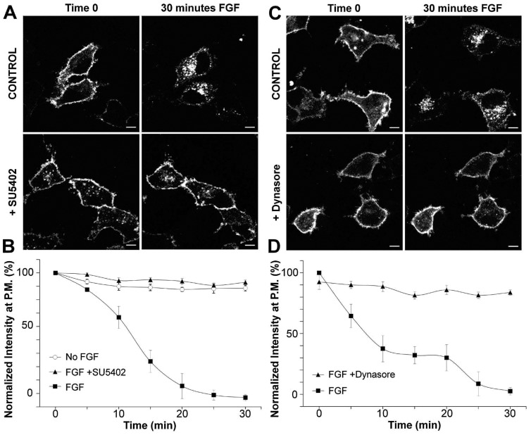
Different pathways have been implicated in the endocytosis of activated RTKs (von Zastrow and Sorkin, 2007; Le Roy and Wrana, 2005). Thus, we first set out to document the mechanism by which FGFR is trafficked following activation. We employed live-cell confocal microscopy to analyse the trafficking of activated FGFR2 in HeLa cells, which express very low levels of endogenous FGFR2 (Ahmed et al., 2008), using a GFP-tagged form of FGFR2 which was previously validated in published studies from John Ladbury's laboratory (Schüller et al., 2008). As depicted in Fig. 1A, live-cell confocal imaging demonstrates that following FGF stimulation, FGFR2–GFP undergoes rapid internalization from the plasma membrane and eventually traffics to a peri-nuclear endocytic compartment. This redistribution away from the cell periphery did not occur in the absence of ligand addition, and SU5402, a potent inhibitor of FGFR kinase activity, completely prevented FGF stimulated internalization (Fig. 1A,B) (Sun et al., 1999). Therefore, the cellular localisation and behaviour of GFP-tagged FGFR2 reflects that of endogenous protein. Thus, we have applied this quantitative and robust assay for trafficking of activated RTKs in live cells to the analysis of the routes of FGFR endocytosis and endocytic trafficking.
Fig. 1.
Active FGFR is internalized through dynamin-dependent endocytosis. HeLa cells transiently transfected with FGFR2-GFP were incubated for 5 minutes in the presence or absence of SU5402 (A) or for 30 minutes in the presence or absence of Dynasore inhibitor (C) and imaged using confocal live-cell microscopy at 37°C for 30 minutes following stimulation with FGF2 + heparin. The first and the last frames are shown, corresponding to the situations before and after FGF2 treatment, respectively. Scale bars: 5 µm. (B,D) The experiments shown in A and C were performed in many cells and the relative FGFR2–GFP intensity in the PM region of the cells was calculated (as described in the Materials and Methods) for each frame of the time-lapse sequence and plotted as a function of time (means ± s.e.m., n = 14 cells).
Several endocytosis pathways have been implicated in the internalization of activated FGFRs (Marchese et al., 1998; Citores et al., 1999; Belleudi et al., 2007; Gleizes et al., 1996; Haugsten et al., 2011). The large GTPase dynamin has been clearly demonstrated to be a crucial regulator of numerous endocytosis pathways (Doherty and McMahon, 2009). Therefore, we assessed whether inhibition of dynamin function with the potent dynamin inhibitor Dynasore affected the ligand induced endocytosis of FGFR (Macia et al., 2006). Importantly, control endocytosis studies verified the efficacy of Dynasore treatment in our systems (supplementary material Fig. S1A). As depicted in Fig. 1C,D, treatment with Dynasore completely prevented internalization of FGFR following FGF stimulation. Therefore, activated FGFRs undergo dynamin-dependent endocytosis.
FGFR is internalized through clathrin-mediated endocytosis
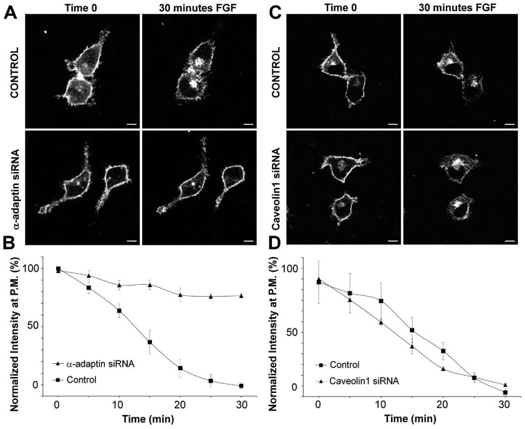
Two of the main endocytosis pathways that are dynamin dependent include clathrin-mediated endocytosis and entry via caveolae (Doherty and McMahon, 2009). We employed an RNAi-based approach utilising previously published siRNA which we have previously employed to silence expression of α-adaptin, a critical component of the clathrin-mediated endocytosis machinery, in HeLa cells (Rappoport and Simon, 2008; Rappoport and Simon, 2009), or caveolin1 (Li et al., 2012), which is required for caveolae formation (Rothberg et al., 1992). Control experiments revealed that α-adaptin siRNA treatment significantly inhibited transferrin endocytosis (supplementary material Fig. S1B) and potently reduced expression of α-adaptin (supplementary material Fig. S1C). Similarly, siRNA targeting caveolin1 significantly inhibited endocytosis of cholera toxin B sub-unit (supplementary material Fig. S2A) and decreased caveolin1 protein levels (supplementary material Fig. S2B). Importantly, these siRNA reagents did not reduce expression of control protein. Therefore, the utility of these inhibitory strategies has been sufficiently validated in our systems for usage in live-cell imaging studies.
As demonstrated in Fig. 2A,B, treatment with α-adaptin siRNA clearly prevented endocytosis of FGFR following FGF treatment. However, silencing of caveolin1 expression had no effect on the internalization of FGFR following stimulation (Fig. 2C,D). Thus, these results show that active FGFR enters cells via clathrin-mediated endocytosis and not through caveolae.
Fig. 2.
Active FGFR enters cells by clathrin-mediated endocytosis and not through caveolae. HeLa cells were co-transfected with FGFR2–GFP and α-adaptin siRNA (A) or caveolin1 siRNA (C) and confocal live-cell microscopy was performed at 37°C for 30 minutes following stimulation with FGF2 + heparin. Scale bars: 5 µm. (B,D) The experiments shown in A and C were performed in many cells and a quantification of FGFR2–GFP intensity in the PM region of the cells as function of time was performed (means ± s.e.m., n = 10 cells).
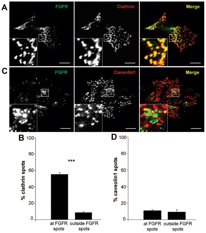
In order to verify the potential for clathrin-mediated endocytosis of activated FGFR we performed a series of colocalisation studies with known markers for these same two endocytosis pathways. Total internal reflection fluorescence (TIRF) microscopy permits selective illumination of the plasma-membrane-associated region with a depth of penetration of only ∼100 nm (Axelrod, 2008; Mattheyses et al., 2010) and has emerged as the technique of choice for imaging trafficking processes at the plasma membrane such as endocytosis (Rappoport, 2008), and the dynamics of activated RTKs (Rappoport and Simon, 2009). As depicted in Fig. 3, TIRF microscopy reveals that following FGF stimulation FGFR2–GFP significantly colocalises with clathrin–dsRed (Fig. 3A,B), a previously validated marker for sites of clathrin-mediated endocytosis (Engqvist-Goldstein et al., 2001). Furthermore, consistent with our RNAi studies (Fig. 2), no significant colocalisation was observed between FGFR and a red-tagged caveolin1 fusion protein known to mark caveolae at the cell surface (Damm et al., 2005). Therefore, these two lines of evidence support the conclusion that activated FGFR enters cells primarily via clathrin-mediated endocytosis. It should be noted though that, as in our previous studies of EGFR trafficking by TIRF microscopy (Rappoport and Simon, 2009), colocalisation of activated FGFR and clathrin at the plasma membrane persists following time points where a majority of the receptor has been internalised. This could either suggest differences between endocytosis kinetics between the adherent and lateral plasma membranes, or reflect the presence of a population of growth factor receptor that does not undergo endocytosis following activation, possibly signalling through clathrin-coated pits (CCPs).
Fig. 3.
Activated FGFR colocalises with clathrin at the plasma membrane but not with caveolin1. HeLa cells co-transfected with FGFR2–GFP and clathrin–dsRed (A) or caveolin1–mRFP (C) were stimulated with FGF2 + heparin for 15 minutes, then fixed and analysed using dual-colour TIRF microscopy. Higher-magnification images (insets) of selected regions of the cells show overlap of FGFR and clathrin/caveolin1 in yellow. Scale bars: 5 µm. (B,D) Quantification of colocalisation was performed by analysing the overlap of each clathrin or cavolin1 spot with an FGFR one. A control of random colocalisation is also shown (means ± s.e.m., n = 24 cells). *P<0.05; **P<0.01; ***P<0.001.
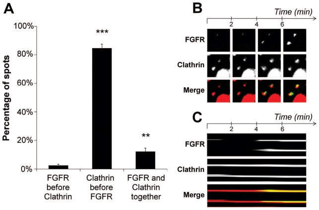
Live-cell TIRF microscopy can be employed to visualise the dynamics of RTKs at the plasma membrane, and our previous results demonstrated that following activation EGFR clusters at pre-formed CCPs prior to endocytosis (Rappoport and Simon, 2009). Three non-mutually exclusive hypothesis could describe the dynamics leading to colocalisation of activated FGFR and clathrin: FGFR could be recruited to CCPs, CCPs could form at the sites of receptor clustering, and clathrin and FGFR could accumulate simultaneously. When two-colour live-cell TIRF was performed on cells expressing FGFR2–GFP and clathrin–dsRed, it was observed that the vast majority of receptor clusters form at the sites of pre-existing clathrin (Fig. 4). In nearly all cases clathrin was observed at the site where subsequently FGFR would cluster. Thus, it seems that recruitment to pre-existing CCPs represents an emerging paradigm for the dynamics of activated RTKs.
Fig. 4.
Activated FGFR clusters at sites of pre-formed clathrin-coated pits. HeLa cells co-expressing FGFR2–GFP and clathrin–dsRed were imaged using simultaneous two-colour TIRF microscopy at 37°C (1 frame/30 sec) after stimulation with FGF2 + heparin. (B) Selected frames from a time-lapse sequence of two FGFR spots (top) and two clathrin spots (middle), and the merged images (bottom) showing the overlap of the two channels in yellow. (C) The kymographic representation of the spots shown in B reveals that FGFR appears in the TIRF field and colocalises with pre-existing clathrin. (A) Quantification of the percentage of clathrin clusters forming at pre-existing FGFR spots, of FGFR spots recruited at pre-formed clathrin clusters or FGFR and clathrin clustering simultaneously at the plasma membrane (means ± s.e.m., n = 33 cells). *P<0.05; **P<0.01; ***P<0.001.
FGFR activation promotes clathrin-mediated endocytosis through Src and Eps8
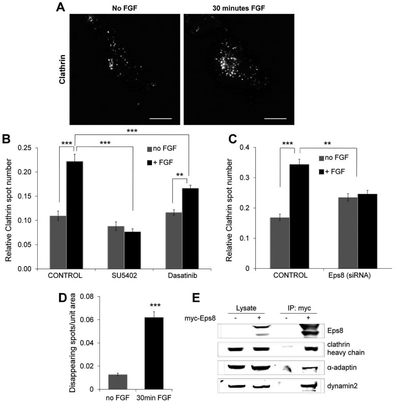
Previous evidence has identified Src as involved in the redistribution of clathrin to the cell periphery following RTK activation (Wilde et al., 1999). Therefore, we assessed whether FGFR stimulation regulated clathrin-mediated endocytosis. As depicted in Fig. 5, stimulation of cells with FGF led to a striking increase in the number of clathrin spots on the plasma membrane (Fig. 5A,B). Furthermore, this also increased the density of endocytosis events as gauged by established TIRF microscopy based criteria (Fig. 5D) (Rappoport, 2008). Interestingly, FGF treatment did not result in an increase in transferrin entry, suggesting that the increased endocytosis observed following FGFR activation may reflect a cargo specific pathway (supplementary material Fig. S3).
Fig. 5.
FGFR activation promotes clathrin-mediated endocytosis via Src and Eps8. (A–C) HeLa cells transiently expressing FGFR2–GFP and clathrin–dsRed and incubated in the presence or absence of SU5402 (5 minutes), Dasatinib inhibitor (30 minutes) or treated with Eps8 siRNA, were analysed using TIRF microscopy before and 30 minutes after stimulation with FGF2 + heparin. (A) Upon FGF stimulation, cells show a significant increase in the number of clathrin spots on the plasma membrane. Scale bars: 5 µm. (B,C) Quantification of the clathrin spots for the different conditions described above reveals that the FGF-dependent increase of clathrin at the plasma membrane is both Src and Eps8 dependent, and requires the full kinase activity of the receptor. (D) HeLa cells expressing FGFR2–GFP and Clathrin–dsRed were imaged using live-cell TIRF microscopy at 37°C for 2 minutes (1 frame/200 msec) before and 30 minutes after stimulation with FGF2 + heparin. Quantification of the clathrin spots disappearing from the TIRF field shows a significant increase in the density of endocytic events upon FGF stimulation (means ± s.e.m., n = 30 cells for each condition in B and C, n = 24 cells in D. *P<0.05; **P<0.01; ***P<0.001. (E) Cellular extracts from HeLa cells transfected with myc–Eps8 (where indicated) were immunoprecipitated with anti-myc, resolved by SDS-PAGE and analysed by immunoblotting for the levels of specified proteins.
The increase in clathrin-mediated endocytosis was a direct consequence of receptor activity, and not an alternative effect of FGF treatment, as SU5402 completely abrogated the ability of FGF addition to increase clathrin spot number at the plasma membrane (Fig. 5B). However, as SU5402 treatment in unstimulated cells did not alter the steady state number of clathrin spots on the cell surface, it can be inferred that basal FGFR signalling does not regulate the baseline level of clathrin-mediated endocytosis. Treatment with the Src family kinase inhibitor Dasatinib, which we have previously shown inhibits Src-dependent FGFR signalling and trafficking events (Sandilands et al., 2007), significantly inhibited the ability of FGFR stimulation to increase clathrin spot numbers (Fig. 5B). However, Dasatinib treatment in unstimulated cells did not reduce the number of CCPs, suggesting that SFK activity is not required for constitutive endocytosis.
Our previously published work identified that Eps8 is phosphorylated in a Src-dependent manner following FGFR activation (Cunningham et al., 2010). Furthermore, Eps8 has been implicated as playing a role in clathrin-mediated endocytosis (Taylor et al., 2011). Consistent with these observations, treatment with Eps8 siRNA completely prevented the increased number of clathrin spots following FGFR activation (Fig. 5C). Importantly, the sequence employed to silencing Eps8 was employed in previously published studies from the laboratory of Giorgio Scita (Disanza et al., 2006), and western blot analysis revealed that siRNA targeting Eps8 potently reduced Eps8 protein levels without affecting expression on negative control protein, tubulin (supplementary material Fig. S4A). Therefore, FGFR signalling through Src to Eps8 appears to be important in recruiting new clathrin at plasma membrane, consequently promoting clathrin-mediated endocytosis. This provides evidence for a biologically relevant link between FGFR-mediated signalling and membrane trafficking mediated through Src and Eps8. Furthermore, we have performed similar studies in LNCap cells (supplementary material Fig. S5), which have previously been shown to endogenously express FGFR2 (Carstens et al., 1997). These studies revealed that in the absence of expression of GFP-tagged FGFR treatment of cells with FGF2 resulted in a significant increase in plasma membrane clathrin spot number. Therefore, these results demonstrate this effect is not cell line specific, and can occur via signalling through endogenous FGFR2.
In order to verify the suggested link between Eps8 and clathrin-mediated endocytosis we performed a series of biochemical studies. Co-immunoprecipitation analyses demonstrated that Eps8 associates with clathrin heavy chain, α-adaptin and dynamin2 (Fig. 5E), all proteins involved in clathrin-mediated endocytosis (Doherty and McMahon, 2009). This suggests that Eps8 is capable of being incorporated into a complex with several members of the endocytosis machinery, further supporting its role in early phases of trafficking following receptor activation. These associations were also observed in similar studies performed in HEK293 cells, the proteomic platform we previously employed to identify the connection between FGFR, Src and Eps8 (Cunningham et al., 2010) (supplementary material Fig. S6). Furthermore, in this model we observed that interactions between Eps8 and the machinery for clathrin-mediated endocytosis occur with and without FGF stimulation.
FGFR early endocytic trafficking requires SFK kinase activity
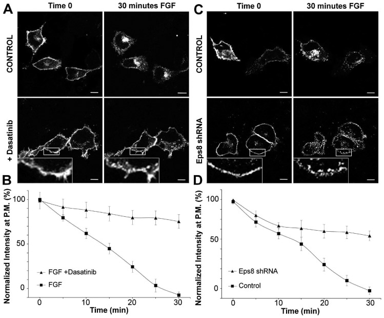
As the above data suggesting a role for Src and Eps8 in the early phases of FGFR signalling and clathrin-mediated endocytosis, we studied the effects of Src inhibition on trafficking of activated FGFR. As depicted in Fig. 6, treatment with Dasatinib resulted in pronounced effects on the behaviour of FGFR2–GFP following FGF stimulation. Almost complete inhibition of redistribution of FGFR from the cell periphery was observed (Fig. 6A,B). However, high magnification imaging of the plasma-membrane-associated region (see Fig. 6A, inset) revealed the presence of numerous peripheral puncta containing FGFR following stimulation. This would support our evidence for a role of Src-kinase-dependent processes in regulating early endocytic trafficking of activated FGFR.
Fig. 6.
Inhibition of Src kinase activity and silencing of Eps8 impair the trafficking of FGFR. (A) HeLa cells transiently expressing FGFR2–GFP were incubated for 30 minutes in the presence or absence of Dasatinib and imaged by confocal live-cell microscopy at 37°C for 30 minutes following stimulation with FGF2 + heparin. (C) Eps8 knockdown cells (Eps8 shRNA) transiently expressing FGFR2–GFP were stimulation with FGF2 + heparin and imaged as in A. The first and the last frame of the time-lapse sequences are shown in A and C. Scale bars: 5 µm. Higher-magnification images of selected regions from the cells (insets) show an atypical peripheral localisation of FGFR-containing vesicles. (B,D) The quantification of FGFR2–GFP intensity in the PM region of the cells as a function of time shows an impaired trafficking of FGFR in both Src-inhibited and Eps8shRNA cells (means ± s.e.m., n = 16 cells).
The role of Eps8 in endocytic trafficking of FGFR
Given the potential role of Eps8 as a downstream effector of Src-kinase-mediated processes, we next studied the consequences of Eps8 depletion on activated FGFR trafficking. In order to assess a role for Eps8 in this process, we made use of a HeLa cell line engineered with stable silencing of Eps8 expression which was previously validated in published studies from the laboratory of Giorgio Scita (Disanza et al., 2006). Western blotting studies confirmed potent and specific silencing of Eps8 in these cells, relative to control (supplementary material Fig. S4B). Similar to our observations in cells treated with the Src inhibitor (Fig. 6A,B), shRNA-mediated reduction in the expression of Eps8 also inhibited trafficking of activated FGFR away from the cellular periphery and towards the perinuclear region (Fig. 6C,D). Furthermore, peripheral FGFR-positive puncta were observed in Eps8 knockdown cells, identical to those observed following Dasatinib treatment (see Fig. 6C, inset). Interestingly, an effect of Eps8 depletion was not apparent at the earliest time points following FGF addition; accumulation of FGFR in a peripheral compartment, rather than direct inhibition of FGFR endocytosis was evident. Thus, inhibition of Src function and silencing of Eps8 result in similar effects on FGFR trafficking further supporting a mechanistic connection between FGFR activation, Src kinase activity and Eps8 function, specifically localised in a peripheral endocytic compartment.
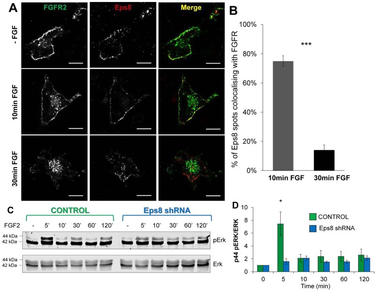
In order to further characterise the potential localisation of Eps8 action on the endocytic trafficking of FGFR we simultaneously visualised both in cells at various times following FGF addition. As depicted in Fig. 7, prior to stimulation both FGFR and Eps8 are observed primarily close to the lateral plasma membrane, consistent with our other observations. However, while FGFR and Eps8 colocalise to a very large degree in peripheral vesicles 10 minutes post-stimulation, once FGFR traffics into the juxtanuclear compartment this coincidence is lost. Quantification revealed that 30 minutes following addition of FGF, Eps8 is retained at the periphery and no longer colocalises with the juxtanuclear pool of FGFR (Fig. 7B). Similarly, Eps8 colocalises with EEA1, a marker for early endosomes (Stenmark et al., 1996) (data not shown). Therefore it appears that Eps8 resides in the early endocytic system, and that activated receptors move through this compartment following endocytosis.
Fig. 7.
During early stages of trafficking Eps8 colocalises with FGFR and is required for signal transduction through the MAPK cascade. HeLa cells transiently expressing FGFR2–GFP and Eps8–mCherry were either stimulated with FGF2 + heparin for 10 or 30 minutes, or not stimulated, then fixed and analysed using confocal microscopy. (A) During the early stages of activation (10 min FGF), FGFR primarily localises in early endosomal compartments, where it colocalises with Eps8. 30 minutes after FGF addition, FGFR redistributes to the perinuclear region, whereas Eps8 still resides in peripheral compartments, just beneath the plasma membrane. Scale bars: 5 µm. (B) Quantification demonstrates significant colocalisation of FGFR and Eps8 only during early phases of receptor activation, where they both localise in early endocytic compartments (means ± s.e.m., n = 14 cells). (C) Eps8 knockdown and vector control HeLa cells transiently expressing FGFR2–GFP were lysed following stimulation with FGF2 + heparin for the indicated times. Cellular extracts where resolved by SDS-PAGE and analysed by immunoblotting for the levels of the specified proteins. The time course of Erk phosphorylation in response to FGF shows a significant attenuation of p44 ERK signal at early times (5 minutes) in Eps8 knockdown cells. (D) Quantification of p44 pERK levels following FGF stimulation in Eps8 knockdown (blue) and vector control HeLa (green) cells (means ± s.e.m., n = 5 experiments) by densitometric scanning of western blots. *P<0.05; **P<0.01; ***P<0.001.
To further elucidate the site of action of Src/Eps8 in FGFR trafficking we performed a series of other colocalisation studies. As depicted in supplementary material Fig. S7, triple colocalisation of activated FGFR, following 10 minutes FGF treatment, Eps8, and transferrin, following a 5 minute incubation places Eps8 in a common early endocytic compartment shared by activated growth factor receptors and transferrin/transferrin receptor. Additionally, in supplementary material Fig. S8 colocalisation between Eps8 and constitutively active Src in the cell periphery, further support our evidence of an early endocytic compartment regulated by Src and Eps8.
The role of Eps8 in FGFR signalling
As we have clearly identified a role for Eps8 in the early endocytic trafficking of FGFR, we next tested whether Eps8 regulated FGFR signalling as well. As depicted in Fig. 7C,D, stimulation with FGF results in a rapid appearance of the p44 form of activated ERK which reaches maximum amplitude at 5 minutes and then declines. In this system, the p42 form of ERK exhibits tonic basal activation and does not respond to FGF stimulation. In fact p42/ERK is apparent in non-transfected HeLa cells subjected to overnight serum starvation (supplementary material Fig. S9). In cells stably silenced for Eps8 expression the FGF-mediated activation of p44 ERK was blunted during the initiating (5 minute) phase of the FGF response. Thus, these findings show that functional Eps8 is required for early phase activation of the Ras/Raf/ERK pathway during the period in which activated FGFRs are recruited away from the plasma membrane. This finding also suggests that activated FGFR can connect to the Ras/Raf/ERK pathway in an Eps8-independent manner which results in lower amplitude signals. Thus this suggests that Eps8 coordinates the signalling and trafficking of FGFR immediately following activation.
The role of Eps8 in trafficking FGFR out of the early endosome
In order to further characterise the role of Eps8 in FGFR trafficking, we performed a series of colocalisation studies with well characterised markers within the endocytic system. As in our previous live-cell imaging studies, 30 minutes following activation FGFR was observed to localise to an intracellular region adjacent to the nucleus (Fig. 8A). Thus, the extent of colocalisation between FGFR and EEA1, was very low 30 minutes post-stimulation (Fig. 8B). However, in the Eps8 knockdown cells the extent of colocalisation between FGFR and EEA1 was significantly higher (Fig. 8B), suggesting that Eps8 functions to permit exit of FGFR from peripheral early endosomes. Similarly, while clear colocalisation was observed between FGFR and Rab11 (Fig. 8C), a marker for the peri-nuclear recycling compartment (Ullrich et al., 1996), in control cells 30 minutes post-stimulation, this was significantly reduced in the Eps8 knockdown cells (Fig. 8D). Finally, we performed similar studies employing Lysotracker as a marker for endocytic degradative compartments (Fig. 8E,F), and found that silencing of Eps8 significantly reduced colocalisation of FGFR and Lysotracker 30 minutes post-stimulation. Finally, super-resolution imaging of FGFR–GFP in Dasatinib treated cells 30 minutes after FGF addition revealed clearly the presence of FGFR in peripheral diffraction limited puncta (supplementary material Fig. S10). Therefore, these results demonstrate that Eps8 is required for the trafficking of activated FGFR out of the early endocytic system and into the peri-nuclear recycling compartment and degradative compartment(s).
Fig. 8.
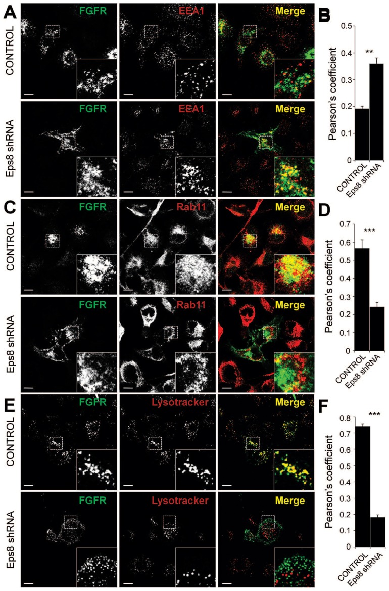
Eps8 is required for the trafficking of activated FGFR out of the early endocytic system and into the peri-nuclear recycling and late degradative compartments. Eps8 knockdown and vector control HeLa cells transiently expressing FGFR2–GFP were fixed following stimulation with FGF2 + heparin for 30 minutes, immunostained for EEA1 (A) or Rab11 (C) or treated with LysoTracker Red (E) and analysed by confocal microscopy. Higher-magnification images of selected regions from the cells (insets) show that in Eps8 knockdown cells, where receptor trafficking is impaired, FGFR is retained in the peripheral compartment (EEA1) and prevented from sorting to the peri-nuclear recycling compartment (Rab11) and to the lysosomal degradative compartment (LysoTracker). Scale bars: 5 µm. (B,D,F) Colocalisation between FGFR and EEA1, Rab11 or LysoTracker was quantified in control cells as 0.19±0.02, 0.57±0.04, and 0.74±0.01, respectively, and in Eps8 knockdown cells as 0.36±0.04, 0.24±0.05 and 0.18±0.01, respectively (Pearson's correlation coefficient; means ± s.e.m., n = 18 cells).
Discussion
Growth factor receptors undergo regulated internalization from the cell surface in response to ligand binding (von Zastrow and Sorkin, 2007; Le Roy and Wrana, 2005). Endocytosis and subsequent degradation represent strategies to attenuate signalling of RTK, and alterations in receptor trafficking have emerged as a mechanism of oncogenic activation (Peschard and Park, 2003). Here we have applied imaging based assays for studying endocytosis and trafficking of FGFRs with cells expressing a previously validated GFP-tagged FGFR2 (Schüller et al., 2008). Through use of this system we have demonstrated the interplay between RTK signalling and trafficking, and identified a mechanistic role for Eps8 in mediating clathrin-mediated endocytosis, endocytic trafficking, and MAP kinase signalling. We have demonstrated that receptor trafficking is regulated through signalling transduction pathways downstream of receptor activation, and that the location of the activated receptor within the endocytic system has significant implications on the amplitude of signalling readouts.
Although FGFs are extremely relevant in both development and cancer progression, very little is known about FGFR trafficking (Marchese et al., 1998; Citores et al., 1999; Belleudi et al., 2007; Gleizes et al., 1996; Haugsten et al., 2011). Our data have shown that internalization of FGFR was severely reduced in cells treated with the dynamin inhibitor Dynasore (Fig. 1), indicating that endocytosis of activated receptor occurs through a dynamin-dependent pathway. Using a siRNA approach we observed a complete block of FGFR endocytosis in cells silenced for α-adaptin, whereas no effect was detected in cells treated with caveolin1 siRNA (Fig. 2). We further confirmed these observations by analysing the colocalisation of FGFR with clathrin or caveolin1 at the plasma membrane using TIRF microscopy. Following stimulation, activated FGFR significantly colocalises with clathrin at the cells surface, while no colocalisation with caveolin1 was observed (Fig. 3). Thus, these results identified clathrin-mediated endocytosis as the major pathway for FGFR internalization. Further analyses via live cell TIRF microscopy demonstrated that, like activated EGFR (Rappoport and Simon, 2009), following stimulation FGFR is recruited into pre-formed CCPs (Fig. 4), suggesting that this may be a mechanism conserved across multiple different RTKs.
The next phase of our studies was to characterise mechanistic links between FGFR signalling and trafficking. Src plays a central role in FGFR signalling pathway and has been previously suggested to regulate FGFR trafficking (Sandilands et al., 2007). Previous analyses from our laboratory identified putative Src targets involved in the regulation of FGFRs signalling and trafficking (Cunningham et al., 2010). In particular the protein Eps8, previously implicated in vesicle trafficking (Lanzetti et al., 2000), was identified as a Src-dependent kinase substrate phosphorylated in response to FGF stimulation. Therefore, we analysed potential roles for Eps8 in the trafficking and signalling of activated FGFR.
Previously it was shown that EGFR activation, signalling through Src, increased the peripheral accumulation of clathrin (Wilde et al., 1999). Our work demonstrates that FGFR activation increases both the number of CCPs and the events of clathrin-mediated endocytosis (Fig. 5). Furthermore, we have demonstrated that Src kinase activity and Eps8 are required for the increase in CCP number following FGFR activation (Fig. 5). Interestingly though, while FGFR activation stimulated clathrin-mediated endocytosis, it did not increase transferrin uptake (supplementary material Fig. S3). Thus, the route of entry for activated RTKs may exist as a cargo specific pathway.
Consistent with live cell imaging studies (Taylor et al., 2011), our biochemical studies demonstrated that Eps8 interacts with the clathrin-mediated endocytosis machinery (Fig. 5). Interestingly, similar studies in HEK293 cells demonstrated these interactions are not FGF2 stimulus dependent (supplementary material Fig. S6). However, we have also identified roles for Src and Eps8 in the further endocytic trafficking of FGFR. Following Dasatinib treatment, FGFR containing vesicles were observed to accumulate at the cell periphery, confirming the role for Src in the early endocytic trafficking of FGFRs (Fig. 6). Similarly, in Eps8 knockdown cells we observed a Dasatinib-like phenotype: a strong inhibition of FGFR trafficking with a pool of receptor localised in a peripheral punctuate compartment (Fig. 6). Thus, this is consistent with our hypothesis that Eps8 is a Src-dependent regulator of FGFR trafficking within the early endocytic system.
Further supporting a role for Eps8 in exit from the early endocytic system, during the early stages of trafficking FGFR significantly colocalises with Eps8 just beneath the plasma membrane (Fig. 7). Furthermore, Eps8 colocalised with FGFR and transferrin in peripheral early endosomes (supplementary material Fig. S7), as well as with constitutively active Src (supplementary material Fig. S8). However, 30 minutes post-stimulation the juxtanuclear pool of FGFR clearly did not colocalise with Eps8 which was retained in a peripheral compartment (Fig. 7). Similarly, exit from the EEA1 early endosome was significantly reduced when Eps8 expression was silenced (Fig. 8). Finally, 30 minutes post-stimulation FGFR was found to localise to the Rab11-positive peri-nuclear recycling compartment, as well as degradative compartments, in stimulated control cells, and this was prevented in Eps8 knockdown cells (Fig. 8). Thus, activated receptors traffics through the Eps8/EEA1-positive peripheral compartment en route to the PNRC and late endosome/lysosome, and in the absence of Eps8 this is prevented.
Finally, we sought to determine if there is a concomitant feedback into FGFR2 signalling mediated through Eps8. Silencing of Eps8 expression had significant effects on a burst in Erk activation through the p44 form immediately following receptor activation (Fig. 7), suggesting that, consistent with recent work with EGFR (Sousa et al., 2012; Brankatschk et al., 2012), FGFR signalling through the MAP kinase pathway can occur prior to/during receptor endocytosis. Importantly, the p42/Erk band revealed in our studies, which was not Eps8 sensitive, is chronically active in HeLa cells (supplementary material Fig. S9). Finally, super-resolution imaging on a Leica gSTED system further elucidated the site of Eps8/Src-dependent FGFR trafficking (supplementary material Fig. S10). In Dasatinib treated cells FGFR clearly resides in peripheral puncta. Therefore, taken together these results identify Eps8 as a key mediator of activated FGFR trafficking and signalling, and identify the peripheral early endocytic compartment as a key crossroads in the regulation of RTKs.
The interplay between receptor tyrosine kinase signalling and trafficking has been emerging an area critical to understanding fundamental aspects of cell biology as well as how mis-regulation of either could potentially lead to pathological conditions, such as developmental defects or chronic disease. Our studies point to a response arc leading from RTK activation through Src via Eps8. Evidence supporting this hypothesis has been gained from a variety of studies, and is based upon work in other signalling systems, suggesting potential widespread significance. In our studies we have demonstrated that following activation FGFR enters the cell via clathrin-mediated endocytosis. FGF stimulation increases the number of CCPs, and events of clathrin-mediated endocytosis. Furthermore, both the FGF-dependent increase in clathrin at the plasma membrane and the early endocytic trafficking of activated FGFR depend upon Src and Eps8. FGFR traffics through an Eps8-positive early endocytic compartment on the way to the PNRC and lysosome, and in the absence of Eps8, FGFR is retained in peripheral early endosomes. Eps8 is also required for early phase pErk activation, placing this event early in the trafficking of activated FGFR. Therefore, FGFR activation induces alterations in signalling and trafficking that are mediated by Src acting through Eps8, and this alters both early endocytic trafficking and downstream MAPK signalling. Thus, the correlation between Src- and Eps8-dependent FGFR signalling and trafficking suggests a direct link between the two. However, validation and elucidation of the direct mechanistic coupling between FGFR signalling and trafficking and the precise role(s) of Src and Eps8 await further work in this area. In conclusion, while our data suggest that signalling through activated FGFR promotes clathrin-mediated endocytosis, and that this effect depends on Src/Eps8, the initial wave of FGFR endocytosis, although clearly clathrin dependant, does not seem to critically rely upon Src/Eps8. Finally, as constitutive endocytosis and the steady state production of clathrin-coated vesicles do not seem to be Src/Eps8 dependant, the endocytic cargo removed from the plasma membrane in FGF treated cells mediated by Src/Eps8 remains presently elusive.
Materials and Methods
Plasmids, siRNA and antibodies
The construct encoding GFP-tagged FGFR2IIIc (FGFR2–GFP) was a gift from J. Ladbury (M.D. Anderson Cancer Center, The University of Texas). DsRed-tagged clathrin light chain A (clathrin-dsRed) was a gift from T. Kirchhausen (Harvard Medical School, Boston, MA). Caveolin1-mRFP was a gift from A. Helenius (ETH Institute of Biochemistry, Zurich, Switzerland). Eps8-mCherry was a gift from Giorgio Scita (IFOM University of Milan, Milan, Italy). Constitutively active Src was provided by Margaret Frame (Edinburgh Cancer Research UK Centre). siRNA oligos targeting α-adaptin (sequence 5′-AAGAGCAUGUGCACGCUGGCCA-3′), caveolin1 (SmartPool l-003467) and Eps8 (sequence 5′-TGCAGACCCTAGTATACCG-3′) were purchased from Dharmacon. Antibodies used were: anti-Rab11 (Invitrogen); anti-EEA1 (Abcam); anti-caveolin1 (BD Biosciences); anti-α-adaptin (Santa Cruz); anti-α-tubulin (Sigma); α-ERK1/2 and α-pERK (Santa Cruz); α-Eps8 (Abcam); anti-phospho-Src family (Tyr416; Cell Signaling); IRDye680 and IRDye800 conjugated secondary antibodies (Odyssey).
Cells and transfection
Eps8 shRNA and control shRNA HeLa cells were a gift from Giorgio Scita (IFOM University of Milan, Milan, Italy). HeLa and LNCaP cells were propagated in DMEM (Invitrogen) supplemented with 10% FBS (Labtech International) in a 5% CO2 atmosphere at 37°C. DNA and siRNA transfection was performed by using Lipofectamine 2000 transfection reagent (Invitrogen) according to the manufacturer's protocol. At 48 hours post-transfection, cells were serum-starved for 30 min and then stimulated with 20 µg/ml Heparin (Sigma) and 50 ng/ml FGF2 for the indicated times. For the inhibition assays cells were either treated with 25 µM SU5402 (Calbiochem) for 5 min, 80 µM Dynasore (Sigma) for 30 min or 50 nM Dasatinib (Sellek Chemicals) for 30 min or treated as above in the absence of drug before FGF stimulation. For live-cell imaging, cells were washed once and the medium removed was replaced with prewarmed (37°C) imaging medium [10 mM HEPES–Hank's balanced salt solution (HBSS; Sigma) pH 7.4].
Laser scanning confocal microscopy and total internal reflection fluorescence microscopy
Confocal laser was performed with an inverted microscope (Eclipse Ti, Nikon A1R) at 37°C using a 60× 1.45 NA oil-immersion objective and 12-bit CCD camera (Ixon 1M EMCCD). GFP constructs were excited with an argon ion 457–514 nm laser, mRFP and dsRed constructs with a Green Diode 561 nm laser. Images were prepared with NIS-Elements Imaging Software version 3.2 (Nikon). TIRF microscopy was performed with the system described above as well as with an inverted microscope (IX81, Olympus) using a 60× 1.49 NA oil-immersion objective and a 12-bit CCD camera (ORCA-R2 C10600, Hamamatsu). GFP constructs were excited with a 491-50 Diode type laser, dsRed constructs with a 561-50 DPSS type laser. Images were analysed and prepared with Xcellence Advanced Lice Cell Imaging System version 1.1 (Olympus). Super-resolution imaging was performed on a gSTED system (Leica) and image stacks were deconvolved using Huygens linked within the Leica software.
Immunofluorescence
Cells grown on coverslips were fixed in 4% paraformaldehyde in TBS for 10 min, washed in TBS/100 mM glycine and permeabilised with TBS/0.1% saponin/20 mM glycine. After blocking with TBS/0.1% saponin/10% FCS, cells were incubated with primary antibodies, then washed and incubated with secondary antibodies. After staining, the coverslips were mounted in Hydromount (National Diagnostic) and imaged by using a Zeiss LSM 710 confocal microscope, a 40× 1.3NA oil-immersion objective and a Transmission-Photomultiplier LSM T-PMT. Each experiment was repeated a minimum of three times and an image that represented the phenotype of most of the cells was selected. For lysosomal staining, cells were incubated with 75 nM LysoTracker Red (Molecular Probes) for 30 minutes and immediately imaged in imaging media at room temperature.
Transferrin and cholera toxin B uptake assay
Following serum-starvation for 30 min, cells were incubated at 37°C in either Alexa-Fluor-546–transferrin (Invitrogen), or Alexa-Fluor-555–cholera-toxin-B (Invitrogen). Cells were then rinsed, fixed and analysed by using a Nikon TE300 inverted epi-fluorescence microscope using an 60× 1.40 NA oil-immersion objective and a cooled CCD camera (Hamamatsu C4742-98-12WRB). For the triple colocalisation, HeLa cells were incubated with Alexa-Fluor-633–transferrin (Invitrogen) for 5 minutes, in order to mark the early endosomes. Cells were then rinsed, fixed and analysed using confocal microscopy.
Quantification analysis
For the trafficking assays FGFR2–GFP intensity in the plasma-membrane-associated region of the cells was calculated for each frame of the time-lapse sequences (1 frame/5 min). From each time point, a background value was subtracted, corresponding to the cytosolic fluorescence intensity before the stimulation with FGF (time = 0 min). Cells were treated with a red membrane stain (data not shown) in order to define the plasma-membrane-associated region. For the colocalisation analysis, each spot of FGFR, clathrin, caveolin1 and Eps8 was identified and a circle was drawn around it. Spots were identified as colocalising when most of the fluorescence intensity of the pixels within the circular regions fitted in both the channels. For quantification of transferrin and cholera toxin uptake, cells randomly located on the coverslips were scanned at fixed intensity settings below pixel-saturation, and the total cellular intensity was determined. Data analysis was performed using either NIS-Elements Imaging Software version 3.2 (Nikon) or Xcellence Advanced Lice Cell Imaging System version 1.1 (Olympus).
Biochemical assays
Cultures of HeLa or Hek293T cells were lysed in lysis buffer (50 mM Tris-HCl pH 7.4, 150 mM NaCl, 1% Triton TX-100, 1 mM sodium orthovanadate, 50 mM sodium fluoride, 25 mM β-glycerophosphate) supplemented with complete mini protease inhibitors cocktail (Roche). The extracts were resolved by a 4–12% NuPAGE Bis-Tris SDS gel (Invitrogen) and transferred to Immobilon PVDF-FL membrane (Millipore). The membrane was blocked in methanol and incubated with primary antibodies overnight at 4°C. After washing in PBS 0.1% Tween-20 the membrane was incubated with secondary antibodies for 1 hour at room temperature followed by washing and detected using an Odyssey infrared imaging system (Li-COR). For the immunoprecipitation assay, cells were lysed and prepared as described. The extracts were immunoprecipitated with dynabeads (Invitrogen) crosslinked with anti-myc antibody. Supernatants were loaded on a 4–12% NuPAGE Bis-Tris SDS gel (Invitrogen) and subjected to immunoblot analysis with indicated antibodies. Quantification by densitometric scanning of western blots was performed using Odyssey Application Software version 3.0 (Li-COR). Since the primary antibodies used in this analysis detected both the 42 kDa and 44 kDa form of ERK/pERK, two bands were visualized and captured. The relative p44 phosphoERK levels shown in Fig. 7D were calculated by normalizing, for each point of the time course, the relative optical density value of the p44 phosphoERKs band to the corresponding total p44 ERK value.
Supplementary Material
Supplementary Material
Acknowledgments
The authors would like to thank Giorgio Scita (IFOM-IEO) for sharing reagents and expertise, and Sylwia Krawczyk and the rest of the Rappoport and Heath laboratories for helpful discussion. The Nikon A1R/TIRF microscope used in this research was obtained through the Birmingham Science City Translational Medicine Clinical Research and Infrastructure Trials Platform, with support from Advantage West Midlands (AWM). The funders had no role in study design, data collection and analysis, decision to publish, or preparation of the manuscript.
Footnotes
Funding
This work was supported by the Biotechnology and Biological Sciences Research Council [new investigator project grant number BB/H002308/1 to J.Z.R.]; Cancer Research UK [programme grant number C80/A10171 to J.K.H.]; and a Ph.D. studentship from the Medical Research Council [training grant reference number G0900175 to G.A.]. Deposited in PMC for release after 6 months.
References
- Ahmed Z., Schüller A. C., Suhling K., Tregidgo C., Ladbury J. E. (2008). Extracellular point mutations in FGFR2 elicit unexpected changes in intracellular signalling. Biochem. J. 413, 37–49 10.1042/BJ20071594 [DOI] [PubMed] [Google Scholar]
- Axelrod D. (2008). Total internal reflection fluorescence microscopy. Methods Cell Biol. Vol. 89 Correia J J, William Detrich H, III, pp. 169–221 10.1016/S0091-679X(08)00607-9 [DOI] [PubMed] [Google Scholar]
- Basilico C., Moscatelli D. (1992). The FGF family of growth factors and oncogenes. Adv. Cancer Res. 59, 115–165 10.1016/S0065-230X(08)60305-X [DOI] [PubMed] [Google Scholar]
- Beenken A., Mohammadi M. (2009). The FGF family: biology, pathophysiology and therapy. Nat. Rev. Drug Discov. 8, 235–253 10.1038/nrd2792 [DOI] [PMC free article] [PubMed] [Google Scholar]
- Beguinot L., Lyall R. M., Willingham M. C., Pastan I. (1984). Down-regulation of the epidermal growth factor receptor in KB cells is due to receptor internalization and subsequent degradation in lysosomes. Proc. Natl. Acad. Sci. USA 81, 2384–2388 10.1073/pnas.81.8.2384 [DOI] [PMC free article] [PubMed] [Google Scholar]
- Belleudi F., Leone L., Nobili V., Raffa S., Francescangeli F., Maggio M., Morrone S., Marchese C., Torrisi M. R. (2007). Keratinocyte growth factor receptor ligands target the receptor to different intracellular pathways. Traffic 8, 1854–1872 10.1111/j.1600-0854.2007.00651.x [DOI] [PubMed] [Google Scholar]
- Brankatschk B., Wichert S. P., Johnson S. D., Schaad O., Rossner M. J., Gruenberg J. (2012). Regulation of the EGF transcriptional response by endocytic sorting. Sci. Signal. 5, ra21 10.1126/scisignal.2002351 [DOI] [PubMed] [Google Scholar]
- Burgess W. H., Maciag T. (1989). The heparin-binding (fibroblast) growth factor family of proteins. Annu. Rev. Biochem. 58, 575–602 10.1146/annurev.bi.58.070189.003043 [DOI] [PubMed] [Google Scholar]
- Carstens R. P., Eaton J. V., Krigman H. R., Walther P. J., Garcia–Blanco M. A. (1997). Alternative splicing of fibroblast growth factor receptor 2 (FGF-R2) in human prostate cancer. Oncogene 15, 3059–3065 10.1038/sj.onc.1201498 [DOI] [PubMed] [Google Scholar]
- Citores L., Wesche J., Kolpakova E., Olsnes S. (1999). Uptake and intracellular transport of acidic fibroblast growth factor: evidence for free and cytoskeleton-anchored fibroblast growth factor receptors. Mol. Biol. Cell 10, 3835–3848 [DOI] [PMC free article] [PubMed] [Google Scholar]
- Cunningham D. L., Sweet S. M., Cooper H. J., Heath J. K. (2010). Differential phosphoproteomics of fibroblast growth factor signaling: identification of Src family kinase-mediated phosphorylation events. J. Proteome Res. 9, 2317–2328 10.1021/pr9010475 [DOI] [PMC free article] [PubMed] [Google Scholar]
- Damm E. M., Pelkmans L., Kartenbeck J., Mezzacasa A., Kurzchalia T., Helenius A. (2005). Clathrin- and caveolin-1-independent endocytosis: entry of simian virus 40 into cells devoid of caveolae. J. Cell Biol. 168, 477–488 10.1083/jcb.200407113 [DOI] [PMC free article] [PubMed] [Google Scholar]
- Di Fiore P. P., De Camilli P. (2001). Endocytosis and signaling. an inseparable partnership. Cell 106, 1–4 10.1016/S0092-8674(01)00428-7 [DOI] [PubMed] [Google Scholar]
- Di Fiore P. P., Scita G. (2002). Eps8 in the midst of GTPases. Int. J. Biochem. Cell Biol. 34, 1178–1183 10.1016/S1357-2725(02)00064-X [DOI] [PubMed] [Google Scholar]
- Disanza A., Mantoani S., Hertzog M., Gerboth S., Frittoli E., Steffen A., Berhoerster K., Kreienkamp H. J., Milanesi F., Di Fiore P. P.et al. (2006). Regulation of cell shape by Cdc42 is mediated by the synergic actin-bundling activity of the Eps8-IRSp53 complex. Nat. Cell Biol. 8, 1337–1347 10.1038/ncb1502 [DOI] [PubMed] [Google Scholar]
- Disanza A., Frittoli E., Palamidessi A., Scita G. (2009). Endocytosis and spatial restriction of cell signaling. Mol. Oncol. 3, 280–296 10.1016/j.molonc.2009.05.008 [DOI] [PMC free article] [PubMed] [Google Scholar]
- Doherty G. J., McMahon H. T. (2009). Mechanisms of endocytosis. Annu. Rev. Biochem. 78, 857–902 10.1146/annurev.biochem.78.081307.110540 [DOI] [PubMed] [Google Scholar]
- Engqvist–Goldstein A. E., Warren R. A., Kessels M. M., Keen J. H., Heuser J., Drubin D. G. (2001). The actin-binding protein Hip1R associates with clathrin during early stages of endocytosis and promotes clathrin assembly in vitro. J. Cell Biol. 154, 1209–1224 10.1083/jcb.200106089 [DOI] [PMC free article] [PubMed] [Google Scholar]
- Gleizes P. E., Noaillac–Depeyre J., Dupont M. A., Gas N. (1996). Basic fibroblast growth factor (FGF-2) is addressed to caveolae after binding to the plasma membrane of BHK cells. Eur. J. Cell Biol. 71, 144–153 [PubMed] [Google Scholar]
- Greenfield C., Hiles I., Waterfield M. D., Federwisch M., Wollmer A., Blundell T. L., McDonald N. (1989). Epidermal growth factor binding induces a conformational change in the external domain of its receptor. EMBO J. 8, 4115–4123 [DOI] [PMC free article] [PubMed] [Google Scholar]
- Grose R., Dickson C. (2005). Fibroblast growth factor signaling in tumorigenesis. Cytokine Growth Factor Rev. 16, 179–186 10.1016/j.cytogfr.2005.01.003 [DOI] [PubMed] [Google Scholar]
- Haugsten E. M., Zakrzewska M., Brech A., Pust S., Olsnes S., Sandvig K., Wesche J. (2011). Clathrin- and dynamin-independent endocytosis of FGFR3 - implications for signalling. PLoS ONE 6, e21708 10.1371/journal.pone.0021708 [DOI] [PMC free article] [PubMed] [Google Scholar]
- Heldin C. H. (1995). Dimerization of cell surface receptors in signal transduction. Cell 80, 213–223 10.1016/0092-8674(95)90404-2 [DOI] [PubMed] [Google Scholar]
- Kholodenko B. N. (2002). MAP kinase cascade signaling and endocytic trafficking: a marriage of convenience? Trends Cell Biol. 12, 173–177 10.1016/S0962-8924(02)02251-1 [DOI] [PubMed] [Google Scholar]
- Lanzetti L., Rybin V., Malabarba M. G., Christoforidis S., Scita G., Zerial M., Di Fiore P. P. (2000). The Eps8 protein coordinates EGF receptor signalling through Rac and trafficking through Rab5. Nature 408, 374–377 10.1038/35042605 [DOI] [PubMed] [Google Scholar]
- Le Roy C., Wrana J. L. (2005). Clathrin- and non-clathrin-mediated endocytic regulation of cell signalling. Nat. Rev. Mol. Cell Biol. 6, 112–126 10.1038/nrm1571 [DOI] [PubMed] [Google Scholar]
- Li W., Liu H., Zhou J. S., Cao J. F., Zhou X. B., Choi A. M., Chen Z. H., Shen H. H. (2012). Caveolin-1 inhibits expression of antioxidant enzymes through direct interaction with nuclear erythroid 2 p45-related factor-2 (Nrf2). J. Biol. Chem. 287, 20922–20930 10.1074/jbc.M112.352336 [DOI] [PMC free article] [PubMed] [Google Scholar]
- Macia E., Ehrlich M., Massol R., Boucrot E., Brunner C., Kirchhausen T. (2006). Dynasore, a cell-permeable inhibitor of dynamin. Dev. Cell 10, 839–850 10.1016/j.devcel.2006.04.002 [DOI] [PubMed] [Google Scholar]
- Marchese C., Mancini P., Belleudi F., Felici A., Gradini R., Sansolini T., Frati L., Torrisi M. R. (1998). Receptor-mediated endocytosis of keratinocyte growth factor. J. Cell Sci. 111, 3517–3527 [DOI] [PubMed] [Google Scholar]
- Mason I. J. (1994). The ins and outs of fibroblast growth factors. Cell 78, 547–552 10.1016/0092-8674(94)90520-7 [DOI] [PubMed] [Google Scholar]
- Mattheyses A. L., Simon S. M., Rappoport J. Z. (2010). Imaging with total internal reflection fluorescence microscopy for the cell biologist. J. Cell Sci. 123, 3621–3628 10.1242/jcs.056218 [DOI] [PMC free article] [PubMed] [Google Scholar]
- Peschard P., Park M. (2003). Escape from Cbl-mediated downregulation: a recurrent theme for oncogenic deregulation of receptor tyrosine kinases. Cancer Cell 3, 519–523 10.1016/S1535-6108(03)00136-3 [DOI] [PubMed] [Google Scholar]
- Polo S., Di Fiore P. P. (2006). Endocytosis conducts the cell signaling orchestra. Cell 124, 897–900 10.1016/j.cell.2006.02.025 [DOI] [PubMed] [Google Scholar]
- Provenzano C., Gallo R., Carbone R., Di Fiore P. P., Falcone G., Castellani L., Alemà S. (1998). Eps8, a tyrosine kinase substrate, is recruited to the cell cortex and dynamic F-actin upon cytoskeleton remodeling. Exp. Cell Res. 242, 186–200 10.1006/excr.1998.4095 [DOI] [PubMed] [Google Scholar]
- Rappoport J. Z. (2008). Focusing on clathrin-mediated endocytosis. Biochem. J. 412, 415–423 10.1042/BJ20080474 [DOI] [PubMed] [Google Scholar]
- Rappoport J. Z., Simon S. M. (2008). A functional GFP fusion for imaging clathrin-mediated endocytosis. Traffic 9, 1250–1255 10.1111/j.1600-0854.2008.00770.x [DOI] [PMC free article] [PubMed] [Google Scholar]
- Rappoport J. Z., Simon S. M. (2009). Endocytic trafficking of activated EGFR is AP-2 dependent and occurs through preformed clathrin spots. J. Cell Sci. 122, 1301–1305 10.1242/jcs.040030 [DOI] [PMC free article] [PubMed] [Google Scholar]
- Rothberg K. G., Heuser J. E., Donzell W. C., Ying Y. S., Glenney J. R., Anderson R. G. (1992). Caveolin, a protein component of caveolae membrane coats. Cell 68, 673–682 10.1016/0092-8674(92)90143-Z [DOI] [PubMed] [Google Scholar]
- Sandilands E., Frame M. C. (2008). Endosomal trafficking of Src tyrosine kinase. Trends Cell Biol. 18, 322–329 10.1016/j.tcb.2008.05.004 [DOI] [PubMed] [Google Scholar]
- Sandilands E., Akbarzadeh S., Vecchione A., McEwan D. G., Frame M. C., Heath J. K. (2007). Src kinase modulates the activation, transport and signalling dynamics of fibroblast growth factor receptors. EMBO Rep. 8, 1162–1169 10.1038/sj.embor.7401097 [DOI] [PMC free article] [PubMed] [Google Scholar]
- Schüller A. C., Ahmed Z., Levitt J. A., Suen K. M., Suhling K., Ladbury J-E. (2008). Indirect recruitment of the signalling adaptor Shc to the fibroblast growth factor receptor 2 (FGFR2). Biochem. J. 416, 189–199 10.1042/BJ20080887 [DOI] [PubMed] [Google Scholar]
- Sorkin A., Von Zastrow M. (2002). Signal transduction and endocytosis: close encounters of many kinds. Nat. Rev. Mol. Cell Biol. 3, 600–614 10.1038/nrm883 [DOI] [PubMed] [Google Scholar]
- Sousa L. P., Lax I., Shen H., Ferguson S. M., De Camilli P., Schlessinger J. (2012). Suppression of EGFR endocytosis by dynamin depletion reveals that EGFR signaling occurs primarily at the plasma membrane. Proc. Natl. Acad. Sci. USA 109, 4419–4424 10.1073/pnas.1200164109 [DOI] [PMC free article] [PubMed] [Google Scholar]
- Stenmark H., Aasland R., Toh B. H., D'Arrigo A. (1996). Endosomal localization of the autoantigen EEA1 is mediated by a zinc-binding FYVE finger. J. Biol. Chem. 271, 24048–24054 10.1074/jbc.271.39.24048 [DOI] [PubMed] [Google Scholar]
- Stoscheck C. M., Carpenter G. (1984). Down regulation of epidermal growth factor receptors: direct demonstration of receptor degradation in human fibroblasts. J. Cell Biol. 98, 1048–1053 10.1083/jcb.98.3.1048 [DOI] [PMC free article] [PubMed] [Google Scholar]
- Sun L., Tran N., Liang C., Tang F., Rice A., Schreck R., Waltz K., Shawver L. K., McMahon G., Tang C. (1999). Design, synthesis, and evaluations of substituted 3-[(3- or 4-carboxyethylpyrrol-2-yl)methylidenyl]indolin-2-ones as inhibitors of VEGF, FGF, and PDGF receptor tyrosine kinases. J. Med. Chem. 42, 5120–5130 10.1021/jm9904295 [DOI] [PubMed] [Google Scholar]
- Taylor M. J., Perrais D., Merrifield C. J. (2011). A high precision survey of the molecular dynamics of mammalian clathrin-mediated endocytosis. PLoS Biol. 9, e1000604 10.1371/journal.pbio.1000604 [DOI] [PMC free article] [PubMed] [Google Scholar]
- Turner N., Grose R. (2010). Fibroblast growth factor signalling: from development to cancer. Nat. Rev. Cancer 10, 116–129 10.1038/nrc2780 [DOI] [PubMed] [Google Scholar]
- Ullrich A., Schlessinger J. (1990). Signal transduction by receptors with tyrosine kinase activity. Cell 61, 203–212 10.1016/0092-8674(90)90801-K [DOI] [PubMed] [Google Scholar]
- Ullrich O., Reinsch S., Urbé S., Zerial M., Parton R. G. (1996). Rab11 regulates recycling through the pericentriolar recycling endosome. J. Cell Biol. 135, 913–924 10.1083/jcb.135.4.913 [DOI] [PMC free article] [PubMed] [Google Scholar]
- von Zastrow M., Sorkin A. (2007). Signaling on the endocytic pathway. Curr. Opin. Cell Biol. 19, 436–445 10.1016/j.ceb.2007.04.021 [DOI] [PMC free article] [PubMed] [Google Scholar]
- Wesche J., Haglund K., Haugsten E. M. (2011). Fibroblast growth factors and their receptors in cancer. Biochem. J. 437, 199–213 10.1042/BJ20101603 [DOI] [PubMed] [Google Scholar]
- Wilde A., Beattie E. C., Lem L., Riethof D. A., Liu S. H., Mobley W. C., Soriano P., Brodsky F. M. (1999). EGF receptor signaling stimulates SRC kinase phosphorylation of clathrin, influencing clathrin redistribution and EGF uptake. Cell 96, 677–687 10.1016/S0092-8674(00)80578-4 [DOI] [PubMed] [Google Scholar]
- Zhang Y., Moheban D. B., Conway B. R., Bhattacharyya A., Segal R. A. (2000). Cell surface Trk receptors mediate NGF-induced survival while internalized receptors regulate NGF-induced differentiation. J. Neurosci. 20, 5671–5678 [DOI] [PMC free article] [PubMed] [Google Scholar]
Associated Data
This section collects any data citations, data availability statements, or supplementary materials included in this article.
Supplementary Materials
Supplementary Material
