Trichilia Monadelpha Bark Extracts Inhibit Carrageenan-Induced Foot-Oedema in the 7-Day Old Chick and the Oedema Associated With Adjuvant-Induced Arthritis in Rats (original) (raw)
Abstract
Trichilia monadelpha (Thonn) JJ De Wilde (Meliaceae) bark extract is used in African traditional medicine for the management of various disease conditions including inflammatory disorders such as arthritis. The present study was undertaken to evaluate the anti-inflammatory properties of aqueous (TWE), alcoholic (TAE) and petroleum ether extract (TPEE) of T. monadelpha using the 7-day old chick-carrageenan footpad oedema (acute inflammation) and the adjuvant-induced arthritis model in rats (chronic inflammation). TWE and TPEE significantly inhibited the chick-carrageenan footpad oedema with maximal inhibitions of 57.79±3.92 and 63.83±12 respectively, but TAE did not. The reference anti-inflammatory drugs (diclofenac and dexamethasone) inhibited the chick-carrageenan-induced footpad oedema, with maximal inhibitions of 64.92±2.03 and 71.85±15.34 respectively. Furthermore, all the extracts and the reference anti-inflammatory agents (diclofenac, dexamethasone, methotrexate) inhibited the inflammatory oedema associated with adjuvant arthritis with maximal inhibitions of 64.41±5.56, 57.04±8.57, 62.18±2.56%, for TWE, TAE and TPEE respectively and 80.28±5.79, 85.75±2.96, 74.68±3.03% for diclofenac, dexamethasone and methotrexate respectively. Phytochemical screening of the plant bark confirmed the presence of a large array of plant constituents such as alkaloids, glycosides, flavonoids, saponins, steroids, tannins and terpenoids, all of which may be potential sources of phyto-antiinflammatory agents. In conclusion, our work suggests that T. monadelpha is a potential source of antiinflammatory agents.
Keywords: Antiinflammatory, Arthritis, Trichilia monadelpha, chick-carrageenan, phyto-antiinflammatory
Introduction
During the past two decades, the use of Traditional Medicine/Complementary and Alternative Medicine (TM/CAM) has expanded worldwide and has gained popularity. It has enjoyed great popularity not only by its continued use for primary health care needs of the poor in developing countries, but has also been used in societies where conventional medicine is predominant in the health care system (Zhang, 2000; WHO, 2002). In Africa, about 80% of the population rely on TM to help meet their health care needs (WHO, 2002). In the recent past, the World Health Organization (WHO) has adopted a deliberate policy to encourage the advancement and utilization of TM in the primary healthcare delivery system, particularly in developing countries. This policy is based on the sound recognition of the role that TM is already playing in the healthcare system of most developing countries, especially in Africa, Asia and Latin America (WHO/ICUN/WWF, 1993; Zhang, 2000; Quick et al., 2002) . Indeed, the greater part of TM therapy involves the use of plant extracts or their active principles (WHO/ICUN/WWF, 1993; Quick et al., 2002; Vickers and Zollman, 1999). Furthermore, conventional drugs such as aspirin (from willow bark), digoxin (from foxglove), quinine (from cinchona bark), and morphine (from the opium poppy) originate from plant sources (Farnsworth, 1988; Vickers and Zollman, 1999). Also, African plants have long been the source of important nutritional and therapeutic products. For example, coffee originates from Ethiopia, Strophanthus species, common to East and West Africa are strong arrow poisons and yield cardenolides for use against cardiac insufficiency, Catharanthus roseus's (the Madagascar periwinkle) alkaloids are well-known antileukaemic agents. This emphasises the need for continuous screening of African plants for medicinal properties (Farnsworth, 1988). Plant medicines have become very useful in the management of chronic disease conditions including asthma, eczema, premenstrual syndrome, migraine, menopausal symptoms, chronic fatigue, irritable bowel syndrome and arthritis (Vickers and Zollman, 1999). Several plant medicines are used in African TM for managing arthritis (Mshana et al., 2000). However, currently, there is insufficient empirical data to provide sound phytopharmacological evidence to validate the ethnomedical uses of some tropical African plants for treatment of arthritis, hence the choice of T. monadelpha as the subject of this investigation.
T. monadelpha (Thonn) JJ De Wilde (Meliaceae) syn. T. heudelotii (Abbiw, 1990) is a medium-sized tree that grows 12–20 m high and up to 0.4 m in girth (Irvine, 1961). The plant is distributed in deciduous and semi-deciduous secondary forests, often in wet places in Cote d'Ivoire, Sierra Leone, Nigeria, Benin, Congo and Ghana (Irvine, 1961). It is commonly called tanduru/tanduro' in Ghana (Abbiw, 1990; Mshana et al., 2000) and ako rere/rere in Nigeria (Odugbemi and Odunayo, 2008). Various parts of the plant, especially the bark, are used traditionally in the management of diverse disease conditions. The bark is used to treat gastrointestinal complaints, cough, gonorrhoea, syphilis (Irvine, 1961; Lemmens, 2008) and skin ulcer (Abbiw, 1990). The bark is also used as an anthelmintic, aphrodisiac, abortifacient, antiplasmodial (Abbiw, 1990; Lemmens, 2008) and as an antiinflammatory and analgesic agent in the management of inflammatory conditions including arthritis (Mshana et al., 2000; Lemmens, 2008). Other parts, such as the leaves and roots have many other medicinal uses (Lemmens, 2008).
To the best of our knowledge, besides its antimalarial (Atindehou et al., 2004) and antimicrobial effects (Aladesanmi and Odediran, 2000; Weiss, 2009) all the other ethnomedical uses have not been validated by any empirical scientific studies. The present study therefore sought to utilise animal models of acute (chick carrageenan) and chronic inflammation (adjuvant arthritis) to pharmacologically provide the first scientific evidence to support the ethnomedical use of T. monadelpha in the management of inflammatory conditions such as arthritis.
Materials and Methods
Plant material
The stem bark of T. monadelpha was collected from Bomaa in the Brong Ahafo region of Ghana in December, 2007. The plant was authenticated by Dr. Kofi Annan of the Department of Pharmacognosy, Faculty of Pharmacy and Pharmaceutical Sciences, KNUST, Kumasi where voucher specimen (FP /07/49) has been kept at the herbarium.
Preparation of extract
The bark was washed with water, chopped into pieces and air-dried for seven days. The dried pieces were pulverized using a hammer mill. The powdered material (1800 g) was divided into three portions of 600 g each. The first portion was macerated with 2.0 L of 70% (v/v) ethanol in a glass-stoppered flask for seventy-two (72) hours. The filtrate from the macerate was concentrated using a rotary evaporator and dried in an oven to give a solid, gummy T. monadelpha extract (referred to as Trichilia alcoholic extract, TAE in this project). The second portion was infused with 3.0 L of water and warmed for 60 minutes at 90 °C. The infusion was filtered to obtain a dark-brown solution, which was evaporated over a hot water-bath and later dried in an oven at 55 °C until a constant weight was obtained. This was finally cooled in a desicator to yield a dark-brown solid, Trichilia water extract (TWE). The third portion (600 g) was defatted with 2.5 L of petroleum ether (60–80 °C) in a glass-stoppered flask for seventy-two (72) hours. The filtrate was evaporated at room temperature (28 °C) to yield a syrupy mass of Trichilia petroleum ether extract (TPEE). The percentage yield of the aqueous, ethanol and petroleum ether extracts were 18.02%, 15.2% and 1.06% respectively. All extracts were kept in a desicator. The various plant extracts were screened using simple qualitative methods described by Evans (2009) and Sofowora (1993).
Drugs and chemicals
Drugs and chemicals used in this research were obtained from the following sources: diclofenac (Troge, Hamburg, Germany), dexamethasone and methotrexate (Pharm-Inter, Brussels, Belgium), carrageenan sodium salt (Sigma Chemicals, St. Louis, MO, USA), liquid paraffin (BDH, Poole, England), heat-killed Mycobacterium tuberculosis (Ministry of Agriculture, Fisheries and Food, U.K.).
Animals
Cockerels (Gallus gallus; strain Shaver 579, Akropong Farms, Kumasi, Ghana) were obtained 1-day post-hatch and were housed in stainless steel cages (34×57×40 cm3) in groups of 12–13 chicks per cage. The chicks had free access to food (Chick Mash, Gafco, Tema, Ghana) and water. Room temperature was maintained at 29 °C, and an overhead incandescent illumination was maintained on a 12-h light-dark cycle. Healthy chicks were selected and tested on day 7. Group sample sizes of five were utilized throughout the study. Male Sprague-Dawley rats (150–200 g) were purchased from Noguchi Memorial Institute for Medical Research, University of Ghana, Legon, Ghana and housed in the animal facility of the Department of Pharmacology, Kwame Nkrumah University of Science and Technology (KNUST). The animals were housed in groups of five in stainless steel cages (34×47×18 cm3) with soft wood shavings as bedding, fed with normal commercial pellet diet (GAFCO, Tema), given water ad libitum and maintained under laboratory conditions (temperature 24–28 °C, relative humidity 60–70%, and 12 hour light-dark cycle). All procedures and techniques used in this study were in accordance with the National Institute of Health Guidelines for the Care and Use of Laboratory Animals (NIH, Department of Health and Human Services publication no. 85-23, revised 1985). All protocols used were approved by the Departmental Ethics Committee.
Carrageenan-induced oedema
The carrageenan-induced oedema in the footpad of chicks (Roach and Sufka, 2003) was used to evaluate the antiinflammatory properties of the extracts (TWE, TAE, and TPEE) compared to reference drugs (diclofenac and dexamethasone). Chicks were randomly divided into groups of five. Chicks were then randomly selected to perform one of the following study groups: control (1 ml tragacanth mucilage); TWE (10, 30, 100 and 300 mg kg−1, p.o.);TAE (10, 30, 100 and 300 mg kg−1, p.o.); TPEE (10, 30, 100 and 300 mg kg−1, p.o.) diclofenac (10, 30 and 100 mg kg−1, i.p.), and dexamethasone (0.1, 1.0 and 1.0 mg kg−1, i.p.). Initial foot volumes were measured by water displacement as described by Fereidoni et al (2000). Inflammation was induced by subplantar injection of carrageenan (10 µl of a 1% carrageenan in normal saline) into the right footpad of the chicks. While the control groups received only the vehicle orally 1 h before carrageenan injection, the drug treated groups were treated with the drugs 30 min for the intraperitoneal route (i.p.) and 1 h for the oral route (p.o.) before carrageenan injection. Foot volumes were measured hourly for 5 hours after carrageenan injection. The oedema component of inflammation was quantified by measuring the difference in foot volume before carrageenan injection and at the various time points. The extract was prepared in 2% tragacanth mucilage. Diclofenac, and dexamethasone, were dissolved in normal saline, and used as positive controls. Test drugs were prepared such that not more than 1 ml of extract and not more than 0.5 ml of reference drug was administered. All drugs were freshly prepared.
Induction of adjuvant-induced arthritis
Adjuvant arthritis was induced as previously described (Pearson, 1956). Baseline values (day 0) for joint thickness were taken by measuring the dorso-ventral diameter of the rat hind paw joint (McCartney-Francis et al., 2003) for both the ipsilateral and contralateral paws using an electronic digital callipers (KTS 150MM, Knighton Tool Supplies, UK) before arthritis was induced by intraplantar injection with 0.1 ml Complete Freund's Adjuvant (CFA) into the right hind paw (ipsilateral paw). The CFA was prepared by triturating heat killed Mycobacterium tuberculosis [strains C, DT and PN (mixed) obtained from the Ministry of Agriculture, Fisheries and Food, U.K]) in paraffin oil to make a 3 mg ml−1 suspension. The edema component of inflammation was quantified by measuring the difference in joint thickness between day 0 and the various time points. Raw scores for the ipsilateral and contralateral joint thickness were individually normalized as percentage of change from their values at day 0, and then averaged for each treatment group. To study the effect of reference drugs [diclofenac (Diclo), dexamethasone (Dexa), methotrexate (MTX)] and the plant extracts (TWE, TAE and TPEE) on established arthritis, drug treatment was started on day 10 with the onset of the polyarthritic phase, and ended on day 28. Rats were randomly grouped (n=5) to receive various treatments as follows:
| Group | Treatment |
|---|---|
| Group 1 | Non-arthritic control/IFA (intraplantar injection of 0.1 ml of IFA) |
| Group 2 | Arthritic control/CFA (intraplantar injection of 0.1 ml CFA) |
| Groups 3–6 | TWE (30, 100, 300 and 1000 mg kg−1 p.o. respectively) daily |
| Groups 7–10 | TAE (30, 100, 300 and 1000 mg kg−1 p.o. respectively) daily |
| Groups 11–14 | TPEE (30–1000 mg kg−1 p.o. respectively) daily, starting from day 10 |
| Groups 15–17 | Dexamethasone (0.1, 0.3 and 1.0 mg kg−1 i.p. respectively) every other day. |
| Groups 18–20 | Diclofenac (10, 30 and 100 mg kg−1 i.p. respectively) every other day. |
| Groups 21–23 | Methotrexate (1.0, 0.3 and 1.0 mg kg−1 i.p. respectively) every four days. |
Plant extracts were suspended in 2% tragacanth mucilage whilst the reference drugs (diclofenac, dexamethasone and methotrexate) were dissolved in normal saline. Test drugs were prepared such that not more than 1 ml of extract and not more than 0.5 ml of reference drug was administered. All drugs were freshly prepared.
Data analysis
To determine treatment effect in the carrageenan-induced oedema and the Adjuvant induced arthritis model, each animal served as its own control. The raw scores for right foot volumes and the ipsilateral hind paw joint thickness were individually normalized as percentage of change from their values at time 0 and day 0, then averaged for each treatment group in the carrageenan-induced oedema and the adjuvant- induced arthritis model respectively. The changes were then presented as % increase in foot volume and % change in joint thickness respectively. Time-course curves were plotted for each model and subjected to two-way (treatment × time) repeated measures analysis of variance (ANOVA) with Bonferroni's post hoc test. The total foot volume (carrageenan-induced oedema) or the total ipsilateral paw joint thickness (Adjuvant-induced arthritis) were calculated in arbitrary units as the area under the time — course curve (AUC) and to determine the percentage inhibition for each treatment, the following equation was used.
% inhibition=(AUCcontrol−AUCtreatmentAUCcontrol)×100
Differences in AUCs were analyzed by ANOVA followed by Student-Newman-Keuls' post hoc test.
For the dose response curves, the oedema response (AUC) was expressed as percentage (%) of the maximum oedema (AUC of control). Doses for 50% of the maximal effect (ED50) for each drug were determined by using an iterative computer least squares method, with the following nonlinear regression (three-parameter logistic) equation:
Y=a+(b−a)(1+10(LogED50−X))
Where, X is the logarithm of dose and Y is the response. Y starts at a (the bottom) and goes to b (the top) with a sigmoid shape. The fitted midpoints (ED50s) of the curves were compared statistically using F test (Miller, 2003; Motulsky and Christopoulos, 2003). GraphPad Prism for Windows version 5.00 (GraphPad Software, San Diego, CA, USA) was used for all statistical analyses and ED50 determinations. Data was presented as means ± standard error of mean (s.e.m), n = 5 and P < 0.05 was considered statistically significant.
Results
The various fractions of the extracts were found to contain plant constituents such as alkaloids, glycosides, flavonoids, saponins, steroids, tannins and terpenoids as listed in table 1.
Table 1.
Phytochemical components of TWE, TAE and TPEE. Key: + = present; − = not present. ++ = present in high concentration.
| alkaloids | tannins | saponins | steroids | flavonoids | terpenoids | glycosides | |
|---|---|---|---|---|---|---|---|
| TWE | + | + | + | − | + | − | + |
| TAE | + | + | + | − | + | + | + |
| TPEE | + | − | − | ++ | − | + | − |
Carrageenan-induced oedema
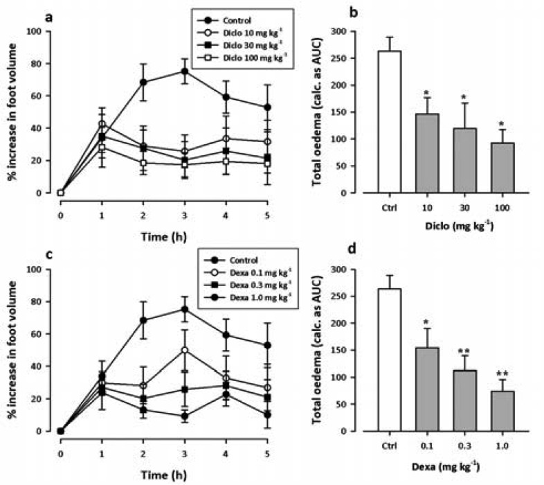
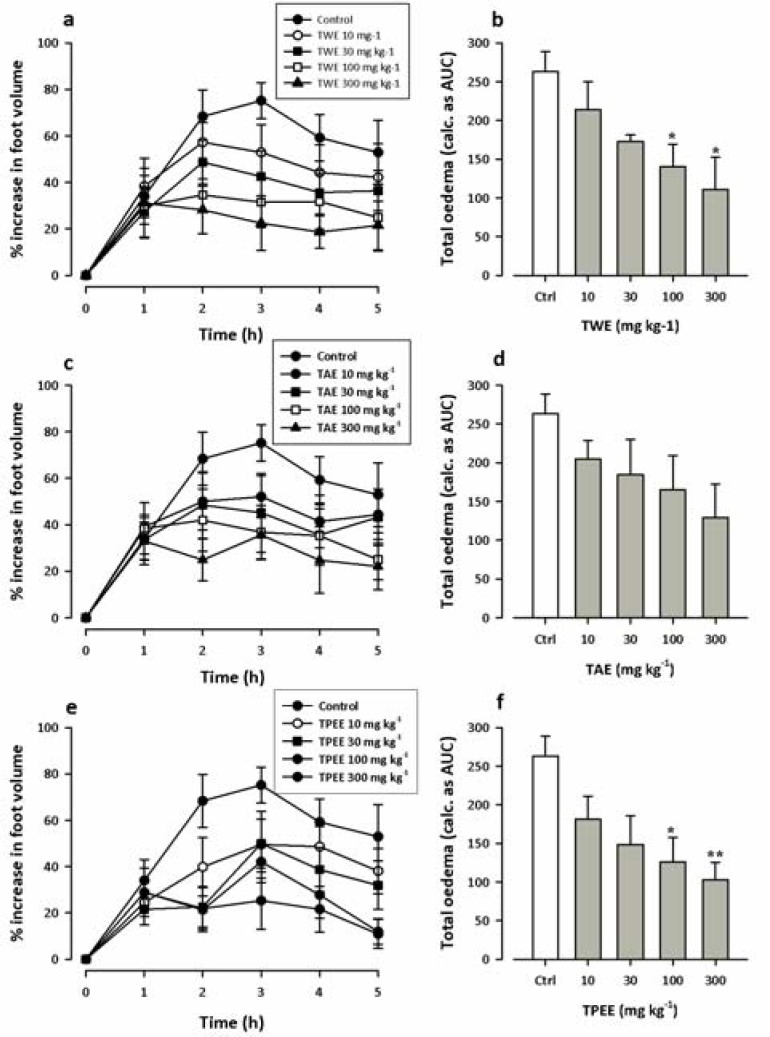
Intradermal injection of 10 µl of 1% carrageenan induced a time-dependent oedema response in the 7-day old chicks that peaked between 2-3 hours post carrageenan injection (Figure 1 and 2) (Roach and Sufka, 2003). Two-way ANOVA (treatment x time) from the time-course curves and one-way ANOVA from the area under the time course curves (AUCs) (Figures 1 and 2) revealed significant antiinflammatory effects for the reference drugs and extracts except the alcoholic extract (TAE), which did not show any significant antiinflammatory effect at the doses tested. The reference antiinflammatory agents diclofenac (_F_3, 96 = 10.74; P < 0.0001) and dexamethasone (_F_3, 96 = 16.81; P < 0.0001) inhibited carregeenan-induced foot oedema at all the doses tested (Figure 1) with maximal inhibition of the total oedema by 64.92±2.03 and 71.85±15.34 at 100 and 1 mg kg−1 for diclofenac and dexamethasone respectively. On the contrary, TWE (_F_4, 120 = 7.47; P < 0.0001) and TPEE (_F_4, 120 = 10.67; P <0.0001) caused inhibition of the total oedema (Figure 2) only at higher doses (100 and 300 mg kg−1) with maximal inhibitory effects of 57.79±3.92% and 63.83±12.84% respectively at 300 mg kg−1. They did not cause significant inhibition of the inflammatory oedema at 10 and 30 mg kg−1.
Figure 1.
Dose-response effects of diclofenac (10–100 mg kg−1, i.p.) (a and b); and dexamethasone (0.1–1.0 mg kg−1, i.p.) (c and d) on carrageenan-induced foot oedema in chicks. Left panels show the time course of effects and the right panels show the total oedema calculated as area under the time — course curve (AUCs) over the 5 h period. Data is presented as means ± s.e.m (n = 5). ** P < 0.01; *P < 0.05 compared to vehicle-treated group (two-way ANOVA followed by Bonferroni's post hoc test).
Figure 2.
Dose-response effects of TWE (10–300 mg kg−1, p.o.) (a and b); TAE (10–300 mg kg−1, p.o.) (c and d) and TPEE (10–300 mg kg−1, p.o.) (e and f) on carrageenan-induced foot oedema in chicks. Left panels show the time course of effects and the right panels show the total oedema calculated as area under the time — course curve (AUCs) over the 5 h period. Data is presented as means ± s.e.m (n = 5). ** P < 0.01; *P < 0.05 compared to vehicle-treated group (two-way ANOVA followed by Bonferroni's post hoc test).
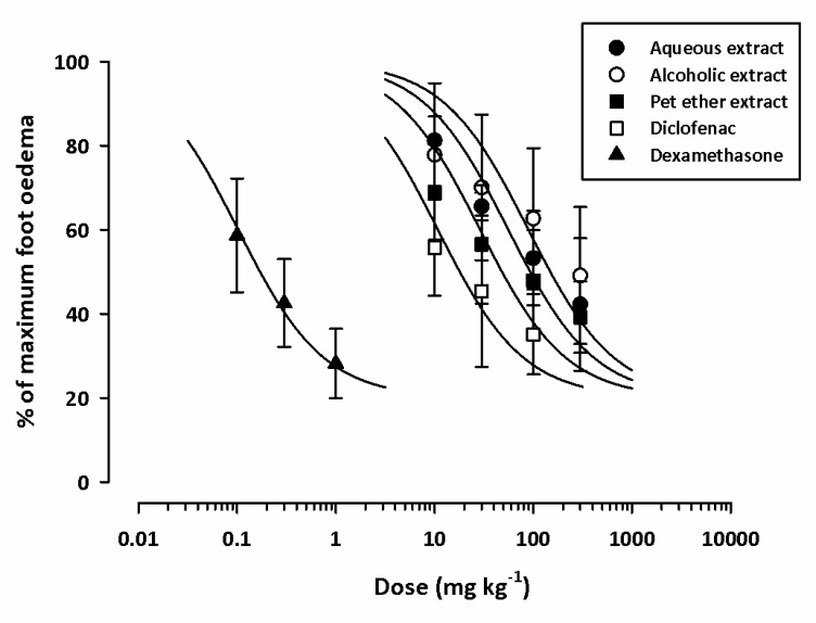
Based on ED50 values (Table 2) obtained from dose-response curves (Figure 3), dexamethasone was the most potent inhibitor of the inflammatory oedema. The rank order of potency was dexamethasone (0.10±0.01 mg kg−1) > diclofenac (10.79±1.57 mg kg−1) > TPEE (28.90±2.67 01 mg kg−1) > TWE (55.78±6.27 mg kg−1) > TAE (98.13±20.10). Comparing ED50 values, TPEE (the most potent plant extract) was about 300 fold less potent than dexamethasone.
Table 2.
ED50 values for drugs. Values are means ± s.e.m. (n=5)
| Drug | ED50 (mg kg−1) | |
|---|---|---|
| Carrageenan-induced oedema | Adjuvant-induced arthritis | |
| Aqueous extract (TWE) | 55.78±6.27. | 79.23±2.70 |
| Alcoholic extract (TAE) | 98.13±20.10 | 93.24±3.43 |
| Pet. Ether extract (TPEE) | 28.90±2.67 | 98.03.±2.49 |
| Diclofenac | 10.79±0.39 | 2.55±0.15 |
| Dexamethasone | 0.10±0.01 | 0.037±0.002 |
| Methotrexate | 0.031±0.002 |
Figure 3.
Dose response curves for dexamethasone (0.1–1 mg kg−1 i.p.), diclofenac (10–100 mg kg−1 i.p.), TWE (10–300 mg kg−1 p.o.), TAE (10–300 mg kg−1 p.o.) and TPEE (10–300 mg kg−1 p.o.) on carrageenan induced foot oedema in 7-day old chicks. Animal were treated 1 hour after carrageenan challenge. Each point represents the mean ± s.e.m. (n = 5).
Adjuvant arthritis
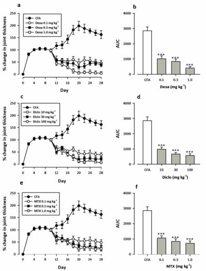
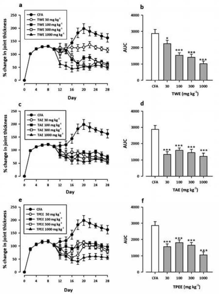
Intraplantar injection of CFA into the right foot pad of rats triggered an inflammatory response characterized by paw swelling in both the ipsilateral (injected) and the contralateral (non-injected) paw. The response on the injected paw was biphasic consisting of an acute phase characterised by unilateral swelling of the ipsilateral paw which peaked 8 days post CFA inoculation, and a subsequent polyarthritic/chronic phase which began by day 12 (Figures 4 and 5) confirmed by the development of inflammatory oedema in the contralateral paw. All the reference antiinflammatory agents - diclofenac (_F_3, 144 = 43.36, P < 0.0001), dexamethasone (_F_3, 144 = 37.07, P < 0.0001) and methotrexate (_F_3, 144 = 30.03, P < 0.0001) caused very significant inhibitions of the polyarthritic phase oedema, as was expected, with maximal inhibitions of 85.75±2.96% at 1 mg kg−1 for dexamethasone, 80.28±5.79% at 100 mg kg−1 for diclofenac and 74.68±3.03% at 1 mg kg−1 for methotrexate (Figure 4). Similarly all the plant extracts. TWE (_F_4, 180 = 14.90 P < 0.0001), TAE (_F_4, 180 = 10.36), and TPEE (_F_4, 180 = 12.08 P < 0.0001) significantly inhibited the polyarthritic phase oedema (Figure 5) with maximal inhibitions of 64.41±5.56% at 100 mg kg−1 for TWE, 57.04±8.57% at 1000 mg kg−1 for TAE and 62.18±2.56% at 1000 mg kg−1 for TPEE. From the AUCs it could be observed that whereas the inhibitory effect of TWE was dose dependent, the effect of TAE and TPEE were not dose dependent (Figure 5).
Figure 4.
Dose-response effects of dexamethasone (0.1–1 mg kg−1, i.p.) (a and b); diclofenac (10–100 mg kg−1, i.p.) (c and d) and methotrexate (0.1–1 mg kg−1, i.p.) (e and f) on ipsilateral joint thickness in the Adjuvant-induced arthritis. Left panels show the time course of effects and the right panels show the total oedema calculated as area under the time — course curve (AUCs) during the polyarthritic phase (days 10–28). Data is presented as means ± s.e.m (n = 5). *** P < 0.001 ** P < 0.01; *P < 0.05 compared to vehicle-treated group (two-way ANOVA followed by Bonferroni's post hoc test).
Figure 5.
Dose-response effects of TWE (30–1000 mg kg−1 p.o.) (a and b); TAE (30–1000 mg kg−1, p.o.) (c and d) and TPEE (30–1000 mg kg−1, p.o.) (e and f) on ipsilateral foot joint thickness in the Adjuvant-induced arthritis. Left panels show the time course of effects and the right panels show the total oedema calculated as area under the time — course curve (AUCs) during the polyarthritic phase (days 10–28). Data is presented as means ± s.e.m (n = 5). *** P < 0.001 ** P < 0.01; *P < 0.05 compared to vehicle-treated group (two-way ANOVA followed by Bonferroni's post hoc test).
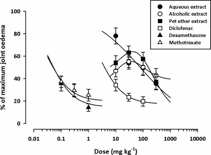
ED50 values (Table 2) obtained from dose-response curves (Figure 6), showed that methotrexate (ED50 = 0.031±0.002) and dexamethasone (ED50 = 0.037±0.002) were equipotent in inhibiting the inflammatory oedema associated with the polyarthritic phase, but diclofenac (ED50 = 2.55±0.15) was about seventy fold less potent in inhibiting the inflammatory oedema compared to dexamethasone and methotrexate. The ED50 values for the extracts (TWE, TAE, and TPEE) did not differ much from each other (79.23±2.70, 93.24±3.43, and 98.03±2.49 respectively) (Table 1), however the water extract (TWE) which is the most potent plant extract in this model was about 2000 fold less potent than methotrexate and dexamethasone
Figure 6.
Dose response curves (inhibition of oedema) for TWE (10–1000 mg kg−1 p.o.) (●), TAE (10–1000 mg kg−1 p.o.) (○), TPEE (10–1000 mg kg−1 p.o.) (■), dexamethasone (0.1–1 mg kg−1 i.p.) (▲), diclofenac (10–100 mg kg−1 i.p.) (□) and methotrexate (0.1–1 mg kg−1 i.p.) (△) on CFA induced arthritis in rats. Each point represents mean ± s.e.m. (n = 5).
Discussion
This study focused on the effect of plant extracts from the bark of T. monadelpha in animal models of acute (chickcarrageenan) and chronic inflammation (adjuvant arthritis). The results from this study clearly demonstrate that extracts from the bark of T. monadelpha has antiinflammatory properties in both acute and chronic inflammation.
Carrageenan-induced acute footpad oedema in laboratory animals first introduced by Winter et al., (1962) is a model of acute inflammation which has been widely used to evaluate non-steroidal antiinflammatory drugs (NSAID) (Di Rosa and Willoughby, 1971). The mediators involved in carrageenan oedema are released in three distinct phases, an initial release of histamine and 5-HT, a second phase mediated by kinins and the third phase highly suspected to be mediated by prostaglandins (Di Rosa et al., 1971). Inhibition of carrageenan-induced inflammation has shown to be highly predictive of antiinflammatory drug activity in human inflammatory disease (Morris, 2003) and remains an acceptable preliminary screening test for antirheumatic effect. Also, studies have shown that intraplantar injection of carrageenan in the 7-day-old chick elicits a quantifiable, reliable and relatively short lasting state of oedema, that is differentially attenuated by the administration of typical antiinflammatory compounds (Roach and Sufka, 2003). The rat adjuvant arthritis on the other hand, is the most frequently used model of chronic inflammation for screening NSAIDs, steroids and immunosuppressive drugs (Weichman, 1989; Crofford et al., 1992; Aota et al., 1996).
In this study, the reference antiinflammatory drugs (diclofenac, dexamethasone and methotrexate) inhibited the oedema associated with carrageenan-induced inflammation and the rat adjuvant arthritis. This clearly demonstrates the effectiveness of the chick-carrageenan, and the rat adjuvant arthritis models as established pharmacological tools for identifying drugs with potential antiinflammatory effects. Diclofenac (an NSAID) is thought to stop inflammation mainly by inhibiting prostaglandin synthesis through the inhibition of cyclooxygenase (COX) (Vane and Botting, 1987; Vane and Botting, 1996) whereas dexamethasone (a steroidal antiinflammatory drug) inhibits inflammation by regulating the expression of corticosteroid-responsive genes resulting in profound effects on the concentration, distribution and function of various inflammatory mediators (e.g. COX, leucocytes, cytokines, prostaglandins, thromboxanes and leucotrienes, vascular endothelium) synthesized by various target tissues (Cronstein and Weissmann, 1995; Vane and Botting, 1996; Schimmer and Parker, 2001). Methothrexate on the other hand, is a folate antagonist and an immunosuppressant, first introduced for the management of malignancies (Cronstein and Weissman, 1995, (Swierkot and Szechinski, 2006). It is presently prescribed at lower doses for the treatment of rheumatoid arthritis (Furst and Kremer, 1988; Swierkot and Szechinski, 2006). Although various biochemical pathways may be involved in its antiinflammatory actions in rheumatoid arthritis, studies to date indicate that the most important actions of low-dose MTX are its effects in increasing adenosine level and reducing the proinflammatory while increasing antiinflammatory cytokine levels (Cronstein and Weissmann, 1995; Swierkot and Szechinski, 2006).
The inhibition of inflammatory oedema by the plant extracts indicates the presence of compounds capable of inhibiting both acute and chronic inflammatory processes. The precise mechanism by which T. monadelpha extracts inhibited inflammation was outside the scope of this study, however, a colossal amount of data from natural product research shows that plant constituents such as alkaloids, flavonoids, glycosides, terpenoids, steroids and many other secondary plant metabolites may exhibit antiinflammatory effects (Calixto et al., 2003; Calixto et al., 2004; Rios, 2008) by their modulatory effects on inflammatory mediators (e.g. arachidonic acid metabolites, peptides, excitatory amino acids, amines), the formation and/or action of second messengers (e.g. cGMP, cAMP, various protein kinases and calcium), the expression of transcription factors (e.g. AP-1, NF-kB), and the expression of key pro-inflammatory molecules such as inducible NO synthase (iNOS), COX, cytokines (e.g. IL-1B, TNF-α), neuropeptides and proteases (Calixto et al., 2003; Calixto et al., 2004; Rios, 2008). Phytochemical studies of the plant bark showed it contains alkaloids, tannins, saponins, steroids, flavonoids, terpenoids and glycosides. These plant metabolites may be responsible for the antiarthritc effects of T. monadelpha extracts, since various plant medicines used for different forms of arthritis have been found to contain similar phytochemicals (Ahmed et al., 2005; Mythilypriya et al., 2008; Puratchikody et al., 2011). Also, pharmacological and phytochemical studies on the stem bark of some members of the family Meliaceae (e.g. Khaya grandifoliola) used in the traditional management of arthritis showed antiinflammatory effects with the major phytochemicals being alkaloids, saponins, and tannins (Abiodun et al., 2009). Furthermore Bridelia ferrugenia (Euphorbiaceae) stem bark, which is ethnomedically used to treat arthritis (Mshana et al., 2000) was found to contain alkaloids, glycosides, steroids and tannins as the major plant constituents (Adebayo and Ishola, 2009). It can therefore be inferred that the presence of such a large array of similar phytochemicals in the stem bark of T. Monadelpha, may be largely responsible for the antiarthritic effect, and extensive ethnomedical uses of the plant.
Nevertheless, whereas it is clear that the T. monadelpha extracts are effective in reducing inflammation in adjuvant arthritis, it remains uncertain whether this is translated into an improvement in indices of joint integrity such as bone and cartilage degradation. Further work is therefore, currently underway to identify the individual molecules responsible for the antiinflammatory properties, their mechanism of action, and also, to ascertain their effect on bone and joint integrity. The ethanolic extract did not show significant antiinflammatory effect in carrageenan oedema, but inhibited the inflammatory oedema associated with adjuvant arthritis. This does not necessarily suggest the absence of compounds with antiinflammatory effect in acute inflammation in the ethanolic extract, but rather since crude extracts contain several compounds, which may exhibit varying pharmacological effects in biological systems, a cancellation effect involving antiiflammatory and proinflammatory components may have occurred (Davicino et al., 2010). It is also possible that the components capable of inhibiting acute inflammation were present in very small concentrations in the alcoholic extract compared with the water and petroleum-ether extracts.
In conclusion, our work suggests that T. monadelpha bark extract is a potential source of antiinflammatory agents and thus provides the first pharmacological evidence to support its use in the management of inflammatory conditions in African TM.
Acknowledgement
The authors are grateful for the technical assistance offered by Messrs Thomas Ansah, Gordon Darku and George Ofei of the Department of Pharmacology, Faculty of Pharmacy and Pharmaceutical Sciences, KNUST, Kumasi.
References
- 1.Abbiw D K. Useful Plants of Ghana. Kew: Intermediate Tecgnology Publications and The Royal Botanic Gardens; 1990. [Google Scholar]
- 2.Abiodun F, Ching F P A, Sunday A A, Odion E. Phytochemical and Antiinflammatory Evaluation of Khaya grandifoliola Stem Bark Extract. Int J PharmTech Res. 2009;1:1061–1064. [Google Scholar]
- 3.Adebayo E A, Ishola O R. Phytochemical and antimicrobial screening of the crude extracts from the root, stem bark and leaves of Bridelia ferruginea. Afr J Biotechnol. 2009;8:650–653. [Google Scholar]
- 4.Ahmed S, Anuntiyo J, Malemud C J, Haqqi T M. Biological Basis for the Use of Botanicals in Osteoarthritis and Rheumatoid Arthritis: A Review. eCAM. 2005;2:301–308. doi: 10.1093/ecam/neh117. [DOI] [PMC free article] [PubMed] [Google Scholar]
- 5.Aladesanmi A J, Odediran S A. Antimicrobial activity of Trichilia heudelotti leaves. Fitoterapia. 2000;71:179–182. doi: 10.1016/s0367-326x(99)00143-4. [DOI] [PubMed] [Google Scholar]
- 6.Aota S, Nakamura T, Suzuki K, Tanaka Y, Okazaki Y, Segawa Y, Miura M, Kikuchi S. Effects of indomethacin administration on bone turnover and bone mass in adjuvant-induced arthritis in rats. Calcif Tissue Int. 1996;59:385–391. doi: 10.1007/s002239900144. [DOI] [PubMed] [Google Scholar]
- 7.Atindehou K K, Schmid C, Brun R, Koné M W, Traore D. Antitrypanosomal and antiplasmodial activity of medicinal plants from Côte d'Ivoire. J Ethnopharmacol. 2004;90:221–227. doi: 10.1016/j.jep.2003.09.032. [DOI] [PubMed] [Google Scholar]
- 8.Calixto J, Otuki M F, Santos A R S. Anti-Inflammatory Compounds of Plant Origin. Part I. Action on Arachidonic Acid Pathway, Nitric Oxide and Nuclear Factor kB (NF-kB) Planta Med. 2003;69:973–983. doi: 10.1055/s-2003-45141. [DOI] [PubMed] [Google Scholar]
- 9.Calixto J B, Campos M M, Otuki M F, Santos A R S. Anti-Inflammatory Compounds of Plant Origin. Part II. Modulation of Pro-Inflammatory cytokines, Chemokines and Adhesion Molecules. Planta Med. 2004;70:93–103. doi: 10.1055/s-2004-815483. [DOI] [PubMed] [Google Scholar]
- 10.Crofford L J, Sano H, Karalis K, Webster E L, Goldmuntz E A, Chrousos G P, Wilder R L. Local secretion of corticotropin-releasing hormone in the joints of Lewis rats with inflammatory arthritis. J Clin Invest. 1992;90:2555–2564. doi: 10.1172/JCI116150. [DOI] [PMC free article] [PubMed] [Google Scholar]
- 11.Cronstein B N, Weissmann G. Targets For Antiinflammatory Drugs. Annu Rev Pharmacol Toxicol. 1995;35:449–462. doi: 10.1146/annurev.pa.35.040195.002313. [DOI] [PubMed] [Google Scholar]
- 12.Davicino R, Mattar A, Casali Y, Anesini C, Micalizzi B. Different activities of Schinus areira L.: anti-inflammatory or pro-inflammatory effect. Immunopharmacol Immunotoxicol. 2010;32:620–627. doi: 10.3109/08923971003657305. [DOI] [PubMed] [Google Scholar]
- 13.Di Rosa M, Giroud J P, Willoughby D A. Studies of the mediators of the acute inflammatory response induced in rats in different sites by carrageenan and turpentine. J Pathol. 1971;104:15–29. doi: 10.1002/path.1711040103. [DOI] [PubMed] [Google Scholar]
- 14.Di Rosa M, Willoughby D A. Screens for anti-inflammatory drugs. J Pharm Pharmacol. 1971;23:297–298. doi: 10.1111/j.2042-7158.1971.tb08661.x. [DOI] [PubMed] [Google Scholar]
- 15.Evans W C. ‘Trease & Evans’ Pharmacognosy. London: Elsevier; 2009. pp. 133–415. [Google Scholar]
- 16.Farnsworth N R. Biodiversity. Wahington, D.C.: E. O. Wilson; 1988. Screening Plants for New Medicines; pp. 83–97. [Google Scholar]
- 17.Fereidoni M, Ahmadiani A, Samnanian S, Javan M. An accurate and simple method for measurement of paw edema. J Pharmacol and Toxicol Methods. 2000;43:11–14. doi: 10.1016/s1056-8719(00)00089-7. [DOI] [PubMed] [Google Scholar]
- 18.Furst D E, Kremer J M. Methotrexate in rheumatoid arthritis. Arthritis Rheum. 1988;31:305–314. doi: 10.1002/art.1780310301. [DOI] [PubMed] [Google Scholar]
- 19.Irvine F R. Woody plants of Ghana. London: Oxford University Press; 1961. p. 528. [Google Scholar]
- 20.Lemmens R H M J. Plant Resources of Tropical Africa. 1. Vol. 7. J. J. de Wilde; 2008. Trichilia Monadelpha (Thonn) Timbers 1. D. Louppe, A. A. Oteng-Amoako and M. Brink, PROTA Foundation Wageningen, Netherlands/Backhuys Publishers, Leiden, Netherlands/CTA, Wageningen, Netherlands: 561–563. [Google Scholar]
- 21.McCartney-Francis N L, Chan J, Wahl S M. Inflammatory Joint Disease. Inflammation Protocols. Totowa, NJ: Humana Press Inc.; 2003. pp. 147–159. [DOI] [PubMed] [Google Scholar]
- 22.Miller J R. GraphPad Version 4.0. Step-by-Step Examples. San Diego, CA: GraphPad Software Inc.; 2003. [Google Scholar]
- 23.Morris C J. Carrageenan-Induced Paw Edema in the Rat and Mouse. Inflammation Protocols. Totowa, NJ: Humana Press Inc.; 2003. pp. 115–121. [DOI] [PubMed] [Google Scholar]
- 24.Motulsky H J, Christopoulos A. Fitting model to biological data using linear and nonlinear regression. A practical guide to curve fitting. San Diego, CA: GraphPad Software Inc.; 2003. [Google Scholar]
- 25.Mshana N R, Abbiw K, Addae-Mensah I, Adjanohoun E, Ahyi M R A, Ekpere J A, Enow-Orock E G, Gbile Z O, Noamesi G K, Odei M A, Odunlami H, Oteng-Yeboah A A, Sarpong K, Soforowa A, Tackie A N. Traditional Medicine and Pharmacopoeia. Contribution to the Revision of Ethnobotanical and Floristic Studies in Ghana. Accra: Organization of African Unity/Scientific, Technical & Research Commision; 2000. pp. 676–677. 618. [Google Scholar]
- 26.Mythilypriya R, Shanthi P, Sachdanandam P. Synergistic Effect of Kalpaamruthaa on Antiarthritic and Antiinflammatory Properties—Its Mechanism of Action. Inflammation. 2008;31:391–398. doi: 10.1007/s10753-008-9090-2. [DOI] [PubMed] [Google Scholar]
- 27.Odugbemi T, Odunayo A. Medicinal Plants According to Family Names. Outlines and Pictures of Medicinal Plants from Nigeria. T. Odugbemi. Lagos: University of Lagos Press; 2008. pp. 117–146. [Google Scholar]
- 28.Pearson C M. Development of arthritis, periarthritis and periostitis in rats given adjuvants. Proc Soc Exp Biol. 1956;91:95–101. doi: 10.3181/00379727-91-22179. [DOI] [PubMed] [Google Scholar]
- 29.Puratchikody A, Yasodha A, Kumar S A, Hari V B N. Preliminary Phytochemical and Anti-arthritic Activity of an Ayuverdic Formulation Yogaraja Gulgulu. J Phytol. 2011;3:31–36. [Google Scholar]
- 30.Quick J, Zhang X, Kasilo O, D'Alessio R, Land S, Graaff P, de Joncheere K, Weerasuriya K, Ken C. WHO Policy Perspectives on Medicines-Traditional Medicine-Growing Needs and Potential. Geneva: E. D. a. M. P. H. T. a. P. Cluster; 2002. pp. 1–6. [Google Scholar]
- 31.Rios J-L. In: Mechanism of action of Anti-inflammatory phytochemicals. Botanical Medicine In Clinical Practice. Watson R R, Preedy V R, editors. Washington D. C.: CAD International; 2008. pp. 524–534. [Google Scholar]
- 32.Roach J T, Sufka K J. Characterisation of the chick carrageenan response. Brain Res. 2003;994:216–225. doi: 10.1016/j.brainres.2003.09.038. [DOI] [PubMed] [Google Scholar]
- 33.Schimmer B P, Parker K L. In: Adrenocorticotropic Hormone; Adrenocortical Steroids And Their Synthetic Analogs; Inhibitors Of The Synthesis And Actions Of Adrenocortical Hormones. Goodman & Gilman's The Pharmacological Basis Of Therapeutics. Hardman J G, Limbird L E, editors. Vol. 10. New York, McGraw-Hill: Medical Publishing Division; 2001. pp. 1649–1677. [Google Scholar]
- 34.Sofowora A. Standardization of Herbal Medicines. Medicinal Plants and Traditional Medicine in Africa. Ibadan: Spectrum Books Ltd.; 1993. pp. 50–63. [Google Scholar]
- 35.Swierkot J, Szechinski J. Methotrexate in rheumatoid arthritis. Pharmacol Rep. 2006;58:473–492. [PubMed] [Google Scholar]
- 36.Vane J, Botting R. Inflammation and the mechanism of action of anti-inflammatory drugs. FASEB J. 1987;1:89–96. [PubMed] [Google Scholar]
- 37.Vane J R, Botting R M. Mechanism of Action of Anti-Inflammatory Drugs. Scand J Rheumatol. 1996;25(Suppl 102):9–21. doi: 10.3109/03009749609097226. [DOI] [PubMed] [Google Scholar]
- 38.Vickers A, Zollman C. ABC of complementary medicine Herbal medicine. BMJ. 1999;319:1050–1053. doi: 10.1136/bmj.319.7216.1050. [DOI] [PMC free article] [PubMed] [Google Scholar]
- 39.Vickers A, Zollman C. ABC of complementary medicine Herbal medicine. Br Med J. 1999;319 doi: 10.1136/bmj.319.7216.1050. [DOI] [PMC free article] [PubMed] [Google Scholar]
- 40.Weichman B M. In: Rat Adjuvant Arthritis: A Model of Chronic Inflammation. Pharmacological Methods in the Control of Inflammation. Chang J Y, Lewis A J, editors. New York: Alan R. Liss, Inc.; 1989. pp. 363–380. [Google Scholar]
- 41.Weiss C. Ethnobotanische und pharmacologische Studen zu Arzneipflanzen der traditionellen Medizin der Elfenbeinkuste. Basel: Uniiversitat Basel; 2009. (Ger). [Google Scholar]
- 42.WHO, author. WHO Traditional Medicine Strategy 2002–2005. Geneva: 2002. [Google Scholar]
- 43.WHO/ICUN/WWF, author. Guidelines on the Conservation of Medicinal Plants. Geneva: The International Union for Conservation of Nature and Natural Resources (IUCN), Gland, Switzerland, in partnership with The World Health Organization (WHO), Geneva, Switzerland, and WWF -World Wide Fund for Nature, Gland, Switzerland; 1993. 1993. [Google Scholar]
- 44.Winter C A, Risley E A, Nuss G W. Carrageenan-induced edema in hind paw of the rat as an assay for anti-inflammatory drugs. Proc Soc Exp Biol. 1962;111:544–547. doi: 10.3181/00379727-111-27849. [DOI] [PubMed] [Google Scholar]
- 45.Zhang X. General guidelines for Methodologies on Research and Evaluation of Traditional Medicine. Geneva: D. o. E. D. a. M. P. (EDM); 2000. WHO/EDM/TRM/2000.1. [Google Scholar]
