Biasing the content of hippocampal replay during sleep (original) (raw)
. Author manuscript; available in PMC: 2015 Mar 10.
Published in final edited form as: Nat Neurosci. 2012 Sep 2;15(10):1439–1444. doi: 10.1038/nn.3203
Abstract
The hippocampus plays an essential role in encoding self-experienced events into memory. During sleep, neural activity in the hippocampus related to a recent experience has been observed to spontaneously reoccur, and this “replay” has been postulated to be important for memory consolidation. Task-related cues can enhance memory consolidation when presented during a post-training sleep session, and if memories are consolidated by hippocampal replay, a specific enhancement for this replay should also be observed. To test this, we have trained rats on an auditory-spatial association task, while recording from neuronal ensembles in the hippocampus. Here we report that during sleep, a task-related auditory cue biases reactivation events towards replaying the spatial memory associated with that cue. These results indicate that sleep replay can be manipulated by external stimulation, and provide further evidence for the role of hippocampal replay in memory consolidation.
The hippocampus is critical for encoding recent episodic experiences into memory1, 2. After an initial encoding phase, a memory is believed to undergo a process of consolidation in which its representation is stabilized in neocortex, allowing future retrieval to be independent of the hippocampus3. The mechanisms underlying memory consolidation are poorly understood, however interactions between the hippocampus and neocortex during sleep have been proposed to be crucial for this process4, 5, 6, 7. In rodents, neurons in hippocampus and cortex that were active during a previous experience have been observed to spontaneously reactivate during non-REM sleep8, 9, 10. Similarly in humans, a reactivation of brain activity related to a previous experience has also been observed in the hippocampus during sleep11.
While “replay” of a neural sequence associated with a previous experience is a neural correlate of memory, its causal role in memory consolidation has not yet been demonstrated. The best evidence supporting replay’s role in memory consolidation has come from the observation that inactivating the hippocampus during replay events produces learning deficits12, 13. However, because in these experiments the hippocampus was electrically stimulated while in a hyper-excitable state, the learning deficits observed could have been due to the disruption of stored memory traces in the hippocampus rather than the blockade of replay events. An alternative approach for investigating replay’s function is to enhance memory consolidation by manipulating which encoded memories are replayed in the hippocampus. Several recent studies in human subjects have paired sensory cues with a hippocampus-dependent memory task, and have observed learning improvements when these sensory cues are presented during non-REM sleep between sessions14, 15, 16. If hippocampal replay drives memory consolidation, presenting a sensory cue during non-REM sleep should be able to bias reactivation events towards replaying the previous experience associated with the cue.
Results
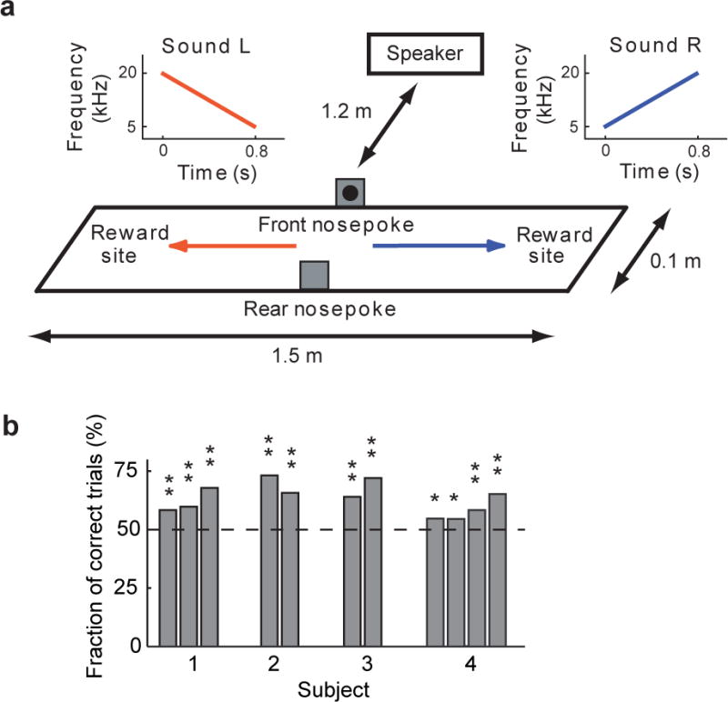
We tested this hypothesis by recording neuronal ensembles10, 17 in the hippocampus (right dorsal CA1) of four rats performing an auditory-spatial association task (Fig. 1a). After initiating a trial with a nosepoke, the rat heard one of two sounds. For Sound R (an upwards frequency sweep) rats had to run to the right end of the track to receive a reward, while Sound L (a downwards frequency sweep) indicated reward delivery at the left end of the track [see Methods]. After rats could perform a significant number of correct trials in a single session (Fig. 1b, P<0.05, Binomial distribution), we recorded place cell activity18, 19 in the hippocampus during the task (Fig. 2a) and a subsequent sleep session (Fig. 2b) where task-related stimuli (Sound R and Sound L) and control stimuli were played in the background in a random order [see Methods]. We did observe a weak correlation between the rat’s position being biased towards the resulting direction traversed on the task (Fig. 2a), with a larger correlation observed for error trials (correct trials, r = 0.09, P < 6.5 × 10⁻⁶, 2474 trials; error trials, r = 0.19, P < 1.5×10⁻¹³, 1446 trials). Given that the rats were not performing at 100%, they were guessing on a subset of the trials, and such a correlation could arise if a position bias influenced their decision when guessing.
Figure 1.
Behavioral taska
a. Behavioral task design. After the rat made a nosepoke, one of two sounds (Sound R or Sound L) was played from a speaker 1.2 m away from the track center (in front of the nosepoke). For a correct trial, the rat has to go the right side of the track for Sound R, and the left side of the track for Sound L.
b. Performance on behavioral task. For the 11 recorded sessions in four subjects, task performance was above chance (* P < 0.05, ** P < 0.001, Binomial distribution).
Figure 2.
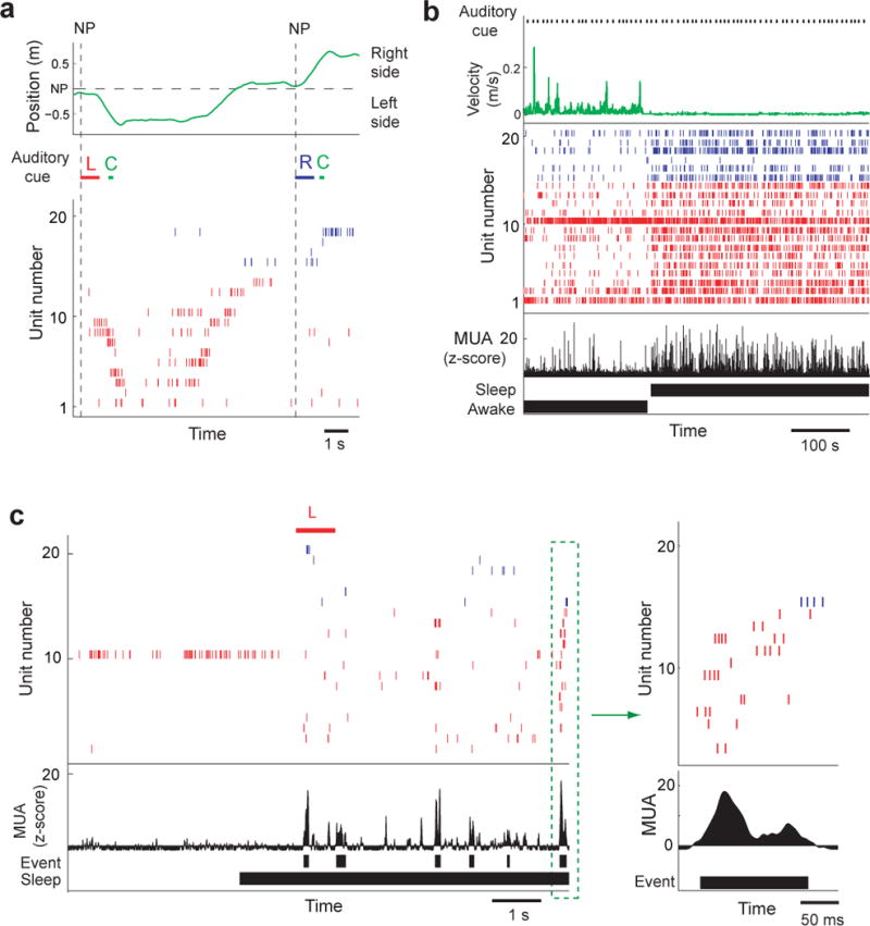
Hippocampal activity during behavior and sleep (Rat 1, session 2)
a. Neuronal ensemble activity during two correct behavioral trials- From top to bottom: (1) the spatial position (green) of the subject on the track, (2) acoustic stimuli- Sound R (R) in blue, a tone indicating a correct trial and reward delivery cue (C) in green, and Sound L (L) in red), (3) raster plot of place cell activity. Nosepokes (NP) are indicated by a vertical dashed line. Sound L was played when a nosepoke was made, and the rat correctly ran to the left side of the track. Having returned and made another nosepoke, the rat heard Sound R and ran to the right side of the track. In the raster plot, spikes from place cells with place fields on the right side of the track are blue, and left-sided place fields in red. Place fields are ordered from top to bottom by their location on the track (right → left side).
b. Neuronal ensemble activity during the onset of sleep while the animal is resting in the sleep box after the behavioral task. From top to bottom (1) the onsets of acoustic stimuli, (2) animal’s velocity (green), (3) a raster plot of place cell activity (same format as in Fig. 2a), (4) multiunit activity, (5) awake/sleep classification. Note the increase in the time scale from Fig. 2a to Fig. 2b.
c. Example of reactivation events occurring after the onset of sleep (from Fig. 2b). Prior to sleep onset, the rat was resting in the sleep chamber. The reactivation event in the green dashed box is shown to the right (same format as Fig. 2a). From top to bottom: (1) acoustic stimulus, (2) place cell activity (red=left-sided place fields, blue=right side place fields), (3) multiunit activity (MUA), (4) candidate reactivation events, (5) sleep classification.
We recorded a total of 409 neurons (4 rats, 11 sessions), of which 199 units had place fields on the track with a minimum peak firing rate greater than 2 spk/s and a mean rate less than 5 spk/s [see Methods]. Reactivation events (Fig. 2c) were detected from large transient increases in multiunit activity17, and analyzed by measuring each place cell’s firing rate and reconstructing the content of replay from the entire ensemble of place cells using Bayesian decoding methods [see Methods].
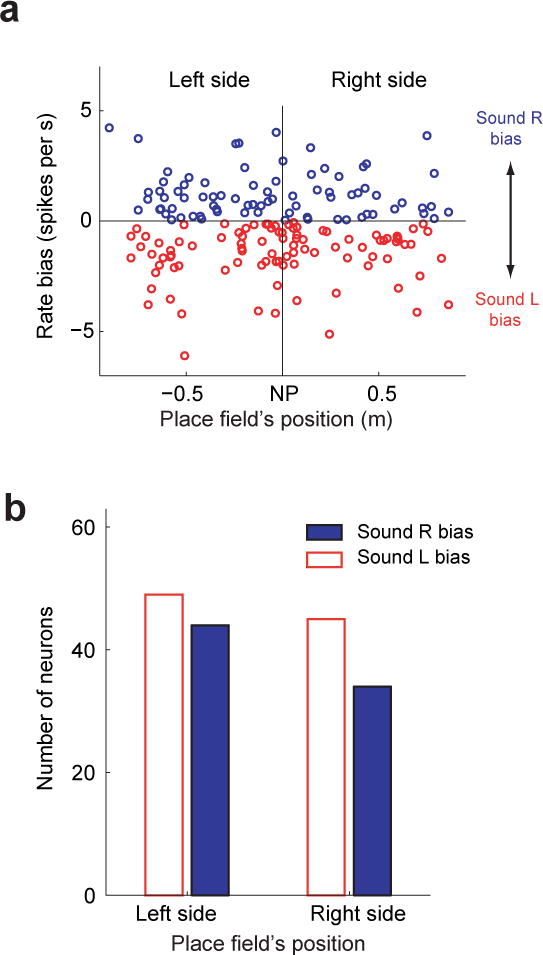
If the content of a replay event can be altered by the presentation of an auditory cue during non-REM sleep, we should observe differences between Sound R and Sound L related replay activity. For each place cell, we first computed the rate bias, for which the mean firing rate during non-REM sleep replay events [see Methods] occurring after Sound L‘s onset and before the onset of the next acoustic stimulus (between 5.8 and 10.8 seconds later) was subtracted from the corresponding events after Sound R. A positive value indicated a Sound R bias, i.e., a place cell had a higher firing rate during replay events that occurred after Sound R compared to Sound L, while a negative value indicated a Sound L bias. We observed that the majority of right-sided place fields had a positive rate bias and left-sided place fields more commonly had a negative rate bias (Fig. 3, Fig. 4a, r = 0.29, P < 1.1 × 10⁻⁴, Pearson correlation, and Fig. 4b, Binomial test, Left-side: P < 1.9 × 10⁻⁴ Right-side: P < 0.003). Furthermore, the mean rate bias was significantly different between right-sided and left-sided place fields (Fig. 4c, first column, P < 7.8 × 10⁻⁵, 1-way ANOVA), with a mean positive bias for right-sided place fields (Sound R preference) and a mean negative bias for left-sided place fields (Sound L preference). We observed a similar significant difference in mean rate bias between right and left-sided place fields using alternative methods of calculating the rate bias (P < 5.0 × 10⁻⁴, 1-way ANOVA, Fig. S1) and each place field’s side-of-the-track preference (P < 1.2 × 10⁻⁴, 1-way ANOVA, Fig. S2). Three out of four subjects showed a significantly greater Sound R bias for right-sided place fields than for left-sided place fields, (P < 0.05, 1-way ANOVA, Fig. S3).
Figure 3.
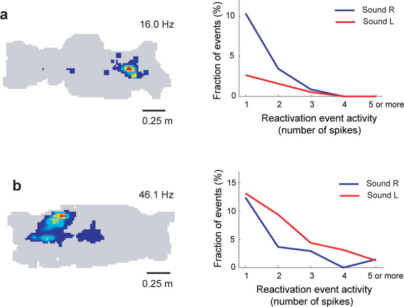
Place cell responses during sleep reactivation events
Place field (left) and plot of place cell activity (right) during reactivation events occurring after sound R (blue) and sound L (red).
a. Rat 4, session 1, cluster 17
b. Rat 1, session 2, cluster 41
Figure 4.
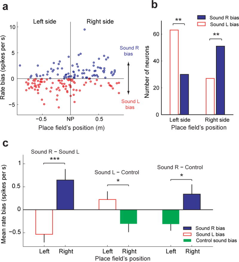
Rate bias during sleep reactivation events
Error bars indicate the standard error of the mean (SEM).
a. Rate bias during sleep replay events of individual place cells with place fields on the left or right side of the nosepoke (n=171). Place cells are ordered along the x-axis according to the location of their place field’s on the track. A positive rate bias indicates that the mean firing rate during replay events occurring after Sound R was higher than after Sound L. The vertical dashed line indicates the center of the track (location of the nosepoke).
b. The number of place cells during sleep replay events with a Sound L or Sound R bias. Place cells are grouped according to the position of their place field along the track (left or right). Left-sided place cells had significantly more units with a Sound L bias than a Sound R bias (** P < 1.9 × 10⁻⁴, Binomial distribution). Opposite to this, right-sided place cells had significantly more units with a Sound R bias than a Sound L bias (** P < 0.003, Binomial distribution).
c. The mean rate bias for left-sided and right-sided place cells during sleep replay events. In the first column, the mean rate bias compares Sound R and Sound L. In the second column the mean rate bias compares Sound L and the two task-related control sounds (reward and error cue). In the third column the mean rate bias compares Sound R and the two task-related control sounds (reward and error cue). The mean rate bias is significantly different between place fields on the left side and right side of the track in all three analyses (1-way ANOVA: Sound R − Sound L, *** P < 7.8 × 10⁻⁵; Sound L − control, * P < 0.04; Sound R − control, * P < 0.02).
Although we observed a significant difference in the mean rate bias between left and right-sided place fields, this analysis does not tell us whether the bias is caused by Sound R, Sound L or a combination of both sounds. Therefore, in addition to playing Sound R and Sound L while the subject was resting/sleeping after the behavioral task, we also played control sounds that were unassociated with a side of the track during the behavioral task [see Methods]. Next, we recalculated the mean rate bias during non-REM sleep by comparing firing rates during replay events that occurred after either Sound R or Sound L to the two task-related control sounds (acoustic cues for correct and incorrect trials). Both Sound R and Sound L independently evoked a significantly different mean rate bias between right and left sided place fields, with Sound L having a positive bias (Sound L > control sounds) on left sided place fields (P < 0.04, 1-way ANOVA, Fig. 4c) and Sound R having a positive bias (Sound R > control sounds) on right sided place fields (P < 0.02, 1-way ANOVA, Fig. 4c). These data indicate that both task-related cues presented during sleep could independently bias hippocampal activity during reactivation events. We found no evidence that acoustic stimulation had an effect on the number of reactivation events that were occurring, as a similar number of replay events occurred for all acoustic stimuli tested (Sound R, Sound L, and control stimuli) (Kruskal-Wallis test, P > 0.05). This indicates that a task-related cue only biases the content of replay events that are spontaneously occurring during sleep.
Sound-evoked responses have been observed in the hippocampus20. We identified 17 out of 199 neurons that had candidate sound-evoked responses during the behavioral task, defined as having a response to sound R that was significantly different (1-way ANOVA, P < 0.05) from sound L, and a firing rate during either sound R’s or sound L’s presentation that was significantly different (1-way ANOVA, P < 0.05) from the discharge rate calculated over the preceding one second of activity (Fig. S4). In order to minimize the influence of a neuron’s place field on our analysis, we directly compared correct and incorrect trials to the same side of the track (for left and right trials), and did this separately for trials initiated from the front and rear nosepoke (candidate auditory evoked responses, front nosepoke: run left=5 neurons, run right=6 neurons; rear nosepoke: run left=2 neurons, run right=5 neurons) (Fig S5a–d). One neuron had a candidate sound evoked response from both the front nosepoke (run right) and the rear nosepoke (run left). While some of these responses appeared to be due to subtle differences in the animal’s behavior that correlated with sound R and sound L trials, this classification of candidate sound evoked responses conservatively estimates the maximum number of neurons that could have sound evoked responses in our experiment. To explore the possibility that our observed cue-evoked biasing of place cell firing during sleep reactivation events was simply a consequence of correlated sound evoked responses in the behavioral apparatus and the sleep chamber, we recalculated the rate bias after excluding all 17 neurons that had candidate sound-evoked responses and observed a similar result (left-sided mean rate bias=−0.47, right-sided mean rate bias=0.62, one-way ANOVA, P < 8.3 × 10⁻⁴). We also calculated the number of units with candidate sound evoked responses while the subject was awake in the sleep box (after the behavioral session). Only two neurons passed our criteria for a candidate sound-evoked response, and neither of these neurons had candidate sound-evoked responses during the behavior task. Furthermore, we did not observe a significant correlation in the sound evoked activity bias (sound R − sound L) between in the two environments (behavioral apparatus and sleep chamber) while the animal was awake (Pearson correlation, front nospoke: right trials vs. sleep box: r = 0.08, P = 0.26, left trials vs sleep box: r = 0.06, P = 0.42, rear nospoke: right trials vs. sleep box: r = −0.13, P = 0.08, left trials vs sleep box: r = −0.03, P = 0.69, Fig. S5e,f). To address any potential bias arising from longer-latency sound evoked responses, we calculated the correlation between the sound evoked activity bias (sound R − sound L) in the two environments while the animal was awake using different analysis windows (0-1 s, 0-5 s, 0-10 s), starting after the offset of the acoustic stimulus. We did not observe a significant correlation in any of these cases (0-1 s: r=−0.03, P=0.63; 0-5 s: r=0.01, P=0.85; 0-10 s: r=−0.01, P=0.87). These results indicate that direct sound-evoked responses are context dependent20, and that any sound-evoked response occurring during behavior will differ from that evoked in the sleep chamber.
In human subjects, the enhancement of memory consolidation by task-related cues is specific to non-REM sleep; no enhancement is observed during REM sleep or when the subject is awake14, 15. In fact, presentation of task-related cues while a subject is awake can be detrimental for memory consolidation if interference is caused by a second behavioral task, potentially a byproduct of reconsolidation16. Therefore, we also computed the mean rate bias using replay events that occurred while the rat was awake in its sleep box. We did not observe a significant difference between the rate bias for right and left-sided place cells (P = 0.82, 1-way ANOVA, Fig. 5, Fig. S6) during awake reactivation events. These data indicate that in rats, the sound-evoked bias in the replay activity of place cells occurs during non-REM sleep, but not in the awake state, in agreement with data previously reported in human subjects.
Figure 5.
Rate bias during awake reactivation events
Error bars indicate the standard error of the mean (SEM).
a. Rate bias of individual place cells during awake reactivation events (n = 172). Format of plot is the same as Fig 4a. Pearson correlation coefficient: r = –0.03, P = 0.74
b. Number of neurons with sound R and sound L biases (Binomial test, left-sided place fields: P=0.27; right-sided place fields: P=0.87)
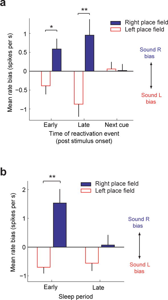
We next investigated the temporal dynamics of the observed bias in non-REM sleep replay events occurring after the onset of a task-related auditory cue. The analysis window starting at the onset of the task-related auditory cue and ending with the onset of the next cue was divided into two equal time windows: Early (0-5.4 s after stimulus onset) and Late (5.4–10.8 s after stimulus onset). The difference in mean rate bias between right-sided and left-sided place fields remained significant for both the early and late portion of this analysis window (1-way ANOVA, Early: P < 0.007, Late: P < 7.4 × 10⁻⁴, Fig. 6a). However, after the onset of the next cue, the previous task-related auditory cue no longer biased the content of reactivation events (1-way ANOVA, P = 0.87, Fig. 6a). These data indicate that although the cue itself only lasts 800 ms, the evoked reactivation bias persists for up to 10 seconds after the offset of the auditory cue (Sound R and Sound L), however after the presentation of a second acoustic stimulus this bias is reset.
Figure 6.
Temporal dynamics of sound-evoked reactivation bias
Error bars indicate the standard error of the mean (SEM).
a. Mean rate bias for events occurring at different time points relative to the onset of the acoustic stimulus. Events were divided into three groups based on when the events occurred, relative to the sound. For the first group (Early), events occurred between 0–5.4 seconds after the onset of the sound (1-way ANOVA, * P < 0.007). For the second group (Late), events occurred between 5.4–10.8 seconds after the onset of the sound, but before the onset of the next acoustic stimulus (1-way ANOVA, ** P < 7.4 × 10⁻⁴). For the third group, events occurred after the onset of the next acoustic stimulus (1-way ANOVA, P = 0.87).
b. In each experiment, the sleep session consisting of one or more sleep/wake cycles was concatenated, and divided into two periods, first half (Early) and second half (Late) of the total time spent asleep. The mean rate bias was calculated using all reactivation events in the Early and Late sleep periods. A significant mean rate bias difference between right and left sided place fields was observed during the Early period of sleep (1-way ANOVA, **P< 1.7 × 10⁻⁵).
We next examined the temporal dynamics of the observed bias in non-REM sleep replay events over the entire sleep session in each experiment, comparing the early vs. late portion of the total time spent asleep (across one or more sleep/wake cycles) [see Methods]. For both the early and late sleep, right-sided place fields had a positive rate bias (Sound R preference) and left-sided place fields had a negative rate bias (Sound L preference) (Fig. 6b). However, the auditory cue was more effective at biasing the content of reactivation in the beginning of the sleep session, as only during the earlier portion of sleep (first half), was the mean rate bias significantly different between right-sided and left-sided place fields (1-way ANOVA, P < 1.7 × 10⁻⁵).
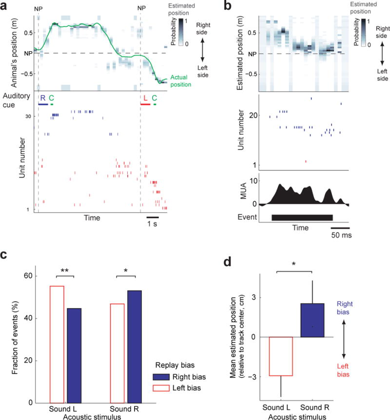
Thus far we have examined how task-related sounds bias the firing rates of individual place cells during replay events. The content of replay is determined by the structured activity from an ensemble of place cells during a reactivation event, and as such it is important to demonstrate that the decoded content of a replay event is actually biased by task-related sounds. In order to do this, we employed a Bayesian method of decoding the rat’s spatial position (Fig. 7a) based on neural ensemble activity17, 21. By computing the conditional probability of each place cell’s response given the rat’s position on the track while performing the behavioral task, we could use Bayes’ rule to measure the estimated likelihood of the rat’s virtual position on the track based on neural ensemble activity during a non-REM sleep replay event (Fig. 7b, see Methods). Bias in the mean-decoded position on the track during a replay event was computed using a probability-weighted average (see Methods). We observed that replay events occurring after Sound R were more likely to be decoded as right-sided, while replay events occurring after Sound L were more likely to be decoded as left-sided (Sound R: P < 0.05, Sound L: P < 0.002, binomial distribution, Fig. 7c). Similarly, if we calculated the mean decoded position bias on the track across all replay events, we found a positive bias for Sound R replay events (right-sided bias) and a negative bias for Sound L replay events (left-sided bias) (P < 0.006, Wilcoxon rank sum test, Fig. 7d). These results indicate that in addition to firing rates of individual place cells being modulated by task-related sounds, the content of a reactivation event is biased towards replaying the portion of the track associated with the task-related cue.
Figure 7.
Bias in Bayesian decoded position during sleep reactivation events
a. Example of Bayesian decoding during two behavioral trials- From top to bottom: (1) the estimated spatial position (Bayesian reconstruction) and actual position (green) of the subject on the track, (2) acoustic stimuli- Sound R (R) in blue, a tone indicating a correct trial and reward delivery cue (C) in green, and Sound L (L) in red), (3) raster plot of place cell activity. Nosepokes (NP) are indicated by a vertical dashed line. All place fields with peak firing rates greater then 0.5 spk/s are displayed in the raster plot, and subsequently used for the reconstruction. Sound R was played when a nosepoke was made, and the rat correctly ran to the right side of the track. Having returned and made another nosepoke, the rat heard Sound L and ran to the left side of the track. In the raster plot, spikes from place cells with place fields on the right side of the track are blue, and left-sided place fields in red. Place fields are ordered from top to bottom by their location on the track (right → left side).
b. Example of Bayesian decoding of a reactivation event during sleep (Rat 2, session 1). From top to bottom: (1) the estimated spatial position of the replay (Bayesian reconstruction), (2) place cell activity (red=left-sided place fields, blue=right side place fields), (3) multiunit activity (MUA), (4) reactivation event detection.
c. Fraction of replay events occurring after Sound R and Sound L with a mean decoded position with a right or left-sided bias. Sound L had significantly more replay events with a left-sided bias (** P < 0.002, Binomial distribution), while Sound R was significantly biased towards the right side (* P < 0.05, Binomial distribution).
d. Mean estimated position during replay events occurring after Sound L and Sound R. Sound R replay events have a positive bias (mean position is on the right side of the track) while Sound L replay events have a negative bias (mean position is on the left side of the track). The difference between the mean estimate position for Sound L and Sound R replay events was statistically significant (* P < 0.006, Wilcoxon rank sum test). The mean estimated track position is relative to the center of the track (negative values- left side, positive values- right side). From the track center (location of nosepoke), the track extends 75 cm to each side. Error bars indicate the standard error of the mean (SEM).
Discussion
Here we have shown that hippocampal replay activity can be manipulated by presenting a task-related auditory cue during sleep, such that the content of replay is biased towards the previous experience associated with that cue. This effect was observed at the level of individual place cell responses as well as in the overall content of replay represented by the entire neuronal ensemble. These data support recent findings in human subjects for which memory consolidation was improved by presenting task-related cues during non-REM sleep after a hippocampus-dependent memory task14, 15 and indicate that a cue-evoked bias in replay content is a potential mechanism responsible for the improvement in memory consolidation. Furthermore, these data help establish a causal relationship between sleep replay and memory consolidation.
While a sound-evoked bias of replay events was observed during non-REM sleep, this effect was not observed during replay events occurring while the rat was awake. These data match similar experiments in human, where memory consolidation was enhanced by presenting task-related cues during non-REM sleep (but not when awake or in REM sleep). In laboratory rodents, replay has been observed in an awake (immobile) state22, 23, 24 as well as during both REM25 and non-REM sleep9, 10. We do not know whether replay serves the same function in all of these different behavioral states. Because awake replay occurs on a compressed time-scale similar to replay during non-REM sleep (roughly ten times faster than the real speed observed during behavior), these two forms of replay are more likely to be functionally alike in contrast to REM replay (which happens on a time scale similar to the previous behavioral episode). However, given that cue-biased memory-consolidation and replay are only observed in non-REM sleep, awake replay and non-REM sleep replay may still differ functionally. Whether REM sleep replay is also susceptible to bias by external stimuli remains to be explored in future studies.
How does a task-related sound affect what the hippocampus replays? During non-REM sleep replay occurs during frames, short periods of elevated activity in the cortex and hippocampus related to cortical up-states9. While both cortex and the hippocampus replay similar content in a coordinated manner, replay appears to be initiated by the hippocampus9. However, frame activity shows the opposite trend, with a cortical frame starting roughly 50 ms before the corresponding hippocampal frame. So although replay of sequential event memory may be driven by the hippocampus, the selection of which memories are replayed by the hippocampus could still be biased by cortical activity occurring prior to a replay event. Bi-directional interactions between the hippocampus and neocortex have been proposed to be important for the storage and maintenance of episodic memories in computational models of hippocampal replay26. Furthermore, external manipulation of cortical activity via stimulation or pharmacology has been previously used to improve27 or disrupt28 memory consolidation of a hippocampus-dependent task. Likewise, given that auditory cortex still responds to sound during non-REM sleep29, 30, cortical responses evoked by task-related sounds could also potentially bias hippocampal activity.
We observed that the cue-evoked replay bias decreased in the second half of the sleep session (Fig. 6b). Task-related cues may become less effective in biasing replay as a result of either the latency from sleep onset or the number of preceding cue-biased reactivations. If multiple events need to be consolidated, events not associated with the task-related cue (and biased against in early sleep) may take priority at later stages of the sleep cycle. Repeatedly reactivating the same neural ensemble could result in its depotentiation, acting as a homeostatic mechanism that decreases the neural ensemble’s future involvement in replay within the sleep cycle.
The content of hippocampal replay activity during the first hour of sleep more closely matches the preceding behavioral episode, compared to replay after the first hour of sleep10. Our data further extend these findings, indicating that there is a limited capacity for biasing replay with task-related cues, and it is most effective in early sleep or during a short nap. Human studies using task-related cues during sleep to improve hippocampus-dependent learning have also targeted early sleep14, 15, 16. The consolidation of declarative memories is thought to depend more on the early period of sleep31, at least partly due to non-REM sleep being more prominent earlier in the night.
Our data suggest that a cue-evoked bias in the content of a reactivation event is maintained for up to at least 10 seconds, or until another acoustic stimulus is played. While this bias may be stored locally in auditory cortex, other brain regions connected to hippocampus and auditory cortex (e.g. entorhinal cortex) could also potentially store this bias using persistent activity32. Task-related cues presented during sleep can additionally be used to improve procedural learning33, suggesting that cue-evoked replay bias may also occur for memories not dependent on the hippocampus. How interactions between cortex and hippocampus select the memory replayed during a reactivation event is an open question, however the use of external stimulation to bias memory consolidation provides a model system for further investigation.
Online Methods
All procedures were approved by the Committee on Animal Care and Massachusetts Institute of Technology and followed NIH guidelines.
Behavioral task
Four Long-Evans rats (400–500 g) were initially trained to nosepoke to receive a food pellet reward (45 mg, Bio-Serv). Following this, the behavioral task was modified so that after initiating a trial with a nosepoke, subjects heard one of two sounds. For Sound R (upwards frequency sweep, 5–20 kHz, 800 ms) subjects had to run to the right end of the linear track, while for Sound L (downwards frequency sweep, 20–5 kHz, 800 ms) subjects had to run to the left end of the linear track. On correct trials, subjects heard a brief tone (6 kHz, 200 ms) and received a food pellet, while on error trials subjects heard a brief white noise burst (500 ms) and received a 10 second time out. If the subject did not make a choice (right or left) within 20 seconds, the trial was treated as an error trial. The behavioral apparatus was custom-made, and automated using custom-made software (MATLAB), a USB-based digital I/O module (Measurement Computing), IR sensors (Sharp GP2D15), reward delivery (ENV-203M, Med-Associates), and acoustic stimulation (JobSite A45-X2 2-Channel Stereo Amplifier, M-Audio Audiophile 192 64-Bit PCI Audio Interface). Sounds were delivered at 70 dB SPL from a speaker (Sony SS-FRF7ED) 1.2 meter in front of the subject. The task was performed in a quiet room. The track was 1.5 meters long and 0.1 m wide, with two nosepoke sensors positioned near the center (one nosepoke sensor on each side of the track). The nosepoke closer to the speaker is referred to as the front nosepoke in the text (Fig. 1a). Both nosepoke sensors could be used to initiate a trial, however if the subject used the two nosepoke sensors unevenly, the more frequently used nosepoke sensor was remotely inactivated, so that the subject would be forced to use the other nosepoke sensor. Two nosepoke sensors were used so that the task would reinforce a spatial association with Sound R and Sound L, rather than a motor response (head turn). The mean amount of time (+− standard deviation) to perform 200 trials across all experiments was 51 minutes (+− 13 minutes). The number of sessions to reach learning criterion was: rat 1, 4 sessions; rat 2, 7 sessions; rat 3, 20 sessions; rat 4, 14 sessions.
Microdrive array and data collection
After learning the task and performing a statistically significant number of correct trials within a single session (P < 0.05, binomial distribution), subjects were implanted with a microdrive array containing 16–18 independently moveable tetrodes. Tetrodes were constructed by twisting together four 13 μm diameter nichrome wire (RediOhm-800, Kanthal, Palm Coast, FL), with each microwire electroplated with gold for an impedence of 250–400 kΩ. Microdrive arrays were implanted under isoflurane anesthesia, and positioned over the right dorsal hippocampus (2.5–3 mm lateral and 4 mm posterior to Bregma). Tetrodes were lowered slowly over 2–3 weeks till they reached the pyramidal cell layer of CA1. Prior to recording, subjects were retrained until at least two consecutive behavioral sessions had a statistically significant number of correct trials. Reference tetrodes were lowered into the white matter above dorsal CA1, and a skull screw above the left Cerebellum served as ground. The local field potential (LFP) was filtered between 1 and 475 Hz, and recorded at a sampling rate of 3125 Hz. Video tracking (sampling rate of 30 Hz) of the subject’s head orientation and position was performed using an overhead camera to record the movement of 2 IR diodes connected to the implanted microdrive array.
Post-behavior sleep session
After performing at least 200 trials with a significant number of correct trials (P<0.05, Binomial distribution), rats were placed in a remote location in a quiet room, and allowed to rest/sleep for 2 to 2.5 hours. Five different sounds were played to the rat [Sound R (upward frequency sweep, 5–20 kHz, 800 ms), Sound L (downward frequency sweep, 20–5 kHz, 800 ms), auditory cue for correct trials (6kHz tone, 200 ms), auditory cue for error trials (white noise, 500 ms), complex tone not associated with behavioral task (f0 = 1 kHz, harmonics spanning 5–20 kHz, 800 ms). The last three stimuli were used as control sounds. Sounds were randomly ordered and played with a randomized interstimulus interval ranging between 5–10 seconds (uniform distribution). Acoustic stimuli were played at a sound level of 50 dB SPL.
Reactivation event detection
Multiunit activity (MUA) was measured using a smoothed histogram (1 ms bins, Gaussian kernel, σ=15 ms) to calculate the firing rate of all units (not necessarily isolated) with spikes that had a peak amplitude greater than 100 uV, across all tetrodes. Candidate reactivation events had a minimum zscore of 2 throughout the duration of the event, and a minimum peak zscore of 4. Multiple events less than 50 ms apart were grouped together into a single event. All reactivation events had to be at least 50 ms in duration. MUA was highly correlated with ripple power, but could be measured with greater temporal precision [Davidson et al. 2010]. A total of 409 neurons were recorded in our experiments, with 199 neurons passing our criteria of place fields with peak firing rates greater than 2 spk/s and mean rates less than 5 spk/s (to avoid interneurons). Neurons with place fields centered at the nosepoke and/or neurons unresponsive during all analyzed reactivation events were excluded from our analysis of rate bias, As a result, our analysis of rate bias was performed on 171 out of 199 neurons (sleep replay) and 172 out of 199 neurons (awake replay analysis),
Reactivation data was analyzed for post-behavior sessions with at least 30 reactivation events during awake periods and sleep periods for each acoustic stimulus (Sound R, Sound L, control sounds), occurring after a behavioral session where the subject performed at least 200 trials, with a significant number of correct trials (P < 0.05, binomial distribution). A total of 11 behavioral sessions matched these criteria.
Classification of sleep
Non-REM sleep was defined by time periods with high frame activity (upper quartile) [Ji and Wilson 2007] containing intermittent high MUA (zscore of 4 or greater) and at least 25% of places cells active within a 2 second window, and minimal movement (lower quartile) detected using video tracking. REM sleep was defined by the ratio of the spectral power density in the theta band (6–10 Hz) to the overall power being greater than 0.4 (Lansink et al. 2008). We obtained similar REM sleep detection using an alternative REM sleep detection algorithm (Theta/Delta ratio > 6, Hirase et al. 2001). The mean (std) total time spent sleeping across experiments was 47 minutes (+− 16 min). The mean (std) length of each sleep episode (across experiments) was 12 minutes (+−5 min).
The Early and Late non-REM sleep periods were determined by concatenating all non-REM sleep periods; Early (Late) sleep was the first (second) half of this sleep session.
Data Analysis
Place fields were computed using velocity filtered data (>15 cm/s). Each place cell’s position on the track was computed by a firing-rate weighted mean of the place field. We also used two alternative measurements of the place field’s position on the track.
Method1=(∑Ri/μ)Method2=(∑Ri−∑Li)/(∑Ri+∑Li)
- where Ri = firing rates of place field on right side of track
- Li = firing rates of place field on left side of track
- μ = mean firing rate of place field
The rate bias (Sound R − Sound L) was computed by taking the mean firing rate of a place cell during all replay events occurring after the onset of Sound R (for up to 10.8 sec or the onset of the next sound), and subtracting from this value the mean firing rate of the same place cell during all replay events occurring after Sound L. The mean firing rate during replay was the number of spikes divided by the median replay duration (for the session).
To estimate the subject’s position during both behavior and replay events from hippocampal ensemble activity, we employed a Bayesian reconstruction algorithm [Zhang et al. 1998, Davidson et al. 2010] using the following equation:
P(x|n)=CP(x)(∏i=1Nfi(x)ni)exp(−τ∑i=1Nfi(x))
Where C is a normalization constant, x is the subject’s position, fi(x) is the firing rate of the ith place field at a given location x, n is the number of spikes within the time window τ.
The maximum likelihood of the subject’s position was calculated during the behavioral task using a 250 ms temporal window and during replay using a 10 ms temporal window. The track bias in the reconstructed replay event was calculated by taking a probability weighted average of all reconstructed positions. Place fields with peak firing rates greater than 0.5 spk/s and mean firing rates< 5 spk/s (to exclude interneurons) were used in this analysis. We also required a minimum of five place fields on each side of the nosepoke for reconstruction. One experiment (Subject 3, experiment 2) was excluded from the group analysis for failing to pass this requirement.
Supplementary Material
supplement
Acknowledgments
We thank Michale Fee, Heikki Tanila and members of the Wilson Lab for their helpful comments and suggestions. We would also like to thank Carmen Varela, Jun Yamamoto, Josh Siegle, Elise Kuo, Ahmed Hussain, and Emily Molina for technical assistance. Support provided by a Merck Award/Helen Hay Whitney Postdoctoral Fellowship (D.B.), Charles King Trust Postdoctoral Fellowship (D.B.), NIH 1-K99-DC012321-01 (D.B.) and NIH 5R01MH061976 (M.A.W.).
Footnotes
The authors do not have any competing interests
Author contributions: D.B. designed the experiment, collected and analyzed the data, D.B. and M.A.W. wrote the manuscript. M.A.W. supervised the experiment.
References for Online Methods
- 1.Davidson TJ, Kloosterman F, Wilson MA. Hippocampal replay of extended experience. Neuron. 2009;63(4):497–507. doi: 10.1016/j.neuron.2009.07.027. [DOI] [PMC free article] [PubMed] [Google Scholar]
- 2.Hirase H, Leinekugel X, Czurkó A, Csicsvari J, Buzsáki G. Firing rates of hippocampal neurons are preserved during subsequent sleep episodes and modified by novel awake experience. Proc Natl Acad Sci. 2001;98(16):9386–90. doi: 10.1073/pnas.161274398. [DOI] [PMC free article] [PubMed] [Google Scholar]
- 3.Ji D, Wilson MA. Coordinated memory replay in the visual cortex and hippocampus during sleep. Nat Neurosci. 2007;10(1):100–7. doi: 10.1038/nn1825. [DOI] [PubMed] [Google Scholar]
- 4.Lansink CS, Goltstein PM, Lankelma JV, Joosten RN, McNaughton BL, Pennartz CM. Preferential reactivation of motivationally relevant information in the ventral striatum. J Neurosci. 2008;28(25):6372–82. doi: 10.1523/JNEUROSCI.1054-08.2008. [DOI] [PMC free article] [PubMed] [Google Scholar]
- 5.Zhang K, Ginzburg I, McNaughton BL, Sejnowski TJ. Interpreting neuronal population activity by reconstruction: unified framework with application to hippocampal place cells. J Neurophysiol. 1998;79(2):1017–44. doi: 10.1152/jn.1998.79.2.1017. [DOI] [PubMed] [Google Scholar]
References
- 1.Dupret D, O’Neill J, Pleydell-Bouverie B, Csicsvari J. The reorganization and reactivation of hippocampal maps predict spatial memory performance. Nat Neurosci. 2010;13(8):995–1002. doi: 10.1038/nn.2599. [DOI] [PMC free article] [PubMed] [Google Scholar]
- 2.Squire LR. Memory and the hippocampus: a synthesis from findings with rats, monkeys, and humans. Psychol Rev. 1992;99(2):195–231. doi: 10.1037/0033-295x.99.2.195. [DOI] [PubMed] [Google Scholar]
- 3.Marshall L, Born J. The contribution of sleep to hippocampus-dependent memory consolidation. Trends Cogn Sci. 2007;11(10):442–50. doi: 10.1016/j.tics.2007.09.001. [DOI] [PubMed] [Google Scholar]
- 4.Siapas AG, Wilson MA. Coordinated interactions between hippocampal ripples and cortical spindles during slow-wave sleep. Neuron. 1998;21(5):1123–8. doi: 10.1016/s0896-6273(00)80629-7. [DOI] [PubMed] [Google Scholar]
- 5.Sirota A, Csicsvari J, Buhl D, Buzsáki G. Communication between neocortex and hippocampus during sleep in rodents. Proc Natl Acad Sci U S A. 2003;100(4):2065–9. doi: 10.1073/pnas.0437938100. [DOI] [PMC free article] [PubMed] [Google Scholar]
- 6.Walker MP, Stickgold R. Sleep-dependent learning and memory consolidation. Neuron. 2004;44(1):121–33. doi: 10.1016/j.neuron.2004.08.031. [DOI] [PubMed] [Google Scholar]
- 7.Wierzynski CM, Lubenov EV, Gu M, Siapas AG. State-dependent spike-timing relationships between hippocampal and prefrontal circuits during sleep. Neuron. 2009;61(4):587–96. doi: 10.1016/j.neuron.2009.01.011. [DOI] [PMC free article] [PubMed] [Google Scholar]
- 8.Wilson MA, McNaughton BL. Reactivation of hippocampal ensemble memories during sleep. Science. 1994;265(5172):676–9. doi: 10.1126/science.8036517. [DOI] [PubMed] [Google Scholar]
- 9.Lee AK, Wilson MA. Memory of sequential experience in the hippocampus during slow wave sleep. Neuron. 2002;36(6):1183–94. doi: 10.1016/s0896-6273(02)01096-6. [DOI] [PubMed] [Google Scholar]
- 10.Ji D, Wilson MA. Coordinated memory replay in the visual cortex and hippocampus during sleep. Nat Neurosci. 2007;10(1):100–7. doi: 10.1038/nn1825. [DOI] [PubMed] [Google Scholar]
- 11.Peigneux P, Laureys S, Fuchs S, Collette F, Perrin F, Reggers J, Phillips C, Degueldre C, Del Fiore G, Aerts J, Luxen A, Maquet P. Are spatial memories strengthened in the human hippocampus during slow wave sleep? Neuron. 2004;44(3):535–45. doi: 10.1016/j.neuron.2004.10.007. [DOI] [PubMed] [Google Scholar]
- 12.Girardeau G, Benchenane K, Wiener SI, Buzsáki G, Zugaro MB. Selective suppression of hippocampal ripples impairs spatial memory. Nat Neurosci. 2009;12(10):1222–3. doi: 10.1038/nn.2384. [DOI] [PubMed] [Google Scholar]
- 13.Ego-Stengel V, Wilson MA. Disruption of ripple-associated hippocampal activity during rest impairs spatial learning in the rat. Hippocampus. 2010;20(1):1–10. doi: 10.1002/hipo.20707. [DOI] [PMC free article] [PubMed] [Google Scholar]
- 14.Rasch B, Büchel C, Gais S, Born J. Odor cues during slow-wave sleep prompt declarative memory consolidation. Science. 2007;315(5817):1426–9. doi: 10.1126/science.1138581. [DOI] [PubMed] [Google Scholar]
- 15.Rudoy JD, Voss JL, Westerberg CE, Paller KA. Strengthening individual memories by reactivating them during sleep. Science. 2009;326(5956):1079. doi: 10.1126/science.1179013. [DOI] [PMC free article] [PubMed] [Google Scholar]
- 16.Diekelmann S, Büchel C, Born J, Rasch B. Labile or stable: opposing consequences for memory when reactivated during waking and sleep. Nat Neurosci. 2011;14(3):381–6. doi: 10.1038/nn.2744. [DOI] [PubMed] [Google Scholar]
- 17.Davidson TJ, Kloosterman F, Wilson MA. Hippocampal replay of extended experience. Neuron. 2009;63(4):497–507. doi: 10.1016/j.neuron.2009.07.027. [DOI] [PMC free article] [PubMed] [Google Scholar]
- 18.O’Keefe J, Dostrovsky J. The hippocampus as a spatial map. Preliminary evidence from unit activity in the freely-moving rat. Brain Res. 1971;34(1):171–5. doi: 10.1016/0006-8993(71)90358-1. [DOI] [PubMed] [Google Scholar]
- 19.Moser EI, Kropff E, Moser MB. Place cells, grid cells, and the brain’s spatial representation system. Annu Rev Neurosci. 2008;31:69–89. doi: 10.1146/annurev.neuro.31.061307.090723. [DOI] [PubMed] [Google Scholar]
- 20.Itskov PM, Vinnik E, Honey C, Schnupp JW, Diamond ME. Sound sensitivity of neurons in rat hippocampus during performance of a sound-guided task. J Neurophysiol. doi: 10.1152/jn.00404.2011. (in press) [DOI] [PMC free article] [PubMed] [Google Scholar]
- 21.Zhang K, Ginzburg I, McNaughton BL, Sejnowski TJ. Interpreting neuronal population activity by reconstruction: unified framework with application to hippocampal place cells. J Neurophysiol. 1998;79(2):1017–44. doi: 10.1152/jn.1998.79.2.1017. [DOI] [PubMed] [Google Scholar]
- 22.Foster DJ, Wilson MA. Reverse replay of behavioural sequences in hippocampal place cells during the awake state. Nature. 2006;440(7084):680–3. doi: 10.1038/nature04587. [DOI] [PubMed] [Google Scholar]
- 23.Diba K, Buzsáki G. Forward and reverse hippocampal place-cell sequences during ripples. Nat Neurosci. 2007;10(10):1241–2. doi: 10.1038/nn1961. [DOI] [PMC free article] [PubMed] [Google Scholar]
- 24.Karlsson MP, Frank LM. Awake replay of remote experiences in the hippocampus. Nat Neurosci. 2009;12(7):913–8. doi: 10.1038/nn.2344. [DOI] [PMC free article] [PubMed] [Google Scholar]
- 25.Louie K, Wilson MA. Temporally structured replay of awake hippocampal ensemble activity during rapid eye movement sleep. Neuron. 2001;29(1):145–56. doi: 10.1016/s0896-6273(01)00186-6. [DOI] [PubMed] [Google Scholar]
- 26.Káli S, Dayan P. Off-line replay maintains declarative memories in a model of hippocampal-neocortical interactions. Nat Neurosci. 2004;7:286–294. doi: 10.1038/nn1202. [DOI] [PubMed] [Google Scholar]
- 27.Marshall L, Helgadóttir H, Mölle M, Born J. Boosting slow oscillations during sleep potentiates memory. Nature. 2006;444(7119):610–3. doi: 10.1038/nature05278. [DOI] [PubMed] [Google Scholar]
- 28.Lesburguères E, Gobbo OL, Alaux-Cantin S, Hambucken A, Trifilieff P, Bontempi B. Early tagging of cortical networks is required for the formation of enduring associative memory. Science. 2011;331(6019):924–8. doi: 10.1126/science.1196164. [DOI] [PubMed] [Google Scholar]
- 29.Issa EB, Wang X. Sensory responses during sleep in primate primary and secondary auditory cortex. J Neurosci. 2008;28(53):14467–80. doi: 10.1523/JNEUROSCI.3086-08.2008. [DOI] [PMC free article] [PubMed] [Google Scholar]
- 30.Schabus M, Dang-Vu TT, Heib DP, Boly M, Desseilles M, Vandewalle G, Schmidt C, Albouy G, Darsaud A, Gais S, Degueldre C, Balteau E, Phillips C, Luxen A, Maquet P. The Fate of Incoming Stimuli during NREM Sleep is Determined by. Spindles and the Phase of the Slow Oscillation Front Neurol. 2012;3:40. doi: 10.3389/fneur.2012.00040. [DOI] [PMC free article] [PubMed] [Google Scholar]
- 31.Plihal W, Born J. Effects of early and late nocturnal sleep on priming and spatial memory. Psychophysiology. 1999;36(5):571–82. [PubMed] [Google Scholar]
- 32.Tahvildari B, Fransén E, Alonso AA, Hasselmo ME. Switching between “On” and “Off” states of persistent activity in lateral entorhinal layer III neurons. Hippocampus. 2007;17(4):257–63. doi: 10.1002/hipo.20270. [DOI] [PubMed] [Google Scholar]
- 33.Antony JW, Gobel EW, O’Hare JK, Reber PJ, Paller KA. Cued memory reactivation during sleep influences skill learning. Nat Neurosci. 2012;15:1114–1116. doi: 10.1038/nn.3152. [DOI] [PMC free article] [PubMed] [Google Scholar]
Associated Data
This section collects any data citations, data availability statements, or supplementary materials included in this article.
Supplementary Materials
supplement