Versatile roles of extracellular vesicles in cancer (original) (raw)
Abstract
Numerous studies have shown that non–cell-autonomous regulation of cancer cells is an important aspect of tumorigenesis. Cancer cells need to communicate with stromal cells by humoral factors such as VEGF, FGFs, and Wnt in order to survive. Recently, extracellular vesicles (EVs) have also been shown to be involved in cell-cell communication between cancer cells and the surrounding microenvironment and to be important for the development of cancer. In addition, these EVs contain small noncoding RNAs, including microRNAs (miRNAs), which contribute to the malignancy of cancer cells. Here, we provide an overview of current research on EVs, especially miRNAs in EVs. We also propose strategies to treat cancers by targeting EVs around cancer cells.
Introduction
A seminal study by Valadi et al. in 2007 (1) showed that variable RNAs, microRNAs (miRNAs), long noncoding RNAs (lncRNAs), and mRNAs can be transported between cells in extracellular vesicles (EVs). EVs are small membranous vesicles that are secreted from numerous cell types. They facilitate intercellular communication by transporting intracellular components such as protein and RNA (2). EVs, including exosomes, microvesicles, and other types of membrane vesicles, are found in various body fluids, such as blood, urine, and saliva, and can be recognized by their unique mechanisms of biogenesis and secretion (2, 3). Until the study by Valadi and colleagues was published, the consensus was that miRNAs only functioned intracellularly in their cells of origin; however, Valadi et al. showed that miRNAs may also function as humoral factors involved in intercellular communication. In 2010, three articles showed that these miRNAs can be transferred to immune cells (4), cancer cells (5), or endothelial cells (6) and are able to function within them. All of these articles suggest that RNAs, including miRNAs, serve as novel humoral factors in cell-cell communication. Current studies are focused on the role of miRNAs in EVs during cancer development.
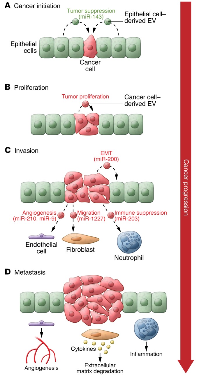
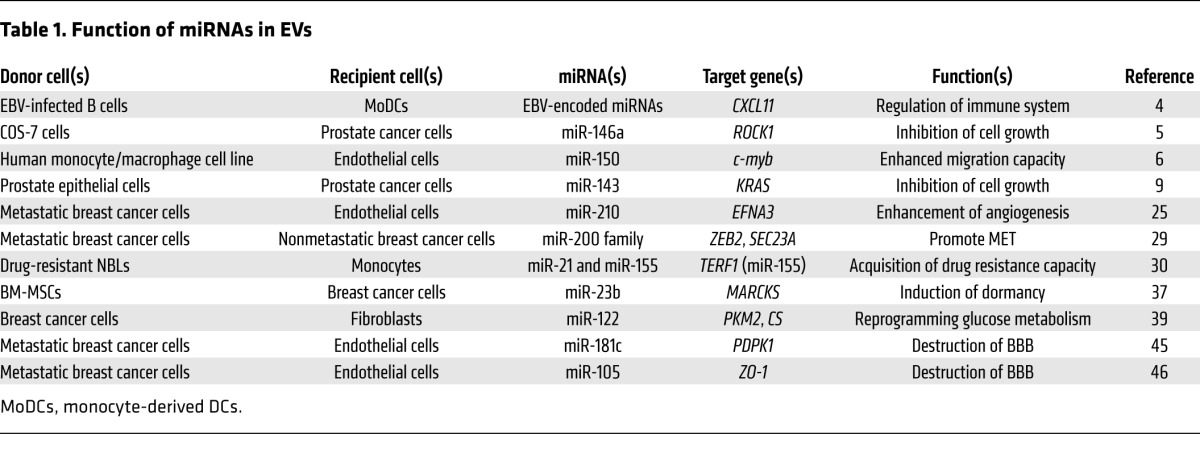
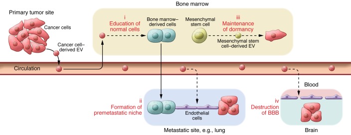
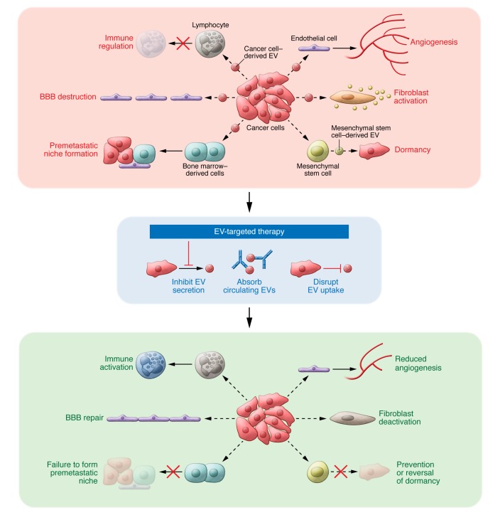
In this review, we summarize the current knowledge regarding the contribution of EV-associated miRNAs to cancer development, including initiation, invasion, metastasis, and recurrence (Figures 1 and 2, and Table 1). Furthermore, we discuss the therapeutic approaches involving EVs and miRNAs, which originate from cancer cells and microenvironmental cells, for the diagnosis and treatment of cancer (Figure 3).
Figure 1. EVs from cancer cells manipulate the cells in their microenvironment.

EVs are involved in every step of cancer development. During cancer initiation (A), normal cells (epithelial cells) attempt to prevent the outgrowth of precancerous cells (or cancer cells) by secreting antiproliferative miRNAs through EVs; however, the cancer cells can circumvent this inhibitory machinery, finally resulting in tumor expansion (B). Cancer cells utilize EVs to mediate horizontal transfer of genes that promote proliferation to cancer cells that do not harbor those genes (B). Cancer cell–derived EVs promote cancer malignancy (i.e., the induction of inflammation by infiltrating neutrophils) (C and D). Additionally, cancer cell–derived EVs activate fibroblasts, leading to extracellular matrix degradation and the induction of cancer-promoting cytokines (C and D). When the tumor microenvironment is hypoxic, cancer cells secrete angiogenesis-inducing EVs that help to overcome oxygen and nutrition deficiency by activating endothelial cells to stimulate vascularization (C and D), contributing to further cancer development, such as metastasis (D).
Figure 2. The roles of cancer cell–derived EVs and their development.
EVs derived from cancer cells infiltrate BM cells (i), leading to the formation of a premetastatic niche (ii). Additionally, cancer cell–derived EVs directly alter the metastatic site to induce angiogenesis. Transfer of EV-associated miRNAs from BM mesenchymal stem cells regulates breast cancer cell dormancy in a metastatic niche (iii). Furthermore, brain metastasis is mediated by EVs triggers the destruction of the BBB (iv).
Table 1. Function of miRNAs in EVs.

Figure 3. Therapeutic strategies against cancer-derived EVs.
EVs are secreted from cancer cells and delivered to recipient cells, modulating their phenotype. For example, EVs from cancer cells are delivered to endothelial cells, which enhances angiogenesis to obtain the oxygen and nutrition required for continued growth of the cancer. We propose the following therapeutic applications: inhibition of cancer cell EV production, disruption of EV uptake by recipient cells, and elimination of circulating cancer cell–derived EVs. These therapeutic strategies will prevent the delivery of EVs from cancer cells to microenvironmental cells, leading to the development of novel antiangiogenic and anticancer drugs.
EV-associated miRNAs both promote and suppress cancer initiation
A number of factors can contribute to tumor formation, including gene amplification, deletion, and mutation; cellular stress; metabolic alterations; and epigenetic changes (7). In addition to these cell-autonomous mechanisms, non-cell-autonomous mechanisms also contribute to cancer initiation (8), including factors that regulate cancer cells or microenvironmental cells such as TGF-β, sonic hedgehog (SHH), Wnt, and EVs.
Recently it has been shown that the EVs from noncancerous neighboring epithelial cells have the capacity to suppress cancer initiation (9). During cancer initiation, there is a conflict between newly transformed cells and surrounding epithelial cells. It is hypothesized that growth-inhibitory miRNAs are actively released from noncancerous cells to kill transformed cells, thereby restoring the tissue to a healthy state. Because abundant healthy cells continuously provide nascent proliferating cells with tumor-suppressive miRNAs for an extended time period, a local concentration of secretory miRNAs can become high enough to restrain tumor initiation. In cancer cells, the expression of tumor-suppressive miRNAs is downregulated (10); consequently, the continuous provision of tumor-suppressive miRNAs via EVs is a homeostatic mechanism that tumor cells must overcome. Once this balance is compromised, the microenvironment will be susceptible to tumor initiation. For example, miR-143 has a higher expression level in normal prostate cell lines compared with cancerous prostate cell lines (11). EVs containing miR-143 in the normal prostate cell line transfer growth-inhibitory signals to cancerous cells both in vitro and in vivo. This competitive biological process has been observed in other disease states, such as between multiple myeloma (MM) and bone marrow mesenchymal stromal cells (BM-MSCs) (12). In this case, EVs isolated from BM-MSCs of patients with MM induced tumor growth in vivo and promoted the dissemination of tumor cells to the BM in an in vivo translational model of MM. The levels of miR-15a, which is downregulated in leukemia (13) and suppresses MM growth (14, 15), were significantly higher in normal BM-MSC–derived EVs compared with MM BM-MSC–derived EVs, suggesting that MSC-derived miR-15a plays a tumor-suppressive role. Conversely, the expression of miR-15a is downregulated in EVs from BM-MSCs that cannot suppress MM expansion. As discussed above, the secretion of miRNAs from noncancerous cells is an effective policing strategy, preventing cells within a given niche from becoming cancerous (Figure 1).
Losing the ability to suppress cancer initiation is not the only reason for oncogenesis. Comorbidity is a major issue affecting the long-term survival of older cancer patients, but the underlying mechanisms are not well understood (16). A pathogenic mechanism that contributes to chronic obstructive pulmonary disease (COPD) is mediated through the regulation of autophagy by EV-associated miR-210 (17). Cigarette smoking alters EV miRNA profiles, potentially controlling airway remodeling in COPD. miR-210 controls the hypoxic response of cancer cells, enabling their survival in hypoxic conditions. The expression of miR-210 increases after exposure to cigarette smoke, promoting adaptation to hypoxia (18). Adaptation to hypoxia is essential for cancer survival, as new cancer cells do not have sufficient access to oxygen and nutrients owing to increased diffusion distances from the existing vascular supply (19). Cigarette smoking also decreases EV packaging of the let-7 miRNA family, suggesting that lung carcinogenesis may be triggered by EV-associated miRNA communication (17). The EV-mediated interaction of pathological cells may underlie the association between lung cancer and COPD. The secretion of altered EVs from pathological cells throughout the body is likely to contribute directly to disease, and the analysis of EVs in each disease may help identify new disease mechanisms.
Regulation of cancer angiogenesis by miRNAs in EVs
Endothelial cells contribute to vascularization, providing oxygen and nutrition — a scarce resource in tumors — to cancer cells. Cytokines such as VEGF and basic FGF (bFGF) are responsible for the communication between endothelial and cancer cells, and EVs from cancer cells contain various molecules, including miRNAs, that promote angiogenesis (20–22). For example, cancer cell–derived, EV-associated miR-9 reduced suppressor of cytokine signaling 5 (SOCS5) levels, activating the JAK/STAT pathway to promote endothelial cell migration and tumor angiogenesis (23). Additionally, miR-210 is secreted in cancer cell–derived EVs to regulate neutral sphingomyelinase 2 (nSMase2) (5, 24) to promote angiogenesis (25). The angiogenic inhibitor ephrin-A3, a miR-210 target gene, is downregulated following EV transfer from cancer cells. miR-210 regulates angiogenesis in endothelial cells (26) and iron homeostasis in cancer cells (18) and is upregulated by hypoxic conditions (18, 26). Furthermore, manipulation of nSMase2 expression alters EV production, which affects metastatic capacity (25). EVs have been shown to contain multiple angiogenic factors (20); however, the exact contributions of these factors have not been elucidated. It will be necessary to clarify the function of each factor as well as demonstrate the collective effect of these factors in order to understand the contribution of angiogenic EVs to cancer development.
Promotion of cancer cell malignancy by EVs
EVs from cancer cells affect not only microenvironmental cells but also other cells in the heterogeneous tumor population, resulting in the transfer of metastatic capability. Most cancer cells release a variety of EV types, which dictate the behavior of the recipient cells for the ultimate benefit of the cancer cells (Figure 1). However, much of the research documenting such effects was done in vitro, and the behavior of EVs in vivo remains to be addressed. The exchange of EVs between tumor cells was recently documented in vivo in a study combining high-resolution intravital imaging with a Cre-LoxP system (27). EVs released by malignant breast cancer cells are taken up by less malignant tumor cells located within the same tumor or within distant tumors. These EVs carry mRNAs involved in migration and metastasis, thereby promoting migratory behavior and metastatic capacity. For example, the miR-200 family, which regulates mesenchymal-epithelial transition (MET) (28), was secreted in EVs from metastatic breast cancer lines to nonmetastatic cancer cells, altering the gene expression of recipient cells and promoting MET (29). miRNA-containing EVs released in the tumor microenvironment can also contribute to the development of drug resistance, as in the case of neuroblastoma (NBL) (30). The expression of EV-associated miRNAs from NBL cell lines identified three highly enriched miRNAs: miR-21 and miR-29a, which can trigger an NF-κB–mediated proinflammatory response in lung cancer (31), and miR-155, which is induced during the macrophage inflammatory response (32). Coculture experiments demonstrated that miR-21 could be transferred from NBL cells to human monocytes and that miR-155 is transferred from human monocytes to NBL cells by EVs. These data demonstrate a unique role for miR-21 and miR-155 in the crosstalk between NBL cells and human monocytes that promotes chemotherapy resistance. EV-mediated transfer of miRNAs is also an important mechanism of intercellular communication in hepatocellular carcinoma (HCC) cells. EV-associated miRNAs in HCC, including miR-584, miR-517c, miR-378, miR-520f, miR-142-5p, miR-451, miR-518d, miR-215, miR-376a*, miR-133b, and miR-367, modulate the expression of TGF-β–activated kinase 1 (TAK1) and associated signaling molecules, resulting in transformation of recipient cells (33).
Control of recurrence by EV-associated miRNAs
Mortality from breast cancer is decreasing due to successful early detection and effective systemic adjuvant therapy; however, breast cancer frequently recurs, typically within 5 years but even up to 10 to 20 years after surgery (34). Importantly, recurrent breast cancer is often more aggressive and untreatable. This is explained by the fact that breast cancer cells survive for a long time in a state of dormancy. Dormant cancer cells cease dividing but survive in a quiescent state while waiting for appropriate environmental conditions to begin proliferating again. Indeed, in early stages of the disease, breast cancer cells can be detected in BM, where they form micrometastases. These cancer cells can later escape dormancy in BM to recirculate and invade other distant organs, thereby giving rise to overt metastases (35, 36).
Cancer cells are maintained in a state of dormancy through interactions with microenvironmental cells; however, little is known about the molecular basis of these interactions. BM mesenchymal stem cells were recently found to play an important role in inducing breast cancer cell dormancy in BM through the transfer of EVs containing cell cycle–inhibitory miRNAs (37). Specifically, miR-23b from BM mesenchymal stem cells promotes dormancy of breast cancer cells by downregulating myristoylated alanine-rich C-kinase substrate (MARCKS), which encodes a protein that promotes cell cycle progression and motility. This raises the obvious question of what miR-23b is doing in EVs under normal physiological conditions. Moreover, what are the molecular mechanisms for escaping dormancy in breast cancer cells? Answering these fundamental questions will help in understanding why breast cancer cells exist in BM and will also enable the development of therapeutic strategies to target these cancer cells.
Long-distance regulation of metastasis by EV-associated miRNAs
Cancer-derived EVs not only affect proximally located cells, but they can also influence cells in distant tissues and organs (Figure 2). For example, highly metastatic melanoma-derived EVs increase the metastasis of primary tumors by educating BM, creating a premetastatic niche (38). Additionally, EV-associated miR-122 from cancer cells downregulates the glycolytic enzyme pyruvate kinase in non-tumor cells located within the premetastatic niche (39). This downregulation suppresses glucose uptake by non-tumor cells in the microenvironment, thereby increasing the nutrient availability for cancer cells in the premetastatic niche.
EVs also appear to be tightly associated with brain metastasis. Brain metastasis is associated with a particularly poor prognosis for cancer patients, but the underlying molecular mechanisms have not been elucidated. One of the key features of brain metastasis is the destruction of the blood-brain barrier (BBB) (40, 41), resulting in the subsequent migration of cancer cells through this interface. Tumor cells recognize and bind to components of the vascular endothelial membrane, thereby initiating extravasation, invasion of cancer cells through the BBB, and new growth at secondary organ sites (42, 43). A number of molecules that putatively promote or prevent BBB destruction have been reported, including some transported in EVs (44). For example, EVs containing miR-181c are transferred from metastatic brain cancer cells to brain endothelial cells, resulting in the destruction of tight junction proteins of the BBB that bind to the primary cytoskeletal protein actin, including claudin-5, occludin, and tight junction protein 1 (ZO-1). miR-181c promotes the destruction of the BBB through delocalization of actin fibers via the downregulation of 3-phosphoinositide–dependent protein kinase–1 (PDPK1), which causes the subsequent degradation of phosphorylated cofilin and the severing of actin filaments. Tight junction proteins are also directly targeted by EV-associated miRNAs. Indeed, miR-105 in breast cancer EVs suppresses ZO-1 expression in endothelial cells, resulting in the loss of cell-cell adhesion, thereby promoting metastasis (45). Circulating miR-181c and miR-105 can be detected in the serum of patients with metastatic brain cancer. These findings open up several avenues for both the diagnosis and treatment of brain metastasis.
The emerging role of EVs in cancer immunology
Immune escape is one of the hallmarks of cancer, and many studies have investigated the mechanisms by which tumor cells evade the host immune system. Tumor cells secrete EVs that stimulate or suppress cancer immunity (46, 47). Indeed, various tumor antigens, such as melan A, mesothelin, and carcinoembryonic antigen (CEA), are packaged in tumor-derived EVs (48–50), which can enhance immune stimulation. There is also increasing evidence that tumor-derived EVs can be immunosuppressive. Tumor-derived EVs are enriched for a variety of immunoregulatory molecules, such as FasL, TRAIL, and galactin-9, that can promote cancer immune escape (51–53). Taken together, the evidence indicates that immunologically active tumor-derived EVs are important for cancer immunological communication in the tumor microenvironment. Therefore, the tumor-derived EVs and their contents may constitute crucial targets for cancer immunotherapy.
The development of new therapeutic drugs that disrupt the immune checkpoint inhibitory pathways of cancer cells have been regarded as a breakthrough in cancer treatment. For example, programmed cell death protein–1 (PD-1) and cytotoxic T lymphocyte–associated antigen–4 (CTLA-4) play crucial roles in T cell co-inhibition and exhaustion. The PD-1 ligand PD-L1 is upregulated in many cancers and contributes to poor prognosis by inhibiting cytotoxic T cell activation. Furthermore, CTLA-4 blockade is a negative regulator of T effector cell activation. Immunological checkpoint blockade using antibodies targeting the PD-1/PD-L1 or CTLA-4 pathways have exhibited promising therapeutic effects in a variety of cancers (54–57). So far, the biological interactions between these immunoregulatory molecules and tumor-derived EVs are unclear. Nevertheless, the presence of immunoregulatory molecules on non–immune cell–derived EVs can suppress T cell activation. Indeed, PD-1 is expressed and transported by DC-derived EVs (58). Furthermore, PD-1 is found on placenta-derived EVs and can inhibit both CD4+ and CD8+ T cells (59). Thus, immunoregulatory molecules on tumor-derived EVs inhibit T cell activation and proliferation (60).
It is becoming increasingly clear that cancer-derived miRNAs are able to regulate the immune system and can therefore serve as potential immunotherapeutic targets. Recently, Fujita et al. showed that miR-197 regulates immune checkpoints in non–small cell lung cancers (61). Furthermore, miR-200/ZEB1 has been shown to control the expression of PD-L1 in tumors (62). Remarkably, Zhou et al. have reported that miR-203 in pancreatic cancer EVs regulates the expression of TLR4 in DCs, potentially repressing antitumor immune responses (63). Because these miRNAs are packaged into EVs released from cancer cells, they may be surrogate markers for immunologically active tumor-derived EVs.
Understanding how EV communication enables cancer immune escape may provide a potential breakthrough in cancer immunotherapy. EV-targeted therapy could be combined with immune checkpoint inhibitors in the near future.
EVs as a new diagnostic tool
EVs reflect the physiological state of their cells of origin (64), and almost all cells secrete EVs containing specific proteins and miRNAs into their microenvironment and the general circulation (3, 65). Thus, EVs can be found in various body fluids, such as blood, urine, and saliva (2, 66), providing a rich source of potential biomarkers.
The first indication that circulating RNA could be used as a biomarker came with the discovery of mRNA and miRNAs in EVs. (1). EV-associated miR-21 enriched in the serum of glioblastoma patients was expressed at higher levels in the serum of patients than of normal controls (20). This initial report became the impetus for research into using miRNAs as effective biomarkers of cancer; subsequently, cancer-specific miRNA profiles have been published (67). Importantly, miRNAs can be readily detected in small-volume samples using specific and sensitive quantitative real-time PCR (qRT-PCR) (68). Additionally, EV-associated miRNAs have been used as diagnostic and prognostic markers (20, 69–79). Microarray analyses of miRNAs in EV-enriched fractions of serum from 88 patients with primary colorectal cancer (CRC) and 11 healthy controls revealed that that the levels of 7 miRNAs were significantly higher in the serum EVs of CRC patients compared with serum EVs from healthy donors (74). This approach of isolating serum EVs from patients has been used to establish miRNA profiles of prostate, colorectal, and pancreatic cancer (70, 75, 78). Although some issues remain surrounding the isolation of miRNA in EVs from serum, such as internal controls and detection methods, miRNA in EVs from serum and plasma will be a useful tool for the diagnosis of cancer or monitoring during treatment.
In addition to miRNAs, proteins associated with cancer cell–derived EVs are also attractive as potential biomarkers. However, the identification and quantification of EV-associated proteins, compared with EV miRNAs, in body fluids remain major challenges because EV proteins are highly heterogeneous and are difficult to collect and handle (80–82). Additionally, the methods used for EV isolation in a research setting are unsuitable for clinical practice due to their lack of reliability. Consequently, novel detection methods are required whereby EVs can be bound to specific complementary proteins and specific EV surface proteins can be utilized as cancer biomarkers. One of the first assays for quantification of EVs used an ELISA-based method for selective detection of EVs (83). In this study, EVs double positive for CD63 or caveolin-1 and Rab-5b were employed to detect melanoma. Levels of such EVs were significantly increased in plasma from melanoma patients compared with that from healthy donors. Multiplex microfluidic systems provide an alternative method for EV detection based on μNMR (84). Indeed, this method has enabled the identification of glioblastoma multiforme (GBM) by the detection of non-tumor, host cell–derived EVs. Additionally, a novel, label-free EV detection method (85) revealed that the levels of epithelial cell adhesion molecule (EpCAM) and CD24 in EVs derived from ovarian cancer patient ascites were decreased in patients who responded to treatment, whereas levels of these markers increased in nonresponding patients. Finally, a highly sensitive and rapid analytical technique for profiling circulating EVs directly from patient blood samples has also been established. This technique uses antibodies to capture two specific antigens residing on EVs that are detectable by photosensitizer beads (86). When this system was used, greater numbers of CD147/CD9 double-positive EVs were detected in serum from CRC patients compared with serum from healthy donors.
Although more investigations are needed to validate the suitability of these technologies, the detection of EV proteins has opened up exciting new avenues for the diagnosis of cancer and the monitoring of treatment responses. It is well established that early detection and accurate monitoring of disease are necessary to improve the clinical outcomes of cancer patients. In this respect, the development of novel biomarkers based on EV miRNAs and proteins could provide many benefits including the improvement of clinical outcomes.
Targeting EVs as a new cancer treatment
The contribution of EVs and miRNAs to cancer development is widely documented, and they hold promise for use in new methods for diagnosing cancer and monitoring tumorigenesis. Therapeutic targeting of molecules involved in EV production and secretion from cancer cells may prevent or delay cancer recurrence. Thus far, only a few articles have addressed the targeting of EVs for cancer treatment, with molecules related to EV secretion and production being good candidates for future studies. For example, the suppression of nSMase2 leads to the inhibition of EV production, resulting in the disruption of invasion (87), angiogenesis (25), chemoresistance (88), and metastasis (29) of cancer cells. Many other molecules are involved in EV production and cancer progression, such as RAB27A, RAB27B (38, 89, 90), and RAB22A (91); however, targeting these genes for cancer treatment would have adverse effects on normal cell function. Instead, the molecular basis for the ability of cancer cells to make EVs at a much higher rate compared with non-cancer cells needs to be elucidated. Consequently, the molecules specific to EV production in cancer cells can be effectively targeted for cancer therapy.
One novel method of cancer treatment is the capture of circulating EVs from cancer cells. As discussed above, cancer cells secrete EVs some distance away from the original tumor (38, 39, 44, 45). Complete elimination of circulating EVs could potentially result in major benefits for patients with cancer. For example, a neutralization antibody targeting circulating EVs could prevent metastasis. Indeed, these approaches have already been proposed (92, 93) and entered early-stage clinical trials. For example, the therapeutic antibody trastuzumab (Herceptin), which binds HER2 — a protein overexpressed in breast cancer cells — evokes a broad range of antitumor effects, including direct inhibition of HER2 signaling, induction of antibody-dependent cell cytotoxicity (ADCC) by NK cells, and internalization of HER proteins. However, HER2 is also expressed on the surface of breast cancer EVs, where it binds and sequesters Herceptin, allowing continued tumor proliferation. Thus, methods allowing total elimination of all target EVs from the circulatory system of patients would be highly beneficial for cancer treatment (92, 93). Indeed, one such technology allows EV-binding lectins and antibodies to be immobilized in the outer-capillary space of plasma filtration membranes that are integrated within kidney dialysis machines (93). Consequently, target EVs, such as those expressing HER2, can be effectively filtered from circulating blood to improve the efficiency of immunotherapy. Another potential strategy for targeting cancer EVs is to disturb the absorption of EVs in recipient cells. Based on current reports, there are some tropisms for receiving EVs. For example, EVs from breast cancer brain metastases tend to incorporate into endothelial cells but not into astrocytes or pericytes (44). Elucidating the mechanisms by which EVs are received may provide another way of disturbing cancer progression. Positing alternative ideas for cancer treatment is an important step in overcoming this debilitating and often incurable disease. In addition to standard surgery, radiotherapy, chemotherapy, immune checkpoint blockades, and molecularly targeted drugs, EVs offer another avenue for treating cancer.
Perspectives and conclusion
As described in the preceding sections, the rapid development of EV research has helped to reveal novel mechanisms underlying cancer initiation and progression. Despite this, a number of outstanding questions remain.
First, studies have shown that miRNAs are unable to strongly affect the expression of a particular gene or set of genes but are involved in fine-tuning gene expression (94). Indeed, Chevillet and colleagues quantified both EV number and miRNA molecules in human body fluids and cultured media from cell lines, and found that there was, on average, less than one molecule of a given miRNA per EV (95). This analysis suggests that most individual EVs in standard preparations do not carry biologically significant numbers of miRNAs. Consequently, it is short-sighted to suggest that the transfer of multiple miRNAs together with other molecules, such as mRNA and proteins, collectively has a large effect on cell fate. For example, normal epithelial cells surrounding cancer cells secrete not only tumor-suppressive miRNAs but also tumor-suppressive proteins. Phosphatase and tensin homolog (PTEN), a tumor suppressor commonly lost in human cancer, can be transferred via EVs to other cells in order to reduce phosphorylation of Akt and reduce proliferation of recipient cells (96), suggesting that EVs carrying tumor-suppresive miRNAs and PTEN might contribute to the suppression of cancer cell expansion. Another example is the angiogenic activity of EVs from cancer cells. As discussed above, EVs from cancer cells contain angiogenic miRNAs, such as miR-210, as well as angiogenic proteins, such as angiogenin, IL-6, IL-8, tissue inhibitor of metalloproteinases–1 (TIMP-1), VEGF, and TIMP-2 (22). Furthermore, the quantity of EVs secreted from cells is also important for the function of EV-associated miRNAs. Alexander and colleagues showed that there is on average one copy of miR-146a per EV from primary BM-derived DCs (BMDCs) and that one BMDC is able to produce approximately 500 EVs after 24 hours of culture (97). This suggests that a single cell can release hundreds of copies of miR-146a to be delivered to recipient BMDCs and mediate target knockdown. Thus, a large number of EVs produced by a given cell allows for the loading of low numbers of miRNA molecules to achieve functional relevance. Moreover, the numbers of EV-secreting cells will also affect cell fate determination. For example, the number of normal epithelial cells surrounding cancer cells will be higher than the number of cancer cells in the initiation stage. Therefore, even if the amount of EVs secreted from a single epithelial cell is too low to suppress cancer initiation, the high number of surrounding epithelial cells will be sufficient to disturb the expansion of cancer cells. Taken together, the evidence suggests that the effect of miRNAs on gene expression is dose dependent and that one must consider not only the number of miRNAs within individual EVs but also the number of EVs and EV-secreting cells.
The second issue for future investigation concerns the mechanism by which the mature or precursor miRNAs (pre-miRNAs) are loaded into EVs and how these miRNAs are subsequently loaded into the RNA-induced silencing complex (RISC) of recipient cells. Interestingly, it has been suggested that the exact mechanism of loading miRNAs into EVs depends on whether the miRNA is a mature or pre-miRNA. For example, Melo and colleagues showed that pre-miRNAs in EVs from cancer cells were associated with the RISC loading complex proteins DICER, TRBP, and AGO2, which process the pre-miRNAs to generate mature miRNAs (98). On the other hand, Villarroya-Beltri and colleagues reported that mature miRNAs are loaded into EVs by binding to the protein heterogeneous nuclear ribonucleoprotein A2B1 (hnRNPA2B1) through the recognition of specific sequence motifs (99). In addition, EV-associated hnRNPA2B1 is sumoylated, and sumoylation controls the binding of hnRNPA2B1 to miRNAs. Interestingly, Koppers-Lalic and colleagues found that 3′ end uridylated miRNA isoforms are overrepresented in EVs (100), and this motif was different from the GGAG motif in the 3′ half of the miRNA sequence (99) proposed by Villarroya-Beltri and colleagues. These discrepancies might be due to the different cells used in each study and the platform for assessing miRNA expression in EVs. In addition to the miRNAs themselves, it was reported that miRNA availability for EV secretion is controlled, at least in part, by the cellular levels of their targeted transcripts (101). Although there have been several articles describing the mechanisms of miRNA loading into EVs and incorporation into the RISC complex of recipient cells, these reports have often been contradictory. For example, Melo and colleagues reported the presence of AGO2 in EVs (98), but other groups did not identify AGO2 (102). Moreover, if mature miRNAs are processed from pre-miRNAs in EVs, the passenger strand should accumulate; however, this has not yet been shown. Considering the heterogeneity of EVs, AGO2 may be present in some specific subtypes of EVs. These discrepancies may also arise from differences in the methods for isolating EVs and their component miRNAs. Thus, establishing a universal method to analyze EV-associated miRNAs is essential to answering these outstanding questions.
The third issue is the lack of direct evidence concerning the function of EVs in vivo. As discussed above, the study by Zomer and colleagues provided solid evidence that EVs from highly metastatic cells are able to transfer to less malignant cells, resulting in the acceleration of tumor progression (27). Although this work clearly shows that EVs can be exchanged between tumor cells in vivo, the effect of EVs from cancer cells on other cell types is still unclear. The Cre-LoxP approach adopted by Zomer and colleagues might be helpful in answering these questions. Another way of elucidating the function of EVs in vivo is to utilize convenient in vivo model systems such as Drosophila melanogaster, for which EV transfer between cells has been proven (103, 104).
Finally, given that the EVs released by a specific cell type are highly heterogeneous, it is possible that the recipient cells are able to receive only a specific subset of these EVs through expression of cognate receptors. For example, it is known that several types of EVs are released from cells, but there are few reports explaining the roles of each of these EV subtypes. The difficulty in elucidating the roles of specific EVs stems from the lack of molecules unique to each EV subtype. This analysis is made more problematic by a lack of appropriate EV isolation methods. Consequently, a future direction of research would be to design appropriate techniques for discriminating between different EV subtypes, as well as elucidating the molecules involved in the production and secretion of individual EV subtypes.
In summary, EVs are a versatile communication tool required for the initiation, invasion, and metastasis of cancer cells. An in-depth understanding of the physiology of EVs during cancer development may pave the way for the design of diagnostic and prognostic tools and and therapeutic strategies involving EVs.
Acknowledgments
We thank members of the Molecular and Cellular Medicine laboratory and Anish Dattani for critically reading the manuscript. N. Kosaka is supported by the JSPS. This work was supported in part by a grant-in-aid for the Japan Science and Technology Agency (JST) through the Center of Open Innovation Network for Smart Health (COINS) initiated by the Council for Science; a grant-in-aid for Basic Science and Platform Technology Program for Innovative Biological Medicine; and a grant-in-aid for Project for Development of Innovative Research on Cancer Therapeutics (P-Direct).
Footnotes
Conflict of interest: The authors have declared that no conflict of interest exists.
Reference information:J Clin Invest. 2016;126(4):1163–1172. doi:10.1172/JCI81130.
Contributor Information
Nobuyoshi Kosaka, Email: nkosaka@ncc.go.jp.
Yusuke Yoshioka, Email: yyoshiok@ncc.go.jp.
Yu Fujita, Email: yufujit2@ncc.go.jp.
Takahiro Ochiya, Email: tochiya@ncc.go.jp.
References
- 1.Valadi H, et al. Exosome-mediated transfer of mRNAs and microRNAs is a novel mechanism of genetic exchange between cells. Nat Cell Biol. 2007;9(6):654–659. doi: 10.1038/ncb1596. [DOI] [PubMed] [Google Scholar]
- 2.Raposo G, Stoorvogel W. Extracellular vesicles: exosomes, microvesicles, and friends. J Cell Biol. 2013;200(4):373–383. doi: 10.1083/jcb.201211138. [DOI] [PMC free article] [PubMed] [Google Scholar]
- 3.Kosaka N, Iguchi H, Ochiya T. Circulating microRNA in body fluid: a new potential biomarker for cancer diagnosis and prognosis. Cancer Sci. 2010;101(10):2087–2092. doi: 10.1111/j.1349-7006.2010.01650.x. [DOI] [PMC free article] [PubMed] [Google Scholar]
- 4.Pegtel DM, et al. Functional delivery of viral miRNAs via exosomes. Proc Natl Acad Sci U S A. 2010;107(14):6328–6333. doi: 10.1073/pnas.0914843107. [DOI] [PMC free article] [PubMed] [Google Scholar]
- 5.Kosaka N, Iguchi H, Yoshioka Y, Takeshita F, Matsuki Y, Ochiya T. Secretory mechanisms and intercellular transfer of microRNAs in living cells. J Biol Chem. 2010;285(23):17442–17452. doi: 10.1074/jbc.M110.107821. [DOI] [PMC free article] [PubMed] [Google Scholar]
- 6.Zhang Y, et al. Secreted monocytic miR-150 enhances targeted endothelial cell migration. Mol Cell. 2010;39(1):133–144. doi: 10.1016/j.molcel.2010.06.010. [DOI] [PubMed] [Google Scholar]
- 7.Hanahan D, Weinberg RA. Hallmarks of cancer: the next generation. Cell. 2011;144(5):646–674. doi: 10.1016/j.cell.2011.02.013. [DOI] [PubMed] [Google Scholar]
- 8.Chaffer CL, Weinberg RA. A perspective on cancer cell metastasis. Science. 2011;331(6024):1559–1564. doi: 10.1126/science.1203543. [DOI] [PubMed] [Google Scholar]
- 9.Kosaka N, Iguchi H, Yoshioka Y, Hagiwara K, Takeshita F, Ochiya T. Competitive interactions of cancer cells and normal cells via secretory microRNAs. J Biol Chem. 2012;287(2):1397–1405. doi: 10.1074/jbc.M111.288662. [DOI] [PMC free article] [PubMed] [Google Scholar]
- 10.Iorio MV, Croce CM. MicroRNA dysregulation in cancer: diagnostics, monitoring and therapeutics. A comprehensive review. EMBO Mol Med. 2012;4(3):143–159. doi: 10.1002/emmm.201100209. [DOI] [PMC free article] [PubMed] [Google Scholar]
- 11.Clapé C, et al. miR-143 interferes with ERK5 signaling, and abrogates prostate cancer progression in mice. PLoS One. 2009;4(10): doi: 10.1371/journal.pone.0007542. [DOI] [PMC free article] [PubMed] [Google Scholar]
- 12.Roccaro AM, et al. BM mesenchymal stromal cell–derived exosomes facilitate multiple myeloma progression. J Clin Invest. 2013;123(4):1542–1555. doi: 10.1172/JCI66517. [DOI] [PMC free article] [PubMed] [Google Scholar]
- 13.Cimmino A, et al. miR-15 and miR-16 induce apoptosis by targeting BCL2. Proc Natl Acad Sci U S A. 2005;102(39):13944–13949. doi: 10.1073/pnas.0506654102. [DOI] [PMC free article] [PubMed] [Google Scholar]
- 14.Corthals SL, et al. Micro-RNA-15a and micro-RNA-16 expression and chromosome 13 deletions in multiple myeloma. Leuk Res. 2010;34(5):677–681. doi: 10.1016/j.leukres.2009.10.026. [DOI] [PubMed] [Google Scholar]
- 15.Gao SM, Xing CY, Chen CQ, Lin SS, Dong PH, Yu FJ. miR-15a and miR-16-1 inhibit the proliferation of leukemic cells by down-regulating WT1 protein level. J Exp Clin Cancer Res. 2011;30(1): doi: 10.1186/1756-9966-30-110. [DOI] [PMC free article] [PubMed] [Google Scholar]
- 16.Pal SK, Hurria A. Impact of age, sex, and comorbidity on cancer therapy and disease progression. J Clin Oncol. 2010;28(26):4086–4093. doi: 10.1200/JCO.2009.27.0579. [DOI] [PubMed] [Google Scholar]
- 17.Fujita Y, et al. Suppression of autophagy by extracellular vesicles promotes myofibroblast differentiation in COPD pathogenesis. J Extracell Vesicles. 2015;4: doi: 10.3402/jev.v4.28388. [DOI] [PMC free article] [PubMed] [Google Scholar]
- 18.Yoshioka Y, Kosaka N, Ochiya T, Kato T. Micromanaging iron homeostasis: hypoxia-inducible micro-RNA-210 suppresses iron homeostasis-related proteins. J Biol Chem. 2012;287(41):34110–34119. doi: 10.1074/jbc.M112.356717. [DOI] [PMC free article] [PubMed] [Google Scholar]
- 19.Semenza GL. HIF-1 mediates metabolic responses to intratumoral hypoxia and oncogenic mutations. J Clin Invest. 2013;123(9):3664–3671. doi: 10.1172/JCI67230. [DOI] [PMC free article] [PubMed] [Google Scholar]
- 20.Skog J, et al. Glioblastoma microvesicles transport RNA and proteins that promote tumor growth and provide diagnostic biomarkers. Nat Cell Biol. 2008;10(12):1470–1476. doi: 10.1038/ncb1800. [DOI] [PMC free article] [PubMed] [Google Scholar]
- 21.Nazarenko I, et al. Cell surface tetraspanin Tspan8 contributes to molecular pathways of exosome-induced endothelial cell activation. Cancer Res. 2010;70(4):1668–1678. doi: 10.1158/0008-5472.CAN-09-2470. [DOI] [PubMed] [Google Scholar]
- 22.Grange C, et al. Microvesicles released from human renal cancer stem cells stimulate angiogenesis and formation of lung premetastatic niche. Cancer Res. 2011;71(15):5346–5356. doi: 10.1158/0008-5472.CAN-11-0241. [DOI] [PubMed] [Google Scholar]
- 23.Zhuang G, et al. Tumor-secreted miR-9 promotes endothelial cell migration and angiogenesis by activating the JAK-STAT pathway. EMBO J. 2012;31(17):3513–3523. doi: 10.1038/emboj.2012.183. [DOI] [PMC free article] [PubMed] [Google Scholar]
- 24.Trajkovic K, et al. Ceramide triggers budding of exosome vesicles into multivesicular endosomes. Science. 2008;319(5867):1244–1247. doi: 10.1126/science.1153124. [DOI] [PubMed] [Google Scholar]
- 25.Kosaka N, Iguchi H, Hagiwara K, Yoshioka Y, Takeshita F, Ochiya T. Neutral sphingomyelinase 2 (nSMase2)-dependent exosomal transfer of angiogenic micrornas regulate cancer cell metastasis. J Biol Chem. 2013;288(15):10849–10859. doi: 10.1074/jbc.M112.446831. [DOI] [PMC free article] [PubMed] [Google Scholar]
- 26.Fasanaro P, et al. MicroRNA-210 modulates endothelial cell response to hypoxia and inhibits the receptor tyrosine kinase ligand ephrin-A3. J Biol Chem. 2008;283(23):15878–15883. doi: 10.1074/jbc.M800731200. [DOI] [PMC free article] [PubMed] [Google Scholar]
- 27.Zomer A, et al. In Vivo imaging reveals extracellular vesicle-mediated phenocopying of metastatic behavior. Cell. 2015;161(5):1046–1057. doi: 10.1016/j.cell.2015.04.042. [DOI] [PMC free article] [PubMed] [Google Scholar]
- 28.Gregory PA, et al. The miR-200 family and miR-205 regulate epithelial to mesenchymal transition by targeting ZEB1 and SIP1. Nat Cell Biol. 2008;10(5):593–601. doi: 10.1038/ncb1722. [DOI] [PubMed] [Google Scholar]
- 29.Le MT, et al. miR-200-containing extracellular vesicles promote breast cancer cell metastasis. J Clin Invest. 2014;124(12):5109–5128. doi: 10.1172/JCI75695. [DOI] [PMC free article] [PubMed] [Google Scholar]
- 30.Challagundla KB, et al. Exosome-mediated transfer of microRNAs within the tumor microenvironment and neuroblastoma resistance to chemotherapy. J Natl Cancer Inst. 2015;107(7): doi: 10.1093/jnci/djv135. [DOI] [PMC free article] [PubMed] [Google Scholar]
- 31.Fabbri M, et al. MicroRNAs bind to Toll-like receptors to induce prometastatic inflammatory response. Proc Natl Acad Sci U S A. 2012;109(31):E2110–E2116. doi: 10.1073/pnas.1209414109. [DOI] [PMC free article] [PubMed] [Google Scholar]
- 32.O’Connell RM, Taganov KD, Boldin MP, Cheng G, Baltimore D. MicroRNA-155 is induced during the macrophage inflammatory response. Proc Natl Acad Sci U S A. 2007;104(5):1604–1609. doi: 10.1073/pnas.0610731104. [DOI] [PMC free article] [PubMed] [Google Scholar]
- 33.Kogure T, Lin WL, Yan IK, Braconi C, Patel T. Inter-cellular nanovesicle mediated microRNA transfer: a mechanism of environmental modulation of hepatocellular cancer cell growth. Hepatology. 2011;54(4):1237–1248. doi: 10.1002/hep.24504. [DOI] [PMC free article] [PubMed] [Google Scholar]
- 34.Kurtz JM, Spitalier JM, Amalric R. Late breast recurrence after lumpectomy and irradiation. Int J Radiat Oncol Biol Phys. 1983;9(8):1191–1194. doi: 10.1016/0360-3016(83)90179-7. [DOI] [PubMed] [Google Scholar]
- 35.Braun S, et al. A pooled analysis of bone marrow micrometastasis in breast cancer. N Engl J Med. 2005;353(8):793–802. doi: 10.1056/NEJMoa050434. [DOI] [PubMed] [Google Scholar]
- 36.Pantel K, Alix-Panabières C, Riethdorf S. Cancer micrometastases. Nat Rev Clin Oncol. 2009;6(6):339–351. doi: 10.1038/nrclinonc.2009.44. [DOI] [PubMed] [Google Scholar]
- 37.Ono M, et al. Exosomes from bone marrow mesenchymal stem cells contain a microRNA that promotes dormancy in metastatic breast cancer cells. Sci Signal. 2014;7(332): doi: 10.1126/scisignal.2005231. [DOI] [PubMed] [Google Scholar]
- 38.Peinado H, et al. Melanoma exosomes educate bone marrow progenitor cells toward a pro-metastatic phenotype through MET. Nat Med. 2012;18(6):883–891. doi: 10.1038/nm.2753. [DOI] [PMC free article] [PubMed] [Google Scholar]
- 39.Fong MY, et al. Breast-cancer-secreted miR-122 reprograms glucose metabolism in premetastatic niche to promote metastasis. Nat Cell Biol. 2015;17(2):183–194. doi: 10.1038/ncb3094. [DOI] [PMC free article] [PubMed] [Google Scholar]
- 40.Arshad F, Wang L, Sy C, Avraham S, Avraham HK. Blood-brain barrier integrity and breast cancer metastasis to the brain. Patholog Res Int. 2010;2011: doi: 10.4061/2011/920509. [DOI] [PMC free article] [PubMed] [Google Scholar]
- 41.Bos PD, Nguyen DX, Massagué J. Modeling metastasis in the mouse. Curr Opin Pharmacol. 2010;10(5):571–577. doi: 10.1016/j.coph.2010.06.003. [DOI] [PMC free article] [PubMed] [Google Scholar]
- 42.Nicolson GL. Organ specificity of tumor metastasis: role of preferential adhesion, invasion and growth of malignant cells at specific secondary sites. Cancer Metastasis Rev. 1988;7(2):143–188. doi: 10.1007/BF00046483. [DOI] [PubMed] [Google Scholar]
- 43.Orr FW, Wang HH, Lafrenie RM, Scherbarth S, Nance DM. Interactions between cancer cells and the endothelium in metastasis. J Pathol. 2000;190(3):310–329. doi: 10.1002/(SICI)1096-9896(200002)190:3<310::AID-PATH525>3.0.CO;2-P. [DOI] [PubMed] [Google Scholar]
- 44.Tominaga, et al. Brain metastatic cancer cells release microRNA-181c-containing extracellular vesicles capable of destructing blood–brain barrier. Nat Commun. 2015;6: doi: 10.1038/ncomms7716. [DOI] [PMC free article] [PubMed] [Google Scholar]
- 45.Zhou W, et al. Cancer-Secreted miR-105 destroys vascular endothelial barriers to promote metastasis. Cancer Cell. 2014;25(4):501–515. doi: 10.1016/j.ccr.2014.03.007. [DOI] [PMC free article] [PubMed] [Google Scholar]
- 46.Bhatia A, Kumar Y. Cellular and molecular mechanisms in cancer immune escape: a comprehensive review. Expert Rev Clin Immunol. 2014;10(1):41–62. doi: 10.1586/1744666X.2014.865519. [DOI] [PubMed] [Google Scholar]
- 47.Robbins PD, Morelli AE. Regulation of immune responses by extracellular vesicles. Nat Rev Immunol. 2014;14(3):195–208. doi: 10.1038/nri3622. [DOI] [PMC free article] [PubMed] [Google Scholar]
- 48.Wolfers J, et al. Tumor-derived exosomes are a source of shared tumor rejection antigens for CTL cross-priming. Nat Med. 2001;7(3):297–303. doi: 10.1038/85438. [DOI] [PubMed] [Google Scholar]
- 49.Andre F, et al. Malignant effusions and immunogenic tumor-derived exosomes. Lancet. 2002;360(9329):295–305. doi: 10.1016/S0140-6736(02)09552-1. [DOI] [PubMed] [Google Scholar]
- 50.Dai S, et al. More efficient induction of HLA-A*0201-restricted and carcinoembryonic antigen (CEA) - Specific CTL response by immunization with exosomes prepared from heat-stressed CEA-positive tumor cells. Clin Cancer Res. 2005;11(20):7554–7563. doi: 10.1158/1078-0432.CCR-05-0810. [DOI] [PubMed] [Google Scholar]
- 51.Andreola G. Induction of lymphocyte apoptosis by tumor cell secretion of FasL-bearing microvesicles. J Exp Med. 2002;195(10):1303–1316. doi: 10.1084/jem.20011624. [DOI] [PMC free article] [PubMed] [Google Scholar]
- 52.Huber V, et al. Human colorectal cancer cells induce T-cell death through release of proapoptotic microvesicles: role in immune escape. Gastroenterology. 2005;128(7):1796–1804. doi: 10.1053/j.gastro.2005.03.045. [DOI] [PubMed] [Google Scholar]
- 53.Klibi J, et al. Blood diffusion and Th1-suppressive effects of galectin-9-containing exosomes released by Epstein-Barr virus-infected nasopharyngeal carcinoma cells. Blood. 2009;113(9):1957–1966. doi: 10.1182/blood-2008-02-142596. [DOI] [PubMed] [Google Scholar]
- 54.Brahmer JR, et al. Safety and activity of anti-PD-L1 antibody in patients with advanced cancer. N Engl J Med. 2012;366(26):2455–2465. doi: 10.1056/NEJMoa1200694. [DOI] [PMC free article] [PubMed] [Google Scholar]
- 55.Hamid O, et al. Safety and tumor responses with lambrolizumab (anti-PD-1) in melanoma. N Engl J Med. 2013;369(2):134–144. doi: 10.1056/NEJMoa1305133. [DOI] [PMC free article] [PubMed] [Google Scholar]
- 56.Topalian SL, et al. Safety, activity, and immune correlates of anti-PD-1 antibody in cancer. N Engl J Med. 2012;366(26):2443–2454. doi: 10.1056/NEJMoa1200690. [DOI] [PMC free article] [PubMed] [Google Scholar]
- 57.Snyder A, et al. Genetic basis for clinical response to CTLA-4 blockade in melanoma. N Engl J Med. 2014;371(23):2189–2199. doi: 10.1056/NEJMoa1406498. [DOI] [PMC free article] [PubMed] [Google Scholar]
- 58.Ruffner MA, Kim SH, Bianco NR, Francisco LM, Sharpe AH, Robbins PD. B7-1/2, but not PD-L1/2 molecules, are required on IL-10-treated tolerogenic DC and DC-derived exosomes for in vivo function. Eur J Immunol. 2009;39(11):3084–3090. doi: 10.1002/eji.200939407. [DOI] [PMC free article] [PubMed] [Google Scholar]
- 59.Sabapatha A, Gerçel-Taylor Ç, Taylor DD. Specific isolation of placenta-derived exosomes from the circulation of pregnant women and their immunoregulatory consequences. Am J Reprod Immunol. 2006;56(5–6):345–355. doi: 10.1111/j.1600-0897.2006.00435.x. [DOI] [PubMed] [Google Scholar]
- 60.Taylor DD, Gerçel-Taylor Ç, Lyons KS, Stanson J, Whiteside TL. T-cell apoptosis and suppression of T-cell receptor/CD3-ζ by Fas ligand-containing membrane vesicles shed from ovarian tumors. Clin Cancer Res. 2003;9(14):5113–5119. [PubMed] [Google Scholar]
- 61.Fujita Y, et al. The clinical relevance of the miR-197/CKS1B/STAT3-mediated PD-L1 network in chemoresistant non-small-cell lung cancer. Mol Ther. 2015;23(4):717–727. doi: 10.1038/mt.2015.10. [DOI] [PMC free article] [PubMed] [Google Scholar]
- 62.Chen L, et al. Metastasis is regulated via microRNA-200/ZEB1 axis control of tumor cell PD-L1 expression and intratumoral immunosuppression. Nat Commun. 2014;5: doi: 10.1038/ncomms6241. [DOI] [PMC free article] [PubMed] [Google Scholar]
- 63.Zhou M, Chen J, Zhou L, Chen W, Ding G, Cao L. Pancreatic cancer derived exosomes regulate the expression of TLR4 in dendritic cells via miR-203. Cell Immunol. 2014;292(1-2):65–69. doi: 10.1016/j.cellimm.2014.09.004. [DOI] [PubMed] [Google Scholar]
- 64.Yáñez-Mó M, et al. Biological properties of extracellular vesicles and their physiological functions. J Extracell Vesicles. 2015;4: doi: 10.3402/jev.v4.27066. [DOI] [PMC free article] [PubMed] [Google Scholar]
- 65.Katsuda T, Kosaka N, Ochiya T. The roles of extracellular vesicles in cancer biology: toward the development of novel cancer biomarkers. Proteomics. 2014;14(4–5):412–425. doi: 10.1002/pmic.201300389. [DOI] [PubMed] [Google Scholar]
- 66.Théry C. Exosomes: secreted vesicles and intercellular communications. F1000 Biol Rep. 2011;3: doi: 10.3410/B3-15. [DOI] [PMC free article] [PubMed] [Google Scholar]
- 67.Wittmann J, Jäck HM. Serum microRNAs as powerful cancer biomarkers. Biochim Biophys Acta. 2010;1806(2):200–207. doi: 10.1016/j.bbcan.2010.07.002. [DOI] [PubMed] [Google Scholar]
- 68.Chen C, et al. Real-time quantification of microRNAs by stem-loop RT-PCR. Nucleic Acids Res. 2005;33(20): doi: 10.1093/nar/gni178. [DOI] [PMC free article] [PubMed] [Google Scholar]
- 69.Que R, Ding G, Chen J, Cao L. Analysis of serum exosomal microRNAs and clinicopathologic features of patients with pancreatic adenocarcinoma. World J Surg Oncol. 2013;11: doi: 10.1186/1477-7819-11-219. [DOI] [PMC free article] [PubMed] [Google Scholar]
- 70.Bryant RJ, et al. Changes in circulating microRNA levels associated with prostate cancer. Br J Cancer. 2012;106(4):768–774. doi: 10.1038/bjc.2011.595. [DOI] [PMC free article] [PubMed] [Google Scholar]
- 71.Liu J, et al. Increased exosomal microRNA-21 and microRNA-146a levels in the cervicovaginal lavage specimens of patients with cervical cancer. Int J Mol Sci. 2014;15(1):758–773. doi: 10.3390/ijms15010758. [DOI] [PMC free article] [PubMed] [Google Scholar]
- 72.Wang H, Hou L, Li A, Duan Y, Gao H, Song X. Expression of serum exosomal microRNA-21 in human hepatocellular carcinoma. Biomed Res Int. 2014;2014: doi: 10.1155/2014/864894. [DOI] [PMC free article] [PubMed] [Google Scholar]
- 73.Wang J, et al. Combined detection of serum exosomal miR-21 and HOTAIR as diagnostic and prognostic biomarkers for laryngeal squamous cell carcinoma. Med Oncol. 2014;31(9): doi: 10.1007/s12032-014-0148-8. [DOI] [PubMed] [Google Scholar]
- 74.Ogata-Kawata H, et al. Circulating exosomal microRNAs as biomarkers of colon cancer. PLoS One. 2014;9(4): doi: 10.1371/journal.pone.0092921. [DOI] [PMC free article] [PubMed] [Google Scholar]
- 75.Matsumura T, et al. Exosomal microRNA in serum is a novel biomarker of recurrence in human colorectal cancer. Br J Cancer. 2015;113(2):275–281. doi: 10.1038/bjc.2015.201. [DOI] [PMC free article] [PubMed] [Google Scholar]
- 76.Manterola L, et al. A small noncoding RNA signature found in exosomes of GBM patient serum as a diagnostic tool. Neuro Oncol. 2014;16(4):520–527. doi: 10.1093/neuonc/not218. [DOI] [PMC free article] [PubMed] [Google Scholar]
- 77.Huang X, et al. Exosomal miR-1290 and miR-375 as prognostic markers in castration-resistant prostate cancer. Eur Urol. 2015;67(1):33–41. doi: 10.1016/j.eururo.2014.07.035. [DOI] [PMC free article] [PubMed] [Google Scholar]
- 78.Madhavan B, et al. Combined evaluation of a panel of protein and miRNA serum-exosome biomarkers for pancreatic cancer diagnosis increases sensitivity and specificity. Int J Cancer. 2015;136(11):2616–2627. doi: 10.1002/ijc.29324. [DOI] [PubMed] [Google Scholar]
- 79.Chiam K, et al. Circulating serum exosomal miRNAs as potential biomarkers for esophageal adenocarcinoma. J Gastrointest Surg. 2015;19(7):1208–1215. doi: 10.1007/s11605-015-2829-9. [DOI] [PubMed] [Google Scholar]
- 80.Witwer KW, et al. Standardization of sample collection, isolation and analysis methods in extracellular vesicle research. J Extracell Vesicles. 2013;2: doi: 10.3402/jev.v2i0.20360. [DOI] [PMC free article] [PubMed] [Google Scholar]
- 81.Zhou H, et al. Collection, storage, preservation, and normalization of human urinary exosomes for biomarker discovery. Kidney Int. 2006;69(8):1471–1476. doi: 10.1038/sj.ki.5000273. [DOI] [PMC free article] [PubMed] [Google Scholar]
- 82.Wang D, Sun W. Urinary extracellular microvesicles: isolation methods and prospects for urinary proteome. Proteomics. 2014;14(16):1922–1932. doi: 10.1002/pmic.201300371. [DOI] [PubMed] [Google Scholar]
- 83.Logozzi M, et al. High levels of exosomes expressing CD63 and caveolin-1 in plasma of melanoma patients. PLoS One. 2009;4(4): doi: 10.1371/journal.pone.0005219. [DOI] [PMC free article] [PubMed] [Google Scholar]
- 84.Shao H, et al. Protein typing of circulating microvesicles allows real-time monitoring of glioblastoma therapy. Nat Med. 2012;18(12):1835–1840. doi: 10.1038/nm.2994. [DOI] [PMC free article] [PubMed] [Google Scholar]
- 85.Im H, et al. Label-free detection and molecular profiling of exosomes with a nano-plasmonic sensor. Nat Biotechnol. 2014;32(5):490–495. doi: 10.1038/nbt.2886. [DOI] [PMC free article] [PubMed] [Google Scholar]
- 86.Yoshioka Y, et al. Ultra-sensitive liquid biopsy of circulating extracellular vesicles using ExoScreen. Nat Commun. 2014;5: doi: 10.1038/ncomms4591. [DOI] [PMC free article] [PubMed] [Google Scholar]
- 87.Singh R, Pochampally R, Watabe K, Lu Z, Mo YY. Exosome-mediated transfer of miR-10b promotes cell invasion in breast cancer. Mol Cancer. 2014;13(1): doi: 10.1186/1476-4598-13-256. [DOI] [PMC free article] [PubMed] [Google Scholar]
- 88.Hu Y, et al. Fibroblast-derived exosomes contribute to chemoresistance through priming cancer stem cells in colorectal cancer. Plos One. 2015;10(5): doi: 10.1371/journal.pone.0125625. [DOI] [PMC free article] [PubMed] [Google Scholar]
- 89.Ostrowski M, et al. Rab27a and Rab27b control different steps of the exosome secretion pathway. Nat Cell Biol. 2009;12(1):19–30. doi: 10.1038/ncb2000. [DOI] [PubMed] [Google Scholar]
- 90.Ostenfeld MS, et al. Cellular disposal of miR23b by RAB27-dependent exosome release is linked to acquisition of metastatic properties. Cancer Res. 2014;74(20):5758–5771. doi: 10.1158/0008-5472.CAN-13-3512. [DOI] [PubMed] [Google Scholar]
- 91.Wang T, et al. Hypoxia-inducible factors and RAB22A mediate formation of microvesicles that stimulate breast cancer invasion and metastasis. Proc Natl Acad Sci U S A. 2014;111(31):E3234–E3242. doi: 10.1073/pnas.1410041111. [DOI] [PMC free article] [PubMed] [Google Scholar]
- 92.Ciravolo V, et al. Potential role of HER2-overexpressing exosomes in countering trastuzumab-based therapy. J Cell Physiol. 2012;227(2):658–667. doi: 10.1002/jcp.22773. [DOI] [PubMed] [Google Scholar]
- 93.Marleau AM, Chen CS, Joyce JA, Tullis RH. Exosome removal as a therapeutic adjuvant in cancer. J Transl Med. 2012;10: doi: 10.1186/1479-5876-10-134. [DOI] [PMC free article] [PubMed] [Google Scholar]
- 94.Bartel DP. MicroRNAs: target recognition and regulatory functions. Cell. 2009;136(2):215–233. doi: 10.1016/j.cell.2009.01.002. [DOI] [PMC free article] [PubMed] [Google Scholar]
- 95.Chevillet JR, et al. Quantitative and stoichiometric analysis of the microRNA content of exosomes. Proc Natl Acad Sci U S A. 2014;111(41):14888–14893. doi: 10.1073/pnas.1408301111. [DOI] [PMC free article] [PubMed] [Google Scholar]
- 96.Putz U, et al. The tumor suppressor PTEN is exported in exosomes and has phosphatase activity in recipient cells. Sci Signal. 2012;5(243): doi: 10.1126/scisignal.2003084. [DOI] [PubMed] [Google Scholar]
- 97.Alexander M, et al. Exosome-delivered microRNAs modulate the inflammatory response to endotoxin. Nat Commun. 2015;6: doi: 10.1038/ncomms8321. [DOI] [PMC free article] [PubMed] [Google Scholar]
- 98.Melo SA, et al. Cancer exosomes perform cell-independent microRNA biogenesis and promote tumorigenesis. Cancer Cell. 2014;26(5):707–721. doi: 10.1016/j.ccell.2014.09.005. [DOI] [PMC free article] [PubMed] [Google Scholar]
- 99.Villarroya-Beltri C, et al. Sumoylated hnRNPA2B1 controls the sorting of miRNAs into exosomes through binding to specific motifs. Nat Commun. 2013;4: doi: 10.1038/ncomms3980. [DOI] [PMC free article] [PubMed] [Google Scholar]
- 100.Koppers-Lalic D, et al. Nontemplated nucleotide additions distinguish the small RNA composition in cells from exosomes. Cell Rep. 2014;8(6):1649–1658. doi: 10.1016/j.celrep.2014.08.027. [DOI] [PubMed] [Google Scholar]
- 101.Squadrito ML, et al. Endogenous RNAs modulate microRNA sorting to exosomes and transfer to acceptor cells. Cell Rep. 2014;8(5):1432–1446. doi: 10.1016/j.celrep.2014.07.035. [DOI] [PubMed] [Google Scholar]
- 102.Gibbings DJ, Ciaudo C, Erhardt M, Voinnet O. Multivesicular bodies associate with components of miRNA effector complexes and modulate miRNA activity. Nat Cell Biol. 2009;11(9):1143–1149. doi: 10.1038/ncb1929. [DOI] [PubMed] [Google Scholar]
- 103.Gross JC, Chaudhary V, Bartscherer K, Boutros M. Active Wnt proteins are secreted on exosomes. Nat Cell Biol. 2012;14(10):1036–1045. doi: 10.1038/ncb2574. [DOI] [PubMed] [Google Scholar]
- 104.Corrigan L, et al. BMP-regulated exosomes from Drosophila male reproductive glands reprogram female behavior. J Cell Biol. 2014;206(5):671–688. doi: 10.1083/jcb.201401072. [DOI] [PMC free article] [PubMed] [Google Scholar]

