High-level and high-throughput recombinant protein production by transient transfection of suspension-growing human 293-EBNA1 cells (original) (raw)
Abstract
A scalable transfection procedure using polyethylenimine (PEI) is described for the human embryonic kidney 293 cell line grown in suspension. Green fluorescent protein (GFP) and human placental secreted alkaline phosphatase (SEAP) were used as reporter genes to monitor transfection efficiency and productivity. Up to 75% of GFP-positive cells were obtained using linear or branched 25 kDa PEI. The 293 cell line and two genetic variants, either expressing the SV40 large T-antigen (293T) or the Epstein–Barr virus (EBV) EBNA1 protein (293E), were tested for protein expression. The highest expression level was obtained with 293E cells using the EBV oriP-containing plasmid pCEP4. We designed the pTT vector, an oriP-based vector having an improved cytomegalovirus expression cassette. Using this vector, 10- and 3-fold increases in SEAP expression was obtained in 293E cells compared with pcDNA3.1 and pCEP4 vectors, respectively. The presence of serum had a positive effect on gene transfer and expression. Transfection of suspension-growing cells was more efficient with linear PEI and was not affected by the presence of medium conditioned for 24 h. Using the pTT vector, >20 mg/l of purified His-tagged SEAP was recovered from a 3.5 l bioreactor. Intracellular proteins were also produced at levels as high as 50 mg/l, representing up to 20% of total cell proteins.
INTRODUCTION
Mammalian cells are an established expression system in the biotechnology industry for the production of recombinant proteins (r-proteins). In contrast to lower eukaryotes or prokaryotes, mammalian cells provide active r-proteins that possess relevant post-translational modifications. However, in order to obtain sufficient amounts of protein for structure/activity analyses or high-throughput screenings, one needs to go through the long and tedious process of stable transfectoma isolation and characterization. As an alternative, the small-scale transient transfection of mammalian cells grown in monolayers can generate significant amounts of r-proteins (1–3) but scalability of this process is limited by culture surface availability. The well established calcium phosphate precipitation technique or the recently described cationic polymer polyethylenimine (PEI) (4) provides cost effective vehicles for the introduction of plasmid DNA into mammalian cells. A major breakthrough has recently emerged for the fast production of milligram amounts of recombinant proteins when these gene transfer vehicles were shown to be effective for the large-scale transfection of mammalian cells grown in suspension culture (5–7).
For an optimal large-scale transient transfection and r-protein expression in mammalian cells, four key aspects need to be taken into account, namely (i) the cell line, (ii) the expression vector, (iii) the transfection vehicle and (iv) the culture medium. The human 293 cell line is widely used for r-protein production as it offers many advantages such as high transfection yields with most gene transfer vehicles, is easily grown in suspension culture, and can be adapted to serum-free media. Moreover, two genetic variants, the 293E and 293T cell lines, expressing the Epstein–Barr virus (EBV) nuclear antigen 1 (EBNA1) or the SV40 large-T antigen, allow episomal amplification of plasmids containing the viral EBV (293E) or SV40 (293T) origins of replication. Thus, they are expected to increase r-protein expression levels by permitting more plasmid copies to persist in the transfected cells throughout the production phase (8). The second important issue for high level r-protein expression is to use vectors with promoters that are highly active in the host cell line, such as the human cytomegalovirus (CMV) promoter (9). This promoter is particularly powerful in 293 cells where it has been shown to be strongly transactivated by the constitutively expressed adenovirus E1a protein (10). Moreover, a highly efficient expression cassette using this promoter was recently described that provides adenovirus-mediated transgene expression levels reaching up to 20% of total cell proteins (TCP) (11,12). The third aspect is related to gene transfer reagent efficacy. Even though many highly effective gene transfer reagents are commercially available, only a few are cost effective when considering operations at the multi-liters scale. For large-scale transient transfection applications, these reagents should also be simple to use, effective with suspension-growing cells and have minimal cytotoxic effects. PEI satisfies most of these criteria as it has high gene transfer activity in many cell lines while displaying low cytotoxicity (4), is cost effective and efficiently transfects suspension growing 293 cells (6). This polymer is available as both linear and branched isoforms with a wide range of molecular weights and polydispersities, these physicochemical parameters being critical for efficient gene transfer activity (13). The last key aspect for efficient protein expression by transient transfection deals with the culture medium. Some gene transfer reagents work only in serum-free media whereas others are insensitive to the presence of serum. Also, as the presence of cellular by-products in conditioned medium is associated with poor transfection yield, it is often necessary to perform a complete medium exchange prior to transfection. However, this step does not satisfy the need for a robust large-scale transient transfection process.
In this study, the model proteins, green fluorescent protein (GFP) and secreted alkaline phosphatase (SEAP), were used to design an expression vector and establish transfection parameters in order to reach high expression levels in suspension growing 293E cells using both linear and branched 25 kDa PEI. We also show that this technology is fully adapted for the high-throughput production of r-proteins and will assuredly be useful for structure–function studies and high-throughput screening assays.
MATERIALS AND METHODS
Chemicals
A 25 kDa branched PEI was obtained from Aldrich (Milwaukee, WI) and 25 kDa linear PEI from Polysciences (Warrington, PA). Stock solutions (1 mg ml–1) were prepared in water, neutralized with HCl, sterilized by filtration (0.22 µm), aliquoted and stored at –80°C.
Cell culture
Human embryonic kidney 293S (293) cells (14) and genetic variants stably expressing EBNA1 (293E) (Invitrogen, Carlsbad, CA) or the large-T antigen (293T) (15) were adapted to suspension culture in low-calcium-hybridoma serum-free medium (HSFM) (14) supplemented with 1% bovine calf serum (BCS), 50 µg ml–1 Geneticin (for 293E and 293T cells), 0.1% Pluronic F-68 (Sigma, Oakville, Ontario, Canada) and 10 mM HEPES. For culture in bioreactors, HEPES was omitted from the medium. Cells were cultured in Erlenmeyer flasks (50 or 125 ml) using 15–25% of the nominal volume at 110–130 r.p.m. (Thermolyne’s BigBill orbital shaker; TekniScience Inc., Terrebonne, Québec, Canada) under standard humidified conditions (37°C and 5% CO2).
Vectors
The pIRESpuro/EGFP (pEGFP) and pSEAP basic vectors were obtained from Clontech (Palo Alto, CA), and pcDNA3.1, pcDNA3.1/Myc-(His)6 and pCEP4 vectors were from Invitrogen. The SuperGlo GFP variant (sgGFP) was from Q·Biogene (Carlsbad, CA). Construction of pCEP5 vector was as follows: the CMV promoter and polyadenylation signal of pCEP4 were removed by sequential digestion and self-ligation using _Sal_I and _Xba_I enzymes, resulting in plasmid pCEP4Δ. A _Bgl_II fragment from pAdCMV5 (11) encoding the CMV5-poly(A) expression cassette was ligated in _Bgl_II-linearized pCEP4Δ, resulting in pCEP5 vector. The pTT vector was generated following deletion of the hygromycin (_Bsm_I and _Sal_I excision followed by fill-in and ligation) and EBNA1 (_Cla_I and _Nsi_I excision followed by fill-in and ligation) expression cassettes. The ColE1 origin (_Fsp_I–_Sal_I fragment, including the 3′ end of β-lactamase ORF) was replaced with a _Fsp_I–_Sal_I fragment from pcDNA3.1 containing the pMB1 origin (and the same 3′ end of β-lactamase ORF). A Myc-(His)6 C-terminal fusion tag was added to SEAP (_Hin_dIII–_Hpa_I fragment from pSEAP-basic) following in-frame ligation in pcDNA3.1/Myc-His digested with _Hin_dIII and _Eco_RV. All plasmids were amplified in Escherichia coli (DH5α) grown in LB medium and purified using MAXI prep columns (Qiagen, Mississauga, Ontario, Canada). For quantification, plasmids were diluted in 50 mM Tris–HCl pH 7.4 and the absorbances at 260 and 280 nm measured. Only plasmid preparations with _A_260/_A_280 ratios between 1.75 and 2.00 were used.
Small-scale transient transfections
Three hours before transfection, cells were centrifuged and resuspended in fresh HSFM supplemented with 1% BCS at a density of 1.0 × 10⁶ cells ml–1. Five hundred microliters, or 10 ml, of cell suspension was distributed per well in a 12-well plate, or in a 125 ml shaker flask, respectively. DNA was diluted in fresh serum-free HSFM (in a volume equivalent to one-tenth of the culture to be transfected), PEI was added, and the mixture immediately vortexed and incubated for 10 min at room temperature prior to its addition to the cells. Following a 3 h incubation with DNA–PEI complexes, culture medium was completed to 1 ml (12-well plate) or 20 ml (shaker flask) by the addition of HSFM supplemented with 1% BCS.
Transfection in bioreactors
A 3.5-l bioreactor containing 2.85 l of HSFM supplemented with 1% BCS was seeded with 293E cells to obtain a final cell density of 2.5 × 10⁵ ml–1. Twenty-four hours later, cells were transfected with 150 ml of a mixture of pTT/SEAP:pEGFP plasmids (19:1, 3 mg total) and PEI (6 mg). Agitation was at 70 r.p.m. using a helical ribbon impeller (16). Dissolved oxygen was maintained at 40% by surface aeration using a nitrogen/oxygen mixture (300 ml/min) and pH was maintained at 7.2 by addition of CO2 in the head space and sodium bicarbonate [10% (w/v) in water] injection in the culture medium. The same conditions were used for transfection in 14-l bioreactors.
Flow cytometry
GFP was analyzed by flow cytometry using an EPICS Profile II (Coulter, Hialeah, FL) equipped with a 15-mW argon-ion laser. Only viable cells were analyzed for the expression of GFP. Data are representative of at least two independent experiments. Error bars represent ±SEM of one experiment done in duplicate.
SEAP analysis
Determination of SEAP activity was performed essentially as previously described by Durocher et al. (17). Briefly, culture medium was diluted in water as required (typically 1/50 to 1/1000) and 50 µl was transferred to a 96-well plate. Fifty microliters of SEAP assay solution containing 20 mM paranitrophenylphosphate (pNPP), 1 mM MgCl2, 10 mM l-homoarginine and 1 M diethanolamine pH 9.8 were then added and absorbance read at 410 nm at 1–2 min intervals at room temperature to determine pNPP hydrolysis rates. Data are representative of at least two independent experiments. Error bars represent ±SEM of one experiment done in duplicate. For the bioreactor run, error bars represent ±SEM of two SEAP measurements.
Electrophoresis, western analyses and quantification
Immunodetection of C-terminal Myc-(His)6-tagged SEAP was done using the anti-Myc 9E10 antibody (Santa Cruz). For analysis of intracellular proteins, cells were solubilized in NuPAGE sample buffer (Novex) or extracted with lysis buffer (50 mM HEPES pH 7.4, 150 mM NaCl, 1% Thesit and 0.5% sodium deoxycholate). Insoluble material was removed from lysates by centrifugation at 12 000 g at 4°C for 5 min. Concentrated NuPAGE buffer (4×) was added to cleared lysates. All samples were heated for 3 min at 95°C. Proteins were resolved on 4–12% Bis–Tris or 3–8% Tris–acetate NuPAGE gradient gels as recommended by the manufacturer. GFP and other non-tagged proteins were quantified relative to purified bovine serum albumin (BSA) following electrophoresis and Coomassie blue R250 staining using the Kodak Digital Science Image Station 440cf equipped with the Kodak Digital Science 1D image analysis software version 3.0 (Eastman Kodak, NY). RR1 was quantified by slot-blot relative to a homogeneity-purified RR1 standard detected by using a monoclonal anti-RR1 antibody. Other Myc-(His)6-tagged proteins were quantified relative to purified SEAP-Myc-(His)6.
RESULTS
Transfection with linear and branched 25 kDa PEI
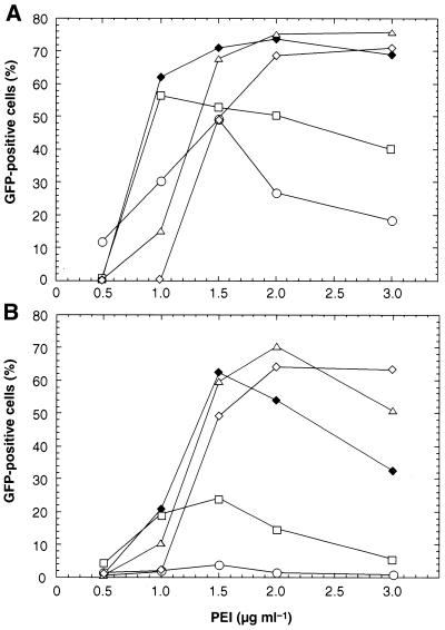
Preliminary results showed that linear and branched 25 kDa PEI were the most effective among various polymers tested (including branched 70 kDa, branched 50–100 kDa and branched 10 kDa; data not shown). Therefore, we optimized transfection of 293E cells with both linear or branched 25 kDa PEI polymers using a plasmid encoding the enhanced GFP (pEGFP). Transfections were performed using cells grown as monolayers in 12-well plates and GFP expression was measured 72 h later by flow cytometry. The effect of DNA to PEI ratios on transfection efficiency is shown in Figure 1 using linear (A) or branched (B) PEI. The indicated amounts of DNA and polymer are for one well containing 5 × 10⁵ cells. Only 0.25 µg of DNA was sufficient to reach a 50% transfection efficiency using linear PEI, whereas a minimum of 1.0 µg was necessary using the branched isoform. Transfection efficiencies of ∼70% were reached with both linear and branched polymers at DNA:PEI (µg:µg) ratios of 1.0:1.5 and 1.5:2.0, respectively. Increasing the amounts of both DNA and PEI did not lead to higher transfection yield.
Figure 1.
Effect of DNA to PEI ratio on transfection efficiency. 293E cells were transfected with linear (A) or branched (B) 25 kDa PEI at various DNA (pEGFP plasmid) concentrations as described in Materials and Methods. DNA concentrations (µg ml–1) used were: 0.25 (circles), 0.50 (squares), 1.0 (closed diamonds), 1.5 (triangles) and 2.0 (open diamonds). Transfection efficiencies were determined by flow cytometry analysis 72 hpt.
Cell line and expression vectors
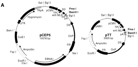
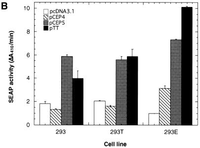
Two commercially available expression vectors containing viral sequences allowing for episomal DNA replication in permissive cell lines were tested. The first vector, pcDNA3.1, contains the SV40 origin of replication that allows cellular polymerases to replicate the DNA up to 10 000 copies in cells expressing the large T antigen (18). The second vector, pCEP4, contains the EBV origin of replication oriP that replicates plasmid DNA up to 90 copies in cells expressing the EBNA1 protein (19). We also generated the pCEP5 vector (Fig. 2A, left) by using an improved CMV expression cassette as described in the adenoviral transfer vector pAdCMV5 (20). This expression cassette has been shown to confer very high levels of r-protein expression in 293 cells (12). The pCEP5 vector was further modified (Materials and Methods) to yield the pTT vector (Fig. 2A, right) that is 4.6 kb smaller, hence providing more space for large cDNA cloning. The cDNA encoding for the reporter protein SEAP was then cloned in each of these four vectors and its expression level monitored following transient transfection in 293, 293T or 293E cells. As shown in Figure 2B, transfection of the 293T cell line with the SV40 ori-containing plasmid pcDNA3.1 did not translate into an increased transgene expression when compared with transfection of the parental 293 cells. However, transfection of 293E cells with the pCEP4 vector resulted in a 2–3-fold increase in SEAP expression compared with transfection of 293 or 293T cells with the same vector. In addition, the use of the pCEP5 vector further increased SEAP expression by a factor of 2–6-fold, depending on the cell line. Finally, the use of the pTT vector in 293E cells resulted in a 33% increase in transgene expression compared with the pCEP5 vector. The overall SEAP expression level in 293E cells was 10-fold higher with the pTT vector compared with the pcDNA3.1 vector.
Figure 2.
Effect of cell lines and vectors on SEAP expression. (A) Genetic maps of pCEP5 (left) and pTT (right) vectors drawn to scale. The pCEP5 vector backbone is identical to pCEP4 vector except for the transgene expression cassette. Construction of the pTT vector is as described in Materials and Methods. TPL, tripartite leader; enh MLP, adenovirus major late promoter enhancer; SD, splice donor; SA, splice acceptor; DS, dyad symmetry; FR, family of repeats. (B) Cells were transfected with 1 µg of DNA and 2 µg of linear PEI and SEAP activity measured 72 hpt. The pEGFP plasmid (0.1 µg) was also added in each condition to monitor for transfection efficiency and SEAP activities were normalized accordingly. Open boxes, pcDNA3.1/SEAP; hatched boxes, pCEP4/SEAP; gray boxes, pCEP5/SEAP; closed boxes, pTT/SEAP.
Effect of serum
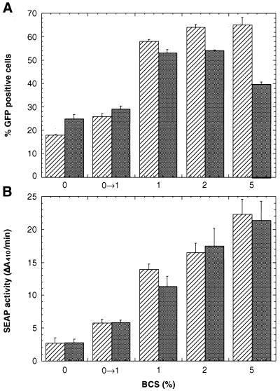
The effect of serum on transfection efficiency (GFP) and r-protein production (SEAP) mediated by both linear and branched PEI was evaluated. Figure 3 shows that when transfection mixture was added to cells in fresh 1% serum-containing medium, a 4–5-fold increase in SEAP activity was obtained compared with its addition to cells in serum-free medium. Increasing serum concentration up to 5% further improved PEI-mediated transfection efficiency and production. When transfection mixture was added to cells in serum-free media followed 3 h later by serum addition to a concentration of 1% (0→1%), a 2-fold increase in transgene expression was obtained; however, this level was only 50% of that obtained in 1% serum.
Figure 3.
Effect of serum on transgene expression. 293E cells were transfected with pTT/sgGFP (A) or pTT/SEAP (B) vectors using 1.0 µg of DNA and 2.0 µg of linear PEI (hatched boxes) or 1.5 and 2.0 µg of branched PEI (gray boxes) in fresh serum-free or serum-supplemented media. In one experiment (0→1%), cells were transfected in serum-free media and serum was added 3 h later to a final concentration of 1%. GFP-positive cells and SEAP activity were measured 72 hpt.
Process optimization for transfection in suspension
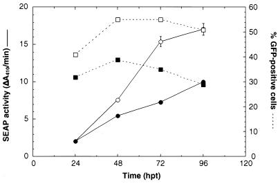
We next evaluated gene transfer efficiency of both linear and branched PEI on suspension-growing 293E cells in 1% BCS-supplemented HSFM. Shaker flask cultures were co-transfected with a mixture of pTT/SEAP:pEGFP (9:1) plasmids (pEGFP was added to monitor transfection efficiency). With both linear and branched PEI, SEAP accumulated in the culture medium for up to 96 hours post-transfection (hpt) (Fig. 4), but gene transfer and expression level were 50% higher using the linear isoform. These results clearly demonstrate that linear, and to a lesser extent branched, PEI are effective for gene transfer in suspension-growing cells. In addition, SEAP expression levels obtained with suspension-growing cells using linear PEI were comparable with those obtained with adherent-growing cells. For all experiments reported below, only linear PEI was used.
Figure 4.
Transfection of suspension growing cells. Cells were resuspended in 10 ml of fresh HSFM containing 1% BCS to a density of 1 × 10⁶ ml–1 in a 125 ml Erlenmeyer flask. Three hours later, 1 ml of the DNA–PEI complexes were added and the culture incubated for an additional 3 h. The volume was then completed to 20 ml with fresh culture medium. The DNA–PEI complexes were as follows: 40 µg of linear or branched PEI was added to 1 ml of HEPES-supplemented HSFM containing 18 µg of pTT/SEAP and 2 µg of pEGFP or 27 µg of pTT/SEAP and 3 µg of pEGFP, respectively. Open symbols, linear PEI; closed symbols: branched PEI.
Our goal was to define a robust, simple and scalable transfection process. In order to reach these objectives, two steps had to be simplified: the 3 h incubation of DNA–PEI complexes with cells in a reduced culture volume, and the medium change 3 h prior to transfection. The first step was performed with the assumption that it would promote interaction of the DNA–PEI complexes with the cells and thus increase transfection efficiency. The second was done according to reports showing a deleterious effect of conditioned medium on transfection efficiency (6,21). Whereas medium exchange is simple to perform on a small scale, this step represents a significant hurdle at scales greater than a few liters.
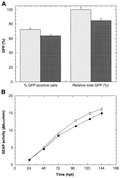
The effect of cell density at time of transfection was first evaluated (Fig. 5A) by transfecting high density (hatched bars; 10 ml at 1 × 10⁶ cells ml–1) or low density cultures (gray bars; 20 ml at 5 × 10⁵ cells ml–1) in shaker flasks. Three hours later, the high cell density flask was diluted to 5 × 10⁵ cells ml–1 with fresh medium, and GFP expression in both flasks monitored 72 h later. This experiment showed that cell concentration prior to transfection could be omitted as only a slight decrease (<10%) in transfection efficiency and a 15% decrease in GFP expression level were observed when cells were transfected in a larger culture volume.
Figure 5.
Effect of cell density and of conditioned medium. (A) Transfection efficiency and relative total GFP expression (in percent) obtained following transfection using standard conditions (hatched bars: 10 ml of cells at 1 × 10⁶ ml–1 followed by addition of 10 ml of fresh medium 3 h after transfection) or using cells at 5 × 10⁵ ml–1 in 20 ml of culture medium (gray bars). GFP was monitored 72 hpt. Relative total GFP was obtained following multiplication of percent GFP-positive cells by the mean fluorescence intensity. (B) Cells were seeded in 20 ml of 1% BCS-supplemented HSFM at a density of 2.5 × 10⁵ ml–1 24 h before transfection. The medium was then left unchanged (conditioned: open circles) or replaced with 20 ml of fresh medium (closed circles). Three hours later, cells were transfected by the addition of 2 ml of DNA–PEI complexes (20 µg of pTT/SEAP and 40 µg of linear PEI).
We next evaluated the effect of conditioned medium on SEAP expression using suspension growing cells. For this study, cells were seeded in shaker flasks at a density of 2.5 × 10⁵ ml–1. Twenty-four hours later, transfection was performed with or without a complete medium exchange. As shown in Figure 5B, no significant difference in SEAP expression was observed when the transfection was carried out in medium conditioned for 24 h, indicating that medium exchange is not necessary.
Transfection in bioreactors
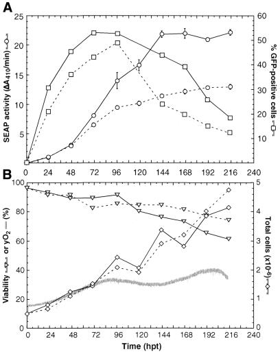
To demonstrate scalability of the process, a 3.5-l bioreactor culture was transfected with a mixture of pTT/SEAP:pEGFP plasmids (19:1). One hour later, a sample (25 ml) was withdrawn and transferred into a shaker flask as a control. In the bioreactor (Fig. 6A, unbroken lines), SEAP (circles) accumulated up to 144 hpt and then reached a plateau whereas accumulation continued up to 216 hpt in the control shaker flask (dashed lines). The percentage of GFP-positive cells (squares) at 96 hpt reached 54 and 50% for the bioreactor and the shaker flask, respectively. At the end of the culture, cell density was 4.1 and 4.7 × 10⁶ ml–1 with a viability of 62 and 72% for the bioreactor and shaker flask, respectively (Fig. 6B). Although viable cell density was 25% lower in the bioreactor compared with the shaker flask, volumetric SEAP productivity was almost 2-fold higher. Similar results were systematically observed in five independent experiments (results not shown), indicating that the productivity of secreted proteins might be increased when using a controlled environment.
Figure 6.
Transient transfection in a 3.5-l bioreactor. (A) 293E cells were seeded at a density of 2.5 × 10⁵ ml–1 in 2.85 l of fresh HSFM supplemented with 1% BCS. Twenty-four hours later, the transfection mixture (6 mg of linear PEI added to 150 ml HSFM containing 2.85 mg pTT/SEAP and 150 µg pEGFP plasmids) was added to the bioreactor (unbroken lines). One hour later, 25 ml of culture was withdrawn from the bioreactor and transferred in a shake flask as a control (dashed lines). SEAP activity (circles) and GFP-positive cells (squares) were determined as described in Materials and Methods. (B) Growth curves (diamonds), viability (triangles) and yO2 (gray line) in the 3.5-l bioreactor (unbroken lines) and shaker flask (dashed lines).
Purification of SEAP and production of other r-proteins
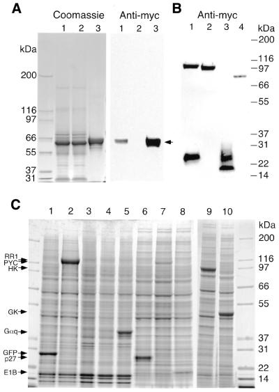
Purification of Myc-(His)6-tagged SEAP harvested from the bioreactor run (Fig. 6) by immobilized metal affinity chromatography (IMAC) is shown in Figure 7A. The left panel shows Coomassie blue-stained protein pattern from the culture medium before loading on the column (lane 1), flow-through (lane 2) and eluted material using 150 mM imidazole (lane 3). The right panel shows immunodetection of SEAP in the same fractions using anti-Myc antibody. This figure shows that all of the His-tagged SEAP was retained on the column whereas very few, if any, serum proteins bound to it (SEAP migrates with an apparent molecular weight slightly higher than BSA). SEAP quantification in the eluted fraction using the Lowry protein assay showed that ∼60 mg of His-tagged SEAP could be recovered by IMAC from the 3-l bioreactor culture. As shown in Figure 7B, high expression levels in bioreactor were also obtained with other secreted r-proteins. Fourteen- (lanes 1, 3 and 4) or 3.5-liter (lane 2) bioreactors were transfected with pTT plasmids encoding for Neuropilin-1 and VEGF (1:1 ratio, lane 1), Tie2 (lane 2), Cripto (lane 3) and c-Met (lane 4). All cultures were harvested 5 days post-transfection. With the exception of Cripto, which has been reported to be highly glycosylated on serine, threonine and asparagine (22), glycosylation of the expressed proteins appeared to be relatively homogeneous as suggested by their migration behavior following SDS–PAGE. High expression levels of intracellular r-proteins were also obtained as shown in Figure 7C. In this experiment, 293E cells were transfected with pTT plasmids encoding for sgGFP (lane 1), herpes simplex virus ribonucleotide reductase 1 (RR1, lane 2), mouse Gαq (lane 5), human p27Kip1 (lane 6), yeast pyruvate carboxylase (PYC, lane 7), adenovirus E1B19K (lane 8), human hexokinase 1 (HK, lane 9) and human glucokinase (GK, lane 10). Three days after transfection, cells were rinsed with PBS, solubilized in sample buffer (GFP, RR1 and Gαq) or extracted with lysis buffer (p27Kip1, PYC, E1B19K, HK and GK), and proteins analyzed by SDS–PAGE. Quantification of r-proteins shown in Figure 7 is summarized in Table 1. In the case of RR1, volumetric production was 50 mg/l, representing 20% of TCP. The mouse Gαq was expressed at 16 mg/l, compared with a barely detectable level (by Commassie staining) when expressed from the pcDNA3.1 vector (lane 4).
Figure 7.
SEAP purification and production of other secreted and intracellular r-proteins. (A) SEAP purification by IMAC. One liter of culture medium from the 3.5-l bioreactor harvest (Fig. 6) was loaded onto a TALON™ IMAC column (10 ml bed volume). Following extensive washing, bound material was eluted with 150 mM imidazole (20 ml). Ten microliters of culture medium (lane 1), flow-through (lane 2) and eluted material (lane 3) were resolved in duplicate on a 3–8% NuPAGE Tris–acetate gradient gel. One half of the gel was directly stained with Coomassie blue R-250 (left panel) whereas the other half was transferred onto a nitrocellulose membrane and probed with anti-Myc antibody (right panel). (B) Expression of secreted C-terminal Myc-(His)6-tagged r-proteins in a 14-l bioreactor. Lane 1, human Neuropilin-1 (1–824; upper band) and VEGF (1–165; lower band) co-transfection in a 1:1 ratio; lane 2, human Tie2 (1–723); lane 3, human Cripto (1–173); lane 4, human c-Met (1–931). Transfections were performed as described in Materials and Methods and culture medium harvested 120 hpt. Fifteen microliters of culture medium was loaded per lane and tagged proteins detected using anti-Myc antibody. (C) Expression of intracellular r-proteins. Lane 1, pTT/sgGFP; lane 2, pTT/RR1; lane 3, pTT empty vector; lane 4, pcDNA3.1/Gαq; lane 5, pTT/Gαq; lane 6, pTT/p27Kip1; lane 7, pTT/PYC; lane 8, pTT/E1B19K; lane 9, pTT/hexokinase; lane 10, pTT/glucokinase. Cells were harvested 72 hpt, rinsed with PBS and solubilized in NuPAGE sample buffer followed by sonication (lanes 1–5) or extracted in lysis buffer (lanes 6–10) as indicated in Materials and Methods. Proteins were resolved on a 4–12% Bis–Tris NuPAGE gradient gel and stained with Coomassie blue R-250.
Table 1. Summary of r-protein expression levels.
| r-Protein | Tag | Localization | Culture mode | Concentration (mg l–1) |
|---|---|---|---|---|
| Human SEAP | Myc-(His)6 | Secreted | 3-l bioreactor | 20a |
| Human Neuropilin-1 | Myc-(His)6 | Secreted | 14-l bioreactor | 8b |
| Human VEGF | Myc-(His)6 | Secreted | 14-l bioreactor | 10b |
| Human Tie2 | Myc-(His)6 | Secreted | 3-l bioreactor | 9 |
| Human Cripto | Myc-(His)6 | Secreted | 14-l bioreactor | 9 |
| Human c-Met | Myc-(His)6 | Secreted | 14-l bioreactor | 1 |
| sgGFP | None | Intracellular | Shaker flask | 20 |
| Herpes virus RR1 | None | Intracellular | Shaker flask | 50 |
| Mouse Gαq | None | Membrane | T-flask | 16 |
| Human p27Kip1 | None | Intracellular | T-flask | 14 |
| Human hexokinase | None | Intracellular | Shaker flask | 40 |
| Human glucokinase | None | Intracellular | Shaker flask | 30 |
| Yeast PYC | None | Intracellular | 1-l bioreactor | 4 |
| Adenovirus E1B19K | None | Intracellular | T-flask | 3 |
DISCUSSION
In this study, we investigated the effects of various parameters on r-protein expression by transient transfection of suspension growing cells using the polycationic polymer PEI. By combining the use of the optimized oriP-containing pTT expression plasmid with the 293E cell line, we reached expression levels of intracellular r-protein representing up to 20% of total cellular proteins. To our knowledge, such high expression levels have never been described in 293 cells using transient transfection, and rival those obtained using virus-mediated transgene expression (12). Expression of the secreted protein SEAP was also considerable, as it was produced at levels exceeding 20 mg/l.
The use of amplifiable expression cassettes in mammalian cells such as the dihydrofolate reductase or glutamine synthetase systems have been shown to result in the isolation of stable cell lines showing very high levels of r-protein expression. As an alternative to these stable amplified systems, vectors with viral-derived elements that allow for episomal replication and amplification, such as the large-T antigen/SV40 ori, or the EBNA1/oriP, are well suited when using transient expression systems (8). Although plasmid DNA containing the SV40 ori was shown to replicate in the large-T antigen expressing 293T cells line (23), we showed that it did not provide higher transgene expression in 293T cells when compared with the 293 parental cell line. In contrast, the use of oriP-containing plasmids in 293E cells significantly increased transgene expression compared with the non-permissive 293 cells. This suggests that the increased transgene expression obtained using EBV replicon-containing plasmids might be mediated by a phenomenon distinct from its ability to support episomal replication. This is further supported by the fact that removal of the DS domain of oriP, which is responsible for initiation of DNA replication in EBNA1 positive cells (24), did not significantly reduce transgene expression (data not shown). One likely mechanism for this oriP-mediated increased expression could arise from the described EBNA1-dependent enhancer activity of oriP (25–27). The EBV oriP contains 24 EBNA1 binding sites (28). As EBNA1 has an efficient nuclear localization signal (29,30), its binding to plasmids bearing an oriP may also increase their nuclear import, thus enhancing transgene expression. Indeed, the most important barrier to transfection seems to be the limited migration of plasmid DNA from the cytoplasm to the nucleus (31). However, contribution of this mechanism to the enhanced transgene expression could be partially hindered when using PEI as the transfection reagent, as this polymer was also shown to actively undergo nuclear localization (32,33).
Whereas linear 25 kDa PEI was reported to efficiently mediate gene transfer in the presence of serum (34), transgene expression mediated by the branched isoform was shown to be reduced by 3-fold in its presence (6). This contrasts with our results showing that gene transfer was also significantly increased using the branched 25 kDa PEI. The mechanism by which serum increases gene delivery and/or transgene expression is not yet clear. Serum might contribute to augment transcriptional activity of the promoter as the CMV immediate early enhancer contains multiple binding sites for serum-activated transcription factors (35,36). However, only a partial recovery of transgene expression was obtained when serum was added to the cells 3 h after their transfection in serum-free medium. This suggests that, in addition to the potential serum-mediated CMV promoter transcriptional activation, some serum component(s) might increase transfection efficacy of DNA–PEI complexes.
A major drawback of gene transfer using polycations or cationic lipids is the inhibitory effect of conditioned medium on gene delivery. In the case of cationic lipids, this inhibition was shown to be mediated by the presence of secreted glycosaminoglycans (21,37), which are expected to efficiently displace DNA from lipid complexes. Whereas it was shown that conditioned medium adversely reduced PEI-mediated transfection of 293E cells (6), no significant effect was observed in our study. The reason for this discrepancy is not clear, but might result from the type of culture medium used, the age of the culture or from the cells themselves. The fact that, in our hands, transfection of cells in their 24 h-conditioned medium does not reduce gene transfer and expression, greatly simplifies process scale-up.
In conclusion, a significant improvement in transgene expression following transient transfection of suspension-growing cells using PEI was obtained by combining optimized parameters such as the pTT expression vector, the 293E cell line, the culture medium and the transfection process. Under these conditions, ∼60 mg of purified SEAP could be obtained from a 3-l culture following a single IMAC purification step. Volumetric expressions of the intracellular proteins GFP and RR1 were, respectively, 20 and 50 mg/l at 72 hpt, representing up to 20% of TCP. As this technology is robust, inexpensive and easy to perform, it is fully adapted for high-throughput production of milligram quantities of r-proteins needed for biochemical or structural studies and high-throughput screenings.
Acknowledgments
ACKNOWLEDGEMENTS
We thank Chunlin Xin, Eric Carpentier, Eric Thibaudeau and Robert Larocque for their technical assistance in cDNA cloning and plasmid purification, Brian Cass for bioreactor runs, and Phuong Lan Pham, Gilles St-Laurent and Cynthia Elias for critical reading of the manuscript. We are grateful to Bernard Massie for providing us with purified RR1, anti-RR1 antibody and RR1 cDNA.
REFERENCES
- 1.Cullen B.R. (1987) Use of eukaryotic expression technology in the functional analysis of cloned genes. Methods Enzymol., 152, 684–704. [DOI] [PubMed] [Google Scholar]
- 2.Blasey H.D., Aubry,J.-P., Mazzei,G. and Bernard,A. (1996) Large scale transient expression with COS cells. Cytotechnology, 18, 183–192. [DOI] [PubMed] [Google Scholar]
- 3.Cachianes G., Ho,C., Weber,R.F., Williams,S.R., Goeddel,D.V. and Leung,D.W. (1993) Epstein–Barr virus-derived vectors for transient and stable expression of recombinant proteins. Biotechniques, 15, 255–259. [PubMed] [Google Scholar]
- 4.Boussif O., Lezoualc’h,F., Zanta,M.A., Mergny,M.D., Scherman,D., Demeneix,B. and Behr,J.P. (1995) A versatile vector for gene and oligonucleotide transfer into cells in culture and in vivo: polyethylenimine. Proc. Natl Acad. Sci. USA, 92, 7297–7301. [DOI] [PMC free article] [PubMed] [Google Scholar]
- 5.Jordan M., Kohne,C. and Wurm,F.M. (1998) Calcium-phosphate mediated DNA transfer into HEK-293 cells in suspension: control of physicochemical parameters allows transfection in stirred media. Cytotechnology, 26, 39–47. [DOI] [PMC free article] [PubMed] [Google Scholar]
- 6.Schlaeger E.-J. and Christensen,K. (1999) Transient gene expression in mammalian cells grown in serum-free suspension culture. Cytotechnology, 30, 71–83. [DOI] [PMC free article] [PubMed] [Google Scholar]
- 7.Wurm F. and Bernard,A. (1999) Large-scale transient expression in mammalian cells for recombinant protein production. Curr. Opin. Biotechnol., 10, 156–159. [DOI] [PubMed] [Google Scholar]
- 8.Van Craenenbroeck K., Vanhoenacker,P. and Haegeman,G. (2000) Episomal vectors for gene expression in mammalian cells. Eur. J. Biochem., 267, 5665–5678. [DOI] [PubMed] [Google Scholar]
- 9.Foecking M.K. and Hofstetter,H. (1986) Powerful and versatile enhancer-promoter unit for mammalian expression vectors. Gene, 45, 101–105. [DOI] [PubMed] [Google Scholar]
- 10.Gorman C.M., Gies,D., McCray,G. and Huang,M. (1989) The human cytomegalovirus major immediate early promoter can be _trans_-activated by adenovirus early proteins. Virology, 171, 377–385. [DOI] [PubMed] [Google Scholar]
- 11.Massie B., Couture,F., Lamoureux,L., Mosser,D.D., Guilbault,C., Jolicoeur,P., Belanger,F. and Langelier,Y. (1998) Inducible overexpression of a toxic protein by an adenovirus vector with a tetracycline-regulatable expression cassette. J. Virol., 72, 2289–2296. [DOI] [PMC free article] [PubMed] [Google Scholar]
- 12.Massie B., Mosser,D., Koutromanis,M., Vitté-Mony,I., Lamoureux,L., Couture,F., Paquet,L., Guilbault,C., Dionne,J., Chahla,D. et al. (1998) New adenovirus vectors for protein production and gene transfer. Cytotechnology, 28, 53–64. [DOI] [PMC free article] [PubMed] [Google Scholar]
- 13.Godbey W.T., Wu,K.K. and Mikos,A.G. (1999) Poly(ethylenimine) and its role in gene delivery. J. Control Release, 60, 149–160. [DOI] [PubMed] [Google Scholar]
- 14.Côté J., Garnier,A., Massie,B. and Kamen,A. (1998) Serum-free production of recombinant proteins and adenoviral vectors by 293SF-3F6 cells. Biotechnol. Bioeng., 59, 567–575. [PubMed] [Google Scholar]
- 15.DuBridge R.B., Tang,P., Hsia,H.C., Leong,P.M., Miller,J.H. and Calos,M.P. (1987) Analysis of mutation in human cells by using an Epstein–Barr virus shuttle system. Mol. Cell. Biol., 7, 379–387. [DOI] [PMC free article] [PubMed] [Google Scholar]
- 16.Kamen A.A., Chavarie,C., André,J. and Archambault,J. (1992) Design parameters and performance of a surface baffled helical ribbon impeller bioreactor for the culture of shear sensitive cells. Chem. Eng. Sci., 27, 2375–2380. [Google Scholar]
- 17.Durocher Y., Perret,S., Thibaudeau,E., Gaumond,M.H., Kamen,A., Stocco,R. and Abramovitz,M. (2000) A reporter gene assay for high-throughput screening of G-protein-coupled receptors stably or transiently expressed in HEK293 EBNA cells grown in suspension culture. Anal. Biochem., 284, 316–326. [DOI] [PubMed] [Google Scholar]
- 18.Chittenden T., Frey,A. and Levine,A.J. (1991) Regulated replication of an episomal simian virus 40 origin plasmid in COS7 cells. J. Virol., 65, 5944–5951. [DOI] [PMC free article] [PubMed] [Google Scholar]
- 19.Yates J.L., Warren,N. and Sugden,B. (1985) Stable replication of plasmids derived from Epstein–Barr virus in various mammalian cells. Nature, 313, 812–815. [DOI] [PubMed] [Google Scholar]
- 20.Massie B., Dionne,J., Lamarche,N., Fleurent,J. and Langelier,Y. (1995) Improved adenovirus vector provides herpes simplex virus ribonucleotide reductase R1 and R2 subunits very efficiently. Biotechnology, 13, 602–608. [DOI] [PubMed] [Google Scholar]
- 21.Ruponen M., Yla-Herttuala,S. and Urtti,A. (1999) Interactions of polymeric and liposomal gene delivery systems with extracellular glycosaminoglycans: physicochemical and transfection studies. Biochim. Biophys. Acta, 1415, 331–341. [DOI] [PubMed] [Google Scholar]
- 22.Schiffer S.G., Foley,S., Kaffashan,A., Hronowski,X., Zichittella,A.E., Yeo,C.Y., Miatkowski,K., Adkins,H.B., Damon,B., Whitman,M. et al. (2001) Fucosylation of Cripto is required for its ability to facilitate nodal signaling. J. Biol. Chem., 276, 37769–37778. [DOI] [PubMed] [Google Scholar]
- 23.Heinzel S.S., Krysan,P.J., Calos,M.P. and DuBridge,R.B. (1988) Use of simian virus 40 replication to amplify Epstein–Barr virus shuttle vectors in human cells. J. Virol., 62, 3738–3746. [DOI] [PMC free article] [PubMed] [Google Scholar]
- 24.Wysokenski D.A. and Yates,J.L. (1989) Multiple EBNA1-binding sites are required to form an EBNA1-dependent enhancer and to activate a minimal replicative origin within oriP of Epstein–Barr virus. J. Virol., 63, 2657–2666. [DOI] [PMC free article] [PubMed] [Google Scholar]
- 25.Reisman D. and Sugden,B. (1986) _trans_-Activation of an Epstein–Barr viral transcriptional enhancer by the Epstein–Barr viral nuclear antigen 1. Mol. Cell. Biol., 6, 3838–3846. [DOI] [PMC free article] [PubMed] [Google Scholar]
- 26.Sugden B. and Warren,N. (1989) A promoter of Epstein–Barr virus that can function during latent infection can be transactivated by EBNA-1, a viral protein required for viral DNA replication during latent infection. J. Virol., 63, 2644–2649. [DOI] [PMC free article] [PubMed] [Google Scholar]
- 27.Gahn T.A. and Sugden,B. (1995) An EBNA-1-dependent enhancer acts from a distance of 10 kilobase pairs to increase expression of the Epstein–Barr virus LMP gene. J. Virol., 69, 2633–2636. [DOI] [PMC free article] [PubMed] [Google Scholar]
- 28.Mackey D. and Sugden,B. (1999) Applications of oriP plasmids and their mode of replication. Methods Enzymol., 306, 308–328. [DOI] [PubMed] [Google Scholar]
- 29.Ambinder R.F., Mullen,M.A., Chang,Y.N., Hayward,G.S. and Hayward,S.D. (1991) Functional domains of Epstein–Barr virus nuclear antigen EBNA-1. J. Virol., 65, 1466–1478. [DOI] [PMC free article] [PubMed] [Google Scholar]
- 30.Längle-Rouault F., Patzel,V., Benavente,A., Taillez,M., Silvestre,N., Bompard,A., Sczakiel,G., Jacobs,E. and Rittner,K. (1998) Up to 100-fold increase of apparent gene expression in the presence of Epstein–Barr virus oriP sequences and EBNA1: implications of the nuclear import of plasmids. J. Virol., 72, 6181–6185. [DOI] [PMC free article] [PubMed] [Google Scholar]
- 31.Zabner J., Fasbender,A.J., Moninger,T., Poellinger,K.A. and Welsh,M.J. (1995) Cellular and molecular barriers to gene transfer by a cationic lipid. J. Biol. Chem., 270, 18997–19007. [DOI] [PubMed] [Google Scholar]
- 32.Pollard H., Remy,J.S., Loussouarn,G., Demolombe,S., Behr,J.P. and Escande,D. (1998) Polyethylenimine but not cationic lipids promotes transgene delivery to the nucleus in mammalian cells. J. Biol. Chem., 273, 7507–7511. [DOI] [PubMed] [Google Scholar]
- 33.Godbey W.T., Wu,K.K. and Mikos,A.G. (1999) Tracking the intracellular path of poly(ethylenimine)/DNA complexes for gene delivery. Proc. Natl Acad. Sci. USA, 96, 5177–5181. [DOI] [PMC free article] [PubMed] [Google Scholar]
- 34.Boussif O., Zanta,M.A. and Behr,J.P. (1996) Optimized galenics improve in vitro gene transfer with cationic molecules up to 1000-fold. Gene Ther., 3, 1074–1080. [PubMed] [Google Scholar]
- 35.Boshart M., Weber,F., Jahn,G., Dorsch-Hasler,K., Fleckenstein,B. and Schaffner,W. (1985) A very strong enhancer is located upstream of an immediate early gene of human cytomegalovirus. Cell, 41, 521–530. [DOI] [PubMed] [Google Scholar]
- 36.Brightwell G., Poirier,V., Cole,E., Ivins,S. and Brown,K.W. (1997) Serum-dependent and cell cycle-dependent expression from a cytomegalovirus-based mammalian expression vector. Gene, 194, 115–123. [DOI] [PubMed] [Google Scholar]
- 37.Belting M. and Petersson,P. (1999) Intracellular accumulation of secreted proteoglycans inhibits cationic lipid-mediated gene transfer. Co-transfer of glycosaminoglycans to the nucleus. J. Biol. Chem., 274, 19375–19382. [DOI] [PubMed] [Google Scholar]
