Rapid and reversible relocalization of heat shock factor 1 within seconds to nuclear stress granules (original) (raw)
Abstract
Heat shock factor 1 (HSF1) is essential for the stress-induced expression of heat shock genes. On exposure to heat shock, HSF1 localizes within seconds to discrete nuclear granules. On recovery from heat shock, HSF1 rapidly dissipates from these stress granules to a diffuse nucleoplasmic distribution, typical of unstressed cells. Subsequent reexposure to heat shock results in the rapid relocalization of HSF1 to the same stress granules with identical kinetics. Although the appearance of HSF1 stress granules corresponds to the hyperphosphorylated, trimeric DNA-binding state of HSF1 and correlates temporally with the inducible transcription of heat shock genes, they are also present in heat-shocked mitotic cells that are devoid of transcription. This finding suggests a role for HSF1 stress granules as a nuclear compartment for the temporal regulation and spatial organization of HSF1 activity and reveals new features of the dynamics of nuclear organization.
Keywords: heat shock, green fluorescent protein, dynamics, nuclear organization, nuclear structures
The molecular response to environmental and physiological stress is the elevated synthesis of the ubiquitous heat shock proteins (HSPs) (reviewed in refs. 1 and 2). The inducible expression of hsp genes is under the control of a family of specific transcription factors, the heat shock factors (HSFs) (reviewed in refs. 3 and 4). Among the three HSFs that have been described in humans, HSF1 is closely associated with the stress-induced expression of heat shock genes. Activation of HSF1 is a multistep process involving trimerization, acquisition of DNA-binding activity, and inducible phosphorylation (5–16).
At the cellular level, HSF1 is predominantly found in a diffuse cytoplasmic and nuclear distribution in unstressed cells, and on heat shock relocates rapidly to form large and irregularly shaped nuclear granules (7). These nuclear structures, referred to as HSF1 stress granules, can be induced by other stresses, such as amino acid analogues or heavy metals, and are detected in different cell types (17). They do not coincide with other previously described nuclear compartments such as nucleoli, coiled bodies, kinetochores, promyelocytic leukemia bodies, or the speckles enriched in splicing factors (17). The HSF1 granules are also distinct from the sites of transcription of the major hsp70 and hsp90 genes (18). However, the number of HSF1 granules correlates with ploidy, which suggests the existence of specific chromosomal targets (18).
In this study, we examined the properties of HSF1 stress granules in living cells and reveal new features on the rapid stress-induced relocalization of HSF1 with implications for the regulation of the heat shock response.
MATERIALS AND METHODS
Cell Culture, Mitotic Arrest, and Induction of the Stress Response.
HeLa cells were grown in DMEM supplemented with 5% fetal bovine serum. For immunofluorescence and fluorescence in situ hybridization (FISH) experiments, cells were grown on two-chamber glass slides (Lab-Tek). For live-cell experiments, cells were grown on 18-mm-circle glass coverslips. Cadmium was used at a final concentration of 30 μM, and azetidine was used at a final concentration of 5 mM. Heat shock was performed by immersion of the slides in a 42°C water bath. Mitotic arrest was induced by treating the cells with 0.01 μg/ml colchicine for 2 hours. For live-cell experiments, cells were transfected with a HSF1–green fluorescent protein (GFP) construct (17) by using the LipoFectamine Plus system (GIBCO/BRL).
Electrophoretic Mobility-Shift Assay and Western Blot.
HSF1 DNA-binding activity was analyzed by using the electrophoretic mobility-shift assay as described (19) with a 32P-labeled double-stranded synthetic heat shock element containing four inverted nGAAn repeats (20). Western blot analysis was performed by using whole-cell extracts and the 10H8 rat monoclonal anti-HSF1 antibody (17). The immune complexes were analyzed by using the ECL detection system (Amersham Pharmacia).
Time-Lapse Microscopy of Living Cells.
Heat shock on living cells was performed by using a temperature controller adapted to the microscope stage (21). Cadmium and azetitine were added directly to the cell medium in the incubation chamber of the stage maintained at 37°C. Time-lapse microscopic images were acquired with a cooled charge-coupled device camera (Photometrics Sensys) mounted on an epifluorescence microscope (Zeiss Axiophot) equipped with a 100 W mercury lamp, by using a ×40 (0.75 numerical aperture) air objective and the appropriate filter for green fluorescence. A total of 20 cells containing a various number of granules was analyzed for each experiment. Fluorescence recovery after photobleaching (FRAP) experiments were performed on a Zeiss confocal microscope (LSM 410), by using the nonattenuated 488 nm laser line and a ×40 (1.0 numerical aperture) oil immersion objective. A total of 20 granules was analyzed by photobleaching.
Immunofluorescence and FISH.
Detection of HSF1 by immunofluorescence was performed on formaldehyde-fixed cells as described (22). Biochemical nuclear extraction was performed before fixation as described by Fey et al. (26). The 10H8 rat monoclonal anti-HSF1 antibody (17) was used at a dilution of 1:300 and detected with a goat anti-rat antibody coupled to fluorescein (Sigma). hsp70 transcripts were detected by FISH as described (22) by using the pH2.3 genomic clone corresponding to the human hsp70 coding sequence (23). DNA was counterstained with 250 ng/ml propidium iodide.
Confocal Microscopy, Optical Sectioning, and Three-Dimensional Reconstructions.
Images of FISH and immunofluorescence experiments were acquired on a Zeiss LSM 410 confocal microscope by using the ×63 (1.4 numerical aperture) oil immersion objective. Optical sections in the z axis of cells were generated every 0.3 μm. The three-dimensional structures of nuclei were reconstructed by using software developed at the University of Grenoble (France) (24, 25).
RESULTS
Three-Dimensional Structure of HSF1 Granules.
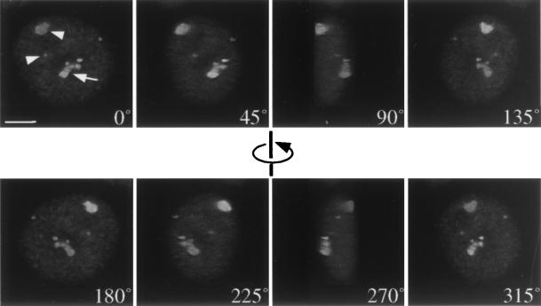
The study of the three-dimensional structure of HSF1 stress granules was achieved by using confocal scanning laser microscopy to generate optical sections through the nuclei of heat-shocked HeLa cells immunostained for HSF1, followed by computerized three-dimensional reconstruction and image analysis (24, 25). HSF1 stress granules were visualized as heterogenous intranuclear structures (Fig. 1). Two different patterns emerged from the analysis of 50 nuclei: single globular granules that can be either small or large (indicated by arrowheads in Fig. 1) or grape-like clusters composed of multiple smaller globular structures that do not appear to be interconnected (arrow in Fig. 1). An average of 19.8 (± 7.4) single globular units was found per nucleus, altogether forming an average of 6–10 HSF1 granules. They varied in size from 0.3 μm to 3 μm, the total volume of the granules per nucleus ranges from 2 μm3 to 17 μm3, and they can occupy up to 1.5% of the total nuclear volume (average: 0.6% ± 0.2%).
Figure 1.
Three-dimensional structure of HSF1 stress granules. Rotated views of the nucleus of a heat-shocked HeLa cell displaying characteristic HSF1 stress granules. Arrowheads correspond to large and small single globular units, and the arrow indicates a grape-like cluster of smaller globular structures. (Bar = 5 μm.)
The comprehensive study of the spatial distribution of HSF1 stress granules revealed that rather than distributed randomly, 50% of the HSF1 stress granules were adjacent to nucleoli and 30% were found in close proximity to the nuclear membrane. These features are evident in the rotated views shown in Fig. 1, in which three of the stress granules are in the periphery of the nucleus, one is centrally positioned, and another is adjacent to the nucleolus.
Biochemical Properties of HSF1 in the Stress Granules.
What is the biochemical state of HSF1 associated with stress granules? Whereas previous studies have established that heat shock and other stresses cause HSF1 to undergo an oligomerization event associated with trimerization and DNA binding (reviewed in refs. 3 and 4), does relocalization of HSF1 to stress granules reflect a specific targeting event in which HSF1 trimers inducibly associate with a specific ligand? To assess the biochemical properties of HSF1 stress granules, we used a differential biochemical extraction assay to investigate the solubility of HSF1 stress granules. After 30 minutes of heat shock at 42°C, HeLa cells were extracted with 0.5% Triton X-100 followed by treatment with 250 mM ammonium sulfate and subsequent digestion by DNase I (26). On fixation and immunostaining for HSF1, the stress granules were retained in >98% of the cells, even after complete digestion of DNA (data not shown). These results reveal that the HSF1 associated with stress granules is neither soluble nor readily extracted. In addition, HSF1 granules cannot form in vitro in nuclear extracts, suggesting that the context of the nucleus is required for HSF1 stress-granule formation (data not shown).
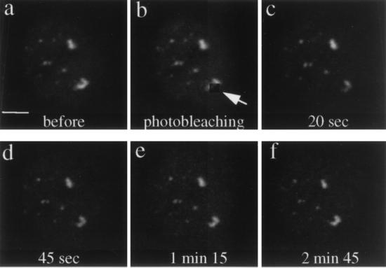
Although the biochemical extraction assay establishes that HSF1 is not released from the stress granules, our results do not exclude the possibility that the stress granules are themselves resistant to the differential extraction protocol. Therefore, to address whether the HSF1 associated with the stress granules is static or mobile, we performed FRAP on living HeLa cells transiently expressing a HSF1–GFP construct and exposed at 42°C for 15 minutes. We selected cells displaying several granules so that at least one can serve as a reference to monitor the specificity of bleaching and the stability of the focal plane. As shown in Fig. 2, a region of a stress granule was photobleached by a 1-min exposure to the 488 nm laser line (Fig. 2b), and averaged images were captured at different time points after photobleaching. Within 20 seconds, HSF1–GFP was detected in the bleached region (Fig. 2c), and within 3 min, both the intensity and the morphology of the granule had recovered completely (Fig. 2 d_–_f). This observation was highly reproducible. The rapid recovery of fluorescence only occurred in living cells and was not detected when the photobleaching was performed on fixed cells (data not shown). These results reveal that the HSF1 protein associated with stress granules is mobile, but cannot be released by differential extraction using nonionic detergents and DNase I digestion.
Figure 2.
The HSF1 protein is highly mobile within the granules. FRAP experiment on living HeLa cells transiently transfected with a HSF1–GFP construct and exposed at 42°C for 15 minutes. A portion of a stress granule was photobleached by a 1-minute exposure to the 488 nm laser line of the confocal microscope. Averaged images were acquired at different time points: before photobleaching (a), just after photobleaching (b), 20 seconds after photobleaching (c), 45 seconds after photobleaching (d), 1 minute 15 seconds after photobleaching (e), and 2 minutes 45 seconds after photobleaching (f). (Bar = 5 μm.)
HSF1 Stress Granules Are Highly Dynamic, Reversible Structures.
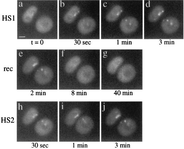
The rapid recovery of fluorescence observed in the FRAP experiments reveals the highly dynamic properties of HSF1 stress granules. Previous studies using indirect immunofluorescence on fixed cells have demonstrated that HSF1 stress granules can be detected within 5 min of heat shock and disappear during attenuation of the heat shock response (17, 18). A more precise kinetics of HSF1 translocation was measured in living HeLa cells transiently expressing a HSF1–GFP construct by using time-lapse microscopy during heat shock at 42°C and recovery. A temperature controller adapted for use on the microscope stage allowed heat shock conditions to be attained within 5 seconds (21). In cells maintained at 37°C, the HSF1–GFP was distributed diffusely throughout the nucleus of the cells, excluding nucleoli (Fig. 3a). HSF1 stress granules were detected within 30 seconds of heat shock (Fig. 3b); with continued exposure to the elevated temperature, the fluorescence intensified and the resolution of the granules sharpened (Fig. 3 c and d). Meanwhile, the diffuse nucleoplasmic staining decreased as HSF1 was recruited to the stress granules (compare Fig. 3 a and d). By 3 minutes after heat exposure, the granules visualized by HSF1–GFP were indistinguishable from the endogenous stress granules (data not shown). The intensity, size, and number of HSF1 granules did not change significantly beyond 3 minutes of heat shock (data not shown).
Figure 3.
HSF1 granules are transient and reversible structures. Time-lapse microscopy during heat shock and recovery on living HeLa cells transiently transfected with a HSF1–GFP construct. Images were acquired before heat shock (a), after 30 seconds (b), 1 minute (c), and 3 minutes (d) of heat shock at 42°C, 2 minutes of recovery (e), 8 minutes of recovery (f), 40 minutes of recovery (g) and after 30 seconds (h), 1 minute (i), and 3 minutes (j) of a second heat shock at 42°C. (Bar = 5 μm.)
When cells were returned to 37°C after the 3-minute heat shock, the intensity and definition of the granules progressively decreased, whereas the diffuse nucleoplasmic staining intensified, and HSF1 stress granules were no longer detected by 7–8 minutes of recovery (Fig. 3 e and f). During recovery from heat shock, the kinetics of disappearance of HSF1 granules depends on the intensity of the original heat shock. For example, the HSF1 stress granules in cells exposed to a 15-minute heat shock persisted for 30 minutes of recovery (data not shown). The disappearance of the granules is temperature-dependent and does not occur if recovery is performed at 4°C, suggesting an ATP-dependent process (data not shown).
Having established that HSF1 stress granules form within seconds of heat shock and regress rapidly on recovery, we asked whether cells exposed to a subsequent heat shock would form HSF1 stress granules at the same subnuclear location. After 40 minutes of recovery from the initial heat shock (Fig. 3g), the cells were exposed to a second heat shock at 42°C, and HSF1 granule reformation was followed. We found that the stress granules reformed with an identical kinetics as during the first heat exposure and at the same position within the nucleus (Fig. 3 h_–_j). This reversible translocation of HSF1 to stress granules can occur repeatedly on multiple cycles of stress and recovery. The movements of the HSF1–GFP were highly reproducible. These results reveal that the number, morphology, and relative intensity of HSF1 within the stress granules are conserved. HSF1 stress granules therefore represent transient and highly dynamic nuclear structures associated with the heat shock response. Their ability to reform at the same location within the nucleus during successive heat exposures suggests a defined structure and perhaps a specific target for the granules.
Kinetics of HSF1 Granule Formation and Activation of the Heat Shock Response.
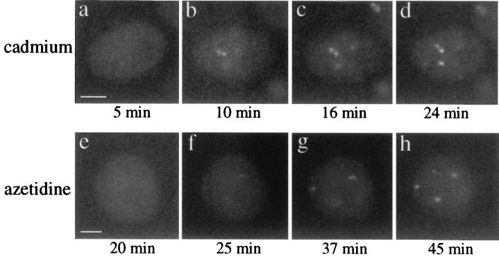
We next examined the kinetics of HSF1 stress granule formation in living HeLa cells transfected with the HSF1–GFP construct and exposed to the heavy metal cadmium or to the amino acid analogue azetidine at 37°C. HSF1 stress granules were detected after 5–10 minutes of treatment with cadmium (Fig. 4a), and steady state was reached after about 24 minutes of cadmium exposure (Fig. 4 b_–_d). During azetidine treatment, granules were detected after 25 minutes (Fig. 4e), and steady state was achieved after 45 minutes (Fig. 4 f_–_h). As described above for heat shock, the intensity and definition of the stress granules increased with time, whereas diffuse nucleoplasmic staining decreased. The kinetics of stress granule formation during azetidine and cadmium treatments are consistent with the delayed activation of HSF1 induced by these two agents (19, 27). The appearance of HSF1 granules did not occur if cadmium or azetidine treatment was performed at 4°C (data not shown). Together, these findings demonstrate that the kinetics of appearance and disappearance of HSF1 granules is directly related to the activation of the heat shock response.
Figure 4.
Kinetics of HSF1 granule formation during azetidine and cadmium treatments. Living HeLa cells transiently expressing a HSF1–GFP construct were exposed to 30 μM cadmium or to 5 mM azetidine, and images were acquired after 5 minutes (a), 10 minutes (b), 16 minutes (c), and 24 minutes (d) of cadmium treatment and after 20 minutes (e), 25 minutes (f), 37 minutes (g) and 45 minutes (h) of azetidine treatment. (Bars = 5 μm.)
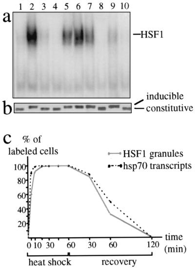
Based on the role of HSF1 in heat shock gene transcription, we next examined the biochemical properties of HSF1 during heat shock and cadmium and azetidine treatments. The DNA-binding properties of HSF1 were analyzed in the different stress conditions by gel mobility-shift assay (Fig. 5a), and the phosphorylation state of HSF1 was determined by using Western blot analysis (Fig. 5b). In control cells, HSF1 was constitutively phosphorylated and lacked DNA-binding activity (lane 1). Exposure of cells to heat shock (lane 2), azetidine (lane 7), or cadmium (lane 9) induces different levels of HSF1 DNA-binding activity and inducible phosphorylation. These two events attenuate during recovery at 37°C (lanes 3 and 4) but persist at 4°C (lanes 5 and 6). Similarly, azetidine or cadmium treatments did not lead to HSF1 activation and hyperphosphorylation when performed at 4°C (lanes 8 and 10). These observations thus confirm that the presence of HSF1 granules correlates with the presence of the inducibly phosphorylated, trimeric, DNA-binding form of the factor.
Figure 5.
HSF1 granules and the activation of the heat shock response. Electrophoretic mobility-shift assay (a) and Western blot analysis (b) were performed on whole-cell extracts prepared from HeLa cells after various treatments: 37°C (lane 1), 1 hour at 42°C (lane 2), 1 hour at 42°C followed by 2 hours recovery at 37°C (lane 3), 1 hour at 42°C followed by 6 hours recovery at 37°C (lane 4), 1 hour at 42°C followed by 2 hours recovery at 4°C (lane 5), 1 hour at 42°C followed by 6 hours recovery at 4°C (lane 6), azetidine (5 mM) for 4 hours at 37°C (lane 7), azetidine (5 mM) for 4 hours at 4°C (lane 8), cadmium (30 μM) for 2 hours at 37°C (lane 9), and cadmium (30 μM) for 2 hours at 4°C (lane 10). (c) HSF1 granules were detected by immunofluorescence in HeLa cells, and hsp70 gene transcription was followed by detection of the nascent hsp70 transcripts by FISH. The percentages of cells displaying HSF1 granules (gray line) and hsp70 transcription sites (dashed line) were measured at different time points during exposure at 42°C and recovery.
We subsequently examined the kinetics of HSF1 stress-granule formation in relation to the transcriptional activation of the hsp70 gene. HSF1 stress granules were detected by using immunofluorescence and hsp70 transcripts by using FISH in HeLa cells (22). The fraction of cells displaying HSF1 granules and hsp70 transcripts were measured during heat shock at 42°C and recovery. The appearance of HSF1 granules closely paralleled activation of hsp70 gene transcription, and this was observed during both activation and attenuation of the heat shock response (Fig. 5c). Similar results were also obtained with hsp90α gene transcription (data not shown).
These findings demonstrate that the presence of HSF1 granules closely correlates with the activation of the stress response as assessed by the activation of HSF1 from the inert monomer to the active trimer and by the activation of hsp genes transcription.
HSF1 Stress Granules Form in Heat-Shocked Mitotic Cells.
The rapid relocalization of HSF1 to the nuclear stress granules is consistent with the dynamic properties of a transcription site. Although this possibility is supported by the close relationship observed between the number of granules and ploidy, HSF1 granules do not colocalize with the sites of transcription of the major hsp70 and hsp90 genes (18). However, these nuclear structures may correspond to the sites of transcription of other hsp or non-hsp genes.
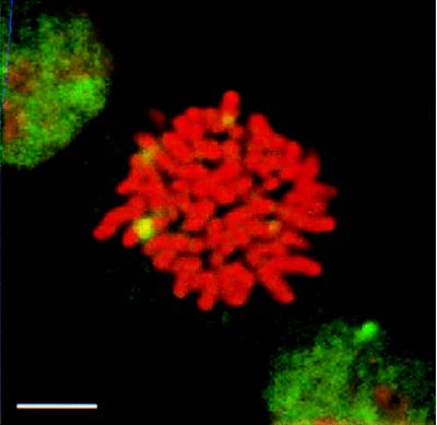
The heat shock transcriptional response occurs during the G1, S, and G2 phases of the cell cycle but not during mitosis, despite activation of HSF1 DNA-binding activity (28, 29). Therefore, we examined the distribution of HSF1 in a population of HeLa cells enriched in mitotic cells by a short treatment with the antimitotic drug colchicine. Whereas HSF1 remains cytoplasmic in unstressed mitotic cells (data not shown), HSF1 stress granules were detected within 5 minutes of heat shock at 42°C (Fig. 6). In contrast to interphase cells in which a substantial cell-to-cell variation in the number of granules is observed, mitotic cells always displayed four HSF1 stress granules that ranged in size from 0.3 to 1.4 μm. Three-dimensional reconstructions of mitotic cells using confocal microscopy and image analysis showed that these granules are associated with the mitotic chromosomes (data not shown). Besides the granules, HSF1 was excluded from the condensed chromosomes, whereas a diffuse staining was observed in the cytoplasm.
Figure 6.
HSF1 granules are present in heat-shocked mitotic cells. HSF1 granules (green) were detected by immunofluorescence in mitotic HeLa cells after 5 minutes of heat shock at 42°C. DNA was counterstained with propidium iodide (red). (Bar = 5 μm.)
Together with the observation by Martinez-Balbás et al. (29) that mitotic HeLa cells are not transcriptionally competent and that heat shock gene transcription cannot be induced by stress despite activation of HSF1 DNA-binding activity, our finding clearly establishes that HSF1 granules form independent of the sites of active transcription.
DISCUSSION
HSF1 stress granules represent a new tool to investigate the effects of physiological stress at the cellular level. The rapid relocalization of HSF1 within seconds to intranuclear stress granules reveals distinctive features and compelling visual evidence that stress signals the dramatic movement of proteins in the living cell. Our analysis of HSF1 stress granules reveals that HSF1 trimers are mobile within the dimensions of the granules, yet the HSF1 factor cannot be extracted from these structures. On recovery from heat shock, HSF1 stress granules are no longer detected, but HSF1 rapidly relocalizes to the same structures during subsequent reexposure to heat shock. Our experiments, however, do not distinguish whether the HSF1 stress granules represent preexisting structures that accumulate the active HSF1 in a stress-dependent manner or whether the granules form de novo as a consequence of stress. The perfect maintenance of stress granules during repeated exposures to heat shock and recovery and in FRAP experiments on living cells may suggest the existence of an underlying structure where HSF1 would temporary and reversibly accumulate during stress. Our experiments, however, do not exclude the possibility that HSF1 stress granules form at specific sites available between chromosomes and other nuclear components.
What is the role of these granules in stressed cells? Although the rapid kinetics of HSF1 granule formation and its correlation with the activation of the stress response are consistent with the dynamic nature of a transcription site, their formation in mitotic cells argues for an alternative role. This conclusion is further supported by experiments based on BrUTP incorporation in which we detected very low levels of incorporation into the granules (data not shown). Because the factor localized within the granules is DNA-binding-competent and active, we propose that HSF1 stress granules define a compartment for the regulation of HSF1 activity. This compartment may function to coordinately regulate the multiple states of HSF1 activity during events of inducible phosphorylation and dephosphorylation (10, 12, 14, 16), association with trans-regulatory chaperones and the negative regulator HSBP1 (30–33) and changes in oligomeric state (6, 7, 34). Rather than regulate the activity of HSF1 at each chromosomal target where the protein is required, HSF1 stress granules may afford central “depots” from which transcriptionally competent HSF1 trimers can be dispensed and cycled. This mechanism would ensure a rapid, tight, and coordinate regulation of heat shock gene transcription. A related situation is observed for splicing factors, which are recruited from their sites of storage, the speckles, to sites of active transcription (35). Alternatively, HSF1 granules may represent sites where the factor is engaged in an activity distinct from transcription such as a structural role in protecting hypersensitive or fragile sites of the genome.
Whatever their role, HSF1 granules represent a new class of discrete, temporary, and reversible nuclear structures. The rapid kinetics of HSF1 granule assembly and disassembly also reveal highly dynamic properties of the nucleus. Therefore, HSF1 stress granules and the cellular response to stress represent a powerful model to study the dynamics of nuclear organization. We provide here evidence that the cell nucleus is able to reorganize its structure within seconds in response to changes in environmental conditions and/or in transcriptional activities. This work thus supports the view that the nucleus is a compartmentalized but nevertheless highly dynamic organelle.
Acknowledgments
We thank Anu Mathew for technical assistance on electrophoretic mobility shift assay, Jan Ellenberg and Sui Huang for their help in time-lapse microscopy, Yan Feng for the temperature-controller device, and Robert Holmgren and Robert Lamb for providing access to the light microscopy facilities. We thank members of the laboratory, Robert Holmgren, and Robert Goldman for their comments on the manuscript. This work was supported by the National Institute of Health Grant GM38109 (R.I.M) and by the Association pour la Recherche contre le Cancer (C.J.). Support for C.J. was provided by the Daniel F. and Ada L. Rice Foundation.
ABBREVIATIONS
HSF1
heat shock factor 1
FISH
fluorescence in situ hybridization
FRAP
fluorescence recovery after photobleaching
HSP
heat shock protein
GFP
green fluorescent protein
Footnotes
This paper was submitted directly (Track II) to the Proceedings Office.
References
- 1.Lindquist S. Annu Rev Biochem. 1986;55:1151–1191. doi: 10.1146/annurev.bi.55.070186.005443. [DOI] [PubMed] [Google Scholar]
- 2.Morimoto R I, Tissières A, Georgopoulos C. The Biology of Heat Shock Proteins and Molecular Chaperones. Plainview, NY: Cold Spring Harbor Lab. Press; 1994. [Google Scholar]
- 3.Wu C. Annu Rev Cell Dev Biol. 1995;11:441–469. doi: 10.1146/annurev.cb.11.110195.002301. [DOI] [PubMed] [Google Scholar]
- 4.Morimoto R I. Genes Dev. 1998;12:3788–3796. doi: 10.1101/gad.12.24.3788. [DOI] [PubMed] [Google Scholar]
- 5.Westwood J T, Clos J, Wu C. Nature (London) 1991;353:822–827. doi: 10.1038/353822a0. [DOI] [PubMed] [Google Scholar]
- 6.Baler R, Dahl G, Voellmy R. Mol Cell Biol. 1993;13:2486–2496. doi: 10.1128/mcb.13.4.2486. [DOI] [PMC free article] [PubMed] [Google Scholar]
- 7.Sarge K D, Murphy S P, Morimoto R I. Mol Cell Biol. 1993;13:1392–1407. doi: 10.1128/mcb.13.3.1392. [DOI] [PMC free article] [PubMed] [Google Scholar]
- 8.Larson J S, Schuetz T J, Kingston R E. Biochemistry. 1995;34:1902–1911. doi: 10.1021/bi00006a011. [DOI] [PubMed] [Google Scholar]
- 9.Zuo J, Rungger D, Voellmy R. Mol Cell Biol. 1995;15:4319–4330. doi: 10.1128/mcb.15.8.4319. [DOI] [PMC free article] [PubMed] [Google Scholar]
- 10.Chu B, Soncin F, Price B D, Stevenson M A, Calderwood S K. J Biol Chem. 1996;271:30847–30857. doi: 10.1074/jbc.271.48.30847. [DOI] [PubMed] [Google Scholar]
- 11.Cotto J J, Kline M, Morimoto R I. J Biol Chem. 1996;271:3355–3358. doi: 10.1074/jbc.271.7.3355. [DOI] [PubMed] [Google Scholar]
- 12.Knauf U, Newton E M, Kyriakis J, Kingston R E. Genes Dev. 1996;10:2782–2793. doi: 10.1101/gad.10.21.2782. [DOI] [PubMed] [Google Scholar]
- 13.Kim J, Nueda A, Meng Y-H, Dynan W S, Mivechi N F. J Cell Biochem. 1997;67:43–54. doi: 10.1002/(sici)1097-4644(19971001)67:1<43::aid-jcb5>3.0.co;2-w. [DOI] [PubMed] [Google Scholar]
- 14.Kline M P, Morimoto R I. Mol Cell Biol. 1997;17:2107–2115. doi: 10.1128/mcb.17.4.2107. [DOI] [PMC free article] [PubMed] [Google Scholar]
- 15.Xia W, Voellmy R. J Biol Chem. 1997;272:4094–4102. doi: 10.1074/jbc.272.7.4094. [DOI] [PubMed] [Google Scholar]
- 16.He B, Meng Y-H, Mivechi N F. Mol Cell Biol. 1998;18:6624–6633. doi: 10.1128/mcb.18.11.6624. [DOI] [PMC free article] [PubMed] [Google Scholar]
- 17.Cotto J J, Fox S G, Morimoto R I. J Cell Sci. 1997;110:2925–2934. doi: 10.1242/jcs.110.23.2925. [DOI] [PubMed] [Google Scholar]
- 18.Jolly C, Morimoto R I, Robert-Nicoud M, Vourc’h C. J Cell Sci. 1997;110:2935–9241. doi: 10.1242/jcs.110.23.2935. [DOI] [PubMed] [Google Scholar]
- 19.Mosser D D, Theodorakis N G, Morimoto R I. Mol Cell Biol. 1988;8:4736–4744. doi: 10.1128/mcb.8.11.4736. [DOI] [PMC free article] [PubMed] [Google Scholar]
- 20.Sarge K D, Zimarino V, Holm K, Wu C, Morimoto R I. Genes Dev. 1991;5:1902–1911. doi: 10.1101/gad.5.10.1902. [DOI] [PubMed] [Google Scholar]
- 21.Feng Y, Wandinger-Ness A. Technical Tips Online. http://tto.trends.com; 1998. , T 01426. [Google Scholar]
- 22.Jolly C, Mongelard F, Robert-Nicoud M, Vourc’h C. J Histochem Cytochem. 1997;45:1585–1592. doi: 10.1177/002215549704501201. [DOI] [PubMed] [Google Scholar]
- 23.Wu B, Hunt C, Morimoto R I. Mol Cell Biol. 1985;5:330–341. doi: 10.1128/mcb.5.2.330. [DOI] [PMC free article] [PubMed] [Google Scholar]
- 24.Parazza F, Humbert C, Usson Y. Comput Med Imaging Graph. 1993;17:189–200. doi: 10.1016/0895-6111(93)90043-m. [DOI] [PubMed] [Google Scholar]
- 25.Parazza F, Bertin E, Wozniak M, Usson Y. J Microsc (Oxford) 1995;178:251–260. [Google Scholar]
- 26.Fey E G, Krochmalnic G, Penman S. J Cell Biol. 1986;102:1654–1665. doi: 10.1083/jcb.102.5.1654. [DOI] [PMC free article] [PubMed] [Google Scholar]
- 27.Williams G T, Morimoto R I. Mol Cell Biol. 1990;10:3125–3146. doi: 10.1128/mcb.10.6.3125. [DOI] [PMC free article] [PubMed] [Google Scholar]
- 28.Milarski K L, Morimoto R I. Proc Natl Acad Sci USA. 1986;83:9517–9521. doi: 10.1073/pnas.83.24.9517. [DOI] [PMC free article] [PubMed] [Google Scholar]
- 29.Martinez-Balbás M A, Dey A, Rabindran S K, Ozato K, Wu C. Cell. 1995;83:29–38. doi: 10.1016/0092-8674(95)90231-7. [DOI] [PubMed] [Google Scholar]
- 30.Mosser D D, Duchaine J, Massie B. Mol Cell Biol. 1993;13:5427–5438. doi: 10.1128/mcb.13.9.5427. [DOI] [PMC free article] [PubMed] [Google Scholar]
- 31.Satyal S H, Chen D, Fox S G, Kramer J M, Morimoto R I. Genes Dev. 1998;12:1962–1974. doi: 10.1101/gad.12.13.1962. [DOI] [PMC free article] [PubMed] [Google Scholar]
- 32.Shi Y, Mosser D D, Morimoto R I. Genes Dev. 1998;12:654–666. doi: 10.1101/gad.12.5.654. [DOI] [PMC free article] [PubMed] [Google Scholar]
- 33.Zou J, Guo Y, Guettouche T, Smith D F, Voellmy R. Cell. 1998;94:471–480. doi: 10.1016/s0092-8674(00)81588-3. [DOI] [PubMed] [Google Scholar]
- 34.Rabindran S K, Haroun R I, Clos J, Wisniewski J, Wu C. Science. 1993;259:230–234. doi: 10.1126/science.8421783. [DOI] [PubMed] [Google Scholar]
- 35.Misteli T, Cáceres J F, Spector D L. Nature (London) 1997;387:523–527. doi: 10.1038/387523a0. [DOI] [PubMed] [Google Scholar]
