Reduced facilitation and vesicular uptake in crustacean and mammalian neuromuscular junction by T-588, a neuroprotective compound (original) (raw)
Abstract
Bath application of compound T-588, a neuroprotective agent, reduced paired-pulse and repetitive-pulse facilitation at mammalian and crustacean neuromuscular junctions. In addition, it reduced voltage-gated sodium and potassium currents in a use-dependent fashion, but had only a small effect on the presynaptic Ca2+ conductance. By contrast, it blocked FM 1–43 vesicular uptake but not its release, in both species. Postsynaptically, T-588 reduced acetylcholine currents at the mammalian junction in a voltage-independent manner, but had no effect on the crayfish glutamate junction. All of these effects were rapidly reversible and were observed at concentrations close to the compound’s acute protective level. We propose that this set of mechanisms, which reduces high-frequency synaptic transmission, is an important contributory factor in the neuroprotective action of T-588.
Keywords: two-photon microscopy, FM 1–43, voltage clamp, ultrastructure
The crustacean and mammalian neuromuscular junctions are useful models to determine the general cellular and molecular basis for synaptic transmission (1, 2). Repetitive and paired-pulse stimulation, which are known to produce powerful facilitation at these junctions (3, 4), were used to investigate the possible actions of T-588 on synaptic facilitation. T-588 is a promising neuroprotective drug (5) as reduction of facilitation could alleviate activity-dependent neuronal death. The results, which include the study of axonal and junctional currents, synaptic facilitation, optical calcium concentration measurements, vesicular recycling (using two-photon laser scanning microscopy), and ultrastructural analysis suggest that T-588 causes a subtle regulation of high-frequency synaptic transmission. These pharmacological attributes, which function as a reversible low pass filter for synaptic transmission, may be responsible for T-588 neuroprotective properties.
Materials and Methods
Dissection Procedures.
Crayfish, Procambarus clarkii, (4–6 cm) were obtained from Carolina Biological Supply. Dissection and motor nerve stimulation were performed according to standard procedures (6) with minor modifications. The motor nerves to the meropodite of the first or second walking legs were isolated and the inhibitory axons were pinched off to stimulate only excitatory fibers via a suction electrode. The inner surface of the claw opener muscle was exposed, and a fine motor nerve running along the surface was selected for intracellular or extracellular recording. The effects of T-588 on sodium and potassium currents were studied in the ventral nerve cord. Both ends of the axons (3 mm length) were tied and pinned on a Sylgard-coated chamber.
During dissection and recording, the tissue was bathed in a saline solution (205 mM NaCl, 5.4 mM KCl, 13.5 mM CaCl2, 2.6 mM MgCl2, 10 mM Hepes, pH 7.3) made according to the method of Van Harreveld (7).
In mice (obtained from Taconic Farms), experiments were carried out on the levatoroauris longus muscle of male Swiss mice weighing 20–30 g. The animals were cared for in accordance with National Guidelines for the Humane Treatment of Laboratory Animals. Mice were anaesthetized with an overdose of 2% tribromoethanol (0.15 ml/10 g body weight) and exsanguinated. The corresponding muscle with its nerve supply was excised and dissected on a Sylgard-coated Petri dish containing a physiological saline solution, (137 mM NaCl, 5 mM KCl, 2 mM CaCl2, 1 mM MgSO4, 12 mM NaHCO3, 1 mM NaH2PO4, and 11 mM glucose), continuously bubbled with 95% O2/5% CO2. The preparation then was transferred to a 1.0- to 2.5-ml recording chamber. Experiments were performed at room temperature (20–23°C).
Electrophysiology.
Facilitation.
In the crayfish, trains of repetitive electrical stimuli were delivered through a suction electrode. Muscle fibers from the claw opener were patch-clamped by using >2 MΩ series resistance electrodes filled with 0.25 M KCl. The muscle fiber membrane potential was held at the resting level and excitatory endplate currents (EPCs) were recorded by using AXOPATCH 200B (Axon Instruments, Foster City, CA). Endplate potentials (EPPs) and presynaptic intracellular or extracellular recording were obtained by using a Dual Channel Intracellular Recording Amplifier (IR-283, Neurodata Instruments, Delaware, PA). Data were digitized/acquired by using a DigiData2000 (Axon Instruments) and analyzed by using pclamp6 (Axon Instruments). Intracellular recordings of presynaptic action potentials were obtained with sharp glass electrodes (≈20 MΩ) filled with 3 M KCl. The axons generally were penetrated at branching points. The direct effect of glutamate at the neuromuscular junction was tested by iontophoretical application from a microelectrode filled with 1 M l-glutamate at pH 8.0 (8). Transmembrane currents in the giant axon of the ventral nerve cord were obtained by using a conventional two-electrode voltage clamp method. Both the current and voltage electrode were coated with silver and Humiseal (Chase, Woodside, NY). The tip resistance of the current electrode was less than 1 MΩ.
In mice, evoked EPPs were recorded intracellularly with conventional glass microelectrodes filled with 3 M KCl (10–30 MΩ). Presynaptic currents were elicited by stimulation of the motor nerve using glass microelectrodes filled with 2 M NaCl (5–10 MΩ) inserted into the perineurial sheath of small diameter nerve bundles (9) and recorded extracellularly. EPCs and spontaneous miniature EPCs (mEPCs) were obtained via voltage, and current electrodes were filled with 3 M KCl and 4 M potassium acetate, respectively (2–5 MΩ). The current electrode was placed within 0.5–1 muscle fiber diameter of the recording electrode. After impaling a muscle fiber, the nerve was continuously stimulated at 0.5 Hz by using either two platinum electrodes or a suction electrode. EPCs were measured in normal saline solution containing 2 μg/ml of μ-conotoxin GIIIB, to avoid muscle contraction. mEPCs were measured in a modified saline solution where 97 mM of [NaCl]o was substituted by sucrose. The recording electrodes were connected to an Axoclamp 2A amplifier (Axon Instruments). Data were acquired by using pclamp6 and analyzed by using axoscope 1.0 (Axon Instruments). origin 3.0 software (Microcal Software, Northampton, MA) was used to represent data.
FM 1–43 dye uptake.
A two-photon laser microscopy (10) was used to determine the localization and degree of activity-dependent vesicular recycling via uptake of the dye FM 1–43 (11, 12). Tissue from crayfish or mice was soaked in a solution containing 2 μM FM 1–43, for 20 min or 10 min, respectively, before stimulation to allow the dye to infiltrate the extracellular matrix. The loading protocol for crayfish comprised 10 min of tetanization at 20 Hz followed by a 15-min loading period. For mice 15 min of tetanization at 20 Hz was followed by 20 min of loading. The preparations then were continuously superfused for 30 min with a physiological saline containing a scavenging compound (β-cyclodextrin sulfobutyl ether, 7 sodium salt, CyDex, Overland Park, KS) to remove the FM 1–43 from the extracellular space (13–16). After this procedure, the presynaptic fluorescent images were recorded by using two-photon laser procedure (Olympus microscope AX70). The laser scanner used a Tsunami Ti:sapphire laser system pumped with Millenia X diode-pumped laser (Spectra-Physics), which enabled a pulse frequency of 80 MHz and a pulse duration of 40–70 fsec. The wavelength was tuned at 900 nm and the laser intensity was <3 mW at an Olympus ×60/0.90. The scanner was driven by a labview-based (version 5, National Instruments, Austin TX) program. Fluorescence was detected with a photomultiplier.
Fluorescence images of each synaptic terminal were captured every 0.5 or 1 μm along the _z_-axis and accumulated to reconstruct a three-dimensional image. During this procedure, 20 μM d-tubocurarine chloride (for mice) or 40 μM nifedipine (for crayfish) was added to prevent muscle twitching. The preparation was destained by tetanic stimulation without dye in the bath, at 20 Hz for 10 min (mice) or 20 min (crayfish). After complete unloading the terminal was reloaded a second time with FM 1–43 in the presence of T-588 to determine its effect on vesicular loading. Once this effect was determined, a second complete unloading procedure was implemented, after which a third reloading of FM 1–43 was implemented in the absence of T-588. This third loading and unloading procedure served as a control for preparation viability and demonstrated the reversibility of T-588 effect.
Calcium imaging.
In crayfish, membrane inpermeant Fura-2 (5 mM) was prepared by using a 800 mM KCl solution and injected at the Y-shape branch of crayfish motoneuron through an intracellular electrode. Fura-2 was defused to synaptic terminals for 20 min. In mice, the preparations were soaked in a solution containing 10 μM Fura-2 AM for 45 min followed by 20-min wash. Fura-2-loaded terminals were imaged with the two-photon microscopy system described above. The wavelength was tuned to 800 nm. The time course of calcium signaling induced by 10 repetitive nerve stimulation at 40 Hz was analyzed by using labview. The EPPs and presynaptic spikes were recorded simultaneously.
Ultrastructure.
Crayfish and mice neuromuscular junctions were soaked in a high potassium (40 mM) solution for 15 min followed by a 20-min rest in a physiological saline. This procedure stimulated vesicular release by presynaptic depolarization during the high potassium regime and vesicular uptake during the rest period. In some experiments all of the solutions contained T-588 and the tissue was soaked in T-588 saline before the high K+ treatment. Each procedure was repeated in two different preparations for each paradigm for a total of n = 8 experiments. The preparations were fixed by immersion in 2% glutaraldehyde, 2% parformaldehyde, and 200 mM CaCl2 in 0.1 M sodium cacodylate buffer pH 7.4 for overnight at 4°C then transferred to 7% sucrose in 0.1 M sodium cacodylate. This procedure was followed by postfixing in 1% osmium tetroxide, in the same buffer, for 1 hr at 4°C, and in-block stained overnight with 2% uranyl acetate in 0.1 M sodium acetate buffer pH 5.0, on ice. After dehydration in ethanol and substitution with propylene oxide, the samples were embedded in Araldite resin (CY212). Longitudinal and cross sections 0.5 mm thick were mounted on glass slides to select the areas with neuromuscular junctions under a light microscope. Thin sections from the chosen areas were collected on single slot, Pyoloform, and carbon-coated grids, contrasted with uranyl acetate and lead citrate, and photographed with a Phillips 208 or a JEOL 100 CX electron microscopes.
Morphometry and quantitative analysis were performed with a Zidas digitizing system (Zeiss) interfaced with a Macintosh G-3 computer. Electron micrographs were taken at an initial magnification of ×20,000 and photographically enlarged to ×50,000 for synaptic vesicle counting. Synaptic vesicle density on the active zone was determined as the number of vesicles per μm2 on an average area of 1.0 μm2 per active zone in the crayfish terminals. For mice terminals, where active zones take the whole extension of the terminals facing the muscle fibers, the total vesicle content was considered for counting. Thirty active zones for crayfish, and the same number of mice terminals, were counted for each group, control and T-588-treated.
Statistical Analysis.
sas (release 6.1, SAS Institute, Cary, NC) was used for statistical analysis. Each statistic we used is shown in Results.
Compounds.
T-588 (Toyama Chemical, Toyama, Japan) was dissolved and perfused in the bathing solution at concentrations of 50, 100, or 300 μM (chemical structure is shown in Fig. 1). l-glutamate monosodium salt, Nifedipine, and d-tubocurarine chloride were purchased from Sigma. Fura-2, Fura-2 AM, and FM 1–43 were purchased from Molecular Probes. β-cyclodextrin sulfobutyl ether, 7 sodium salt, an FM1–43 scavenger, was purchased from CyDex.
Figure 1.
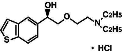
Chemical structure of T-588
Results
Electrophysiology: Crayfish.
Sodium and potassium currents and presynaptic spikes.
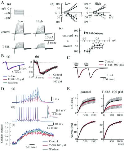
A two-electrode voltage clamp paradigm was implemented to assess the pharmacological effect of T-588 on voltage-gated axonic currents. To assess use-dependence we used two acquisition protocols. One, a low-frequency acquisition used intervals of less than 1 Hz. The other used high-frequency tetanic stimulation (40 Hz). Typical results are shown in Fig. 2Aa, and the current-voltage relationships are plotted in Fig. 2Ab. As illustrated in Fig. 2Ac, at low frequencies, T-588 mildly reduced both inward and outward currents (open symbols) whereas at high frequencies the inward current was more substantially reduced (closed symbols). These results indicate use-dependent reduction of sodium channel current by that T-588.
Figure 2.
T-588 in crayfish neuromuscular junction. (A) Axonic voltage-gated currents. (a) Data acquired at low (<1 Hz) and high (40 Hz) voltage step frequency. (b) I–V relationship of control (○) and T-588 (●) (100 μM). (c) Current reduction by T-588. Inward (circles) and outward current (squares) are shown separately. Data represent the mean ± SE, n = 7 experiments. T-588 significantly reduced the inward current at high frequency (closed symbols) and less at low frequency (open symbols) indicating an inward current use-dependent block. *, P < 0.05; **, P < 0.01 probability by paired t test. (B) Single EPC. (a) Single EPC waveforms (recorded before, during T-588, and after washout) are superimposed. (b) Single EPC amplitudes measured every 0.5 msec and normalized (1.0 as peak amplitude in each experiment). Each point in the graph shows mean value of 15 preparations with SE. Black and red lines represent before and during 100 μM T-588, respectively. No change was observed. (C) Postsynaptic glutamate current. Glutamate current generated by paired pulses (at 400-msec intervals) iontophoresis of l-glutamate to a muscle fiber voltage-clamped at the neuromuscular junction. Average wave forms of 10 successive traces during T-588 (300 μM) and after washout are superimposed. (D) Synaptic facilitation and Ca2+ influx. Example waveform of EPPs (a) and presynaptic spikes (b) evoked by 10 repetitive stimulation at 40 Hz and the simultaneously recorded time course of Ca2+ influx (c) at a particular terminal are shown. Calcium uptake was determined from Fura-2 images acquired with two-photon microscopy as an averaged waveform of successive eight measurements. (E) EPC facilitation evoked by 40-Hz repetitive stimulation. (Upper) Absolute amplitude of EPC through 50th stimulus. (Lower) Amplitude is shown as percent to the maximum response in each group. Black and red before and during T-588, respectively. Each point is shown as a mean value with SE, n = 9 experiments.
In agreement with these results, the amplitude of both presynaptic spikes and the synaptic events were not affected by T-588 at activation frequencies <30 Hz (Fig. 2_Da_). However, as shown in Fig. 2_Db_, T-588 reduced the amplitude and delayed the onset of presynaptic spikes (the 10th is shown) at prolonged stimulation >40 Hz. The average of 10 trains is shown. Similar results were observed in the other experiments (n = 10) using this paradigm.
Synaptic facilitation: single spike synaptic transmission and facilitation.
Given that the first response to a train of presynaptic pulses remained substantially unchanged during T-588 application, we decided to test the effect of this compound on single synaptic events. As shown in Fig. 2Ba, single EPC was not modified by 100 μM T-588. A plot of single EPCs’ amplitude before and after 100 μM T-588 application in 15 synapses indicated little change in the EPC with this drug (Fig. 2Bb).
Classically, repetitive stimulation produces powerful facilitation of EPC in neuromuscular junction of crayfish opener muscle (1, 17). Such facilitation, shown in Fig. 2Da, demonstrates the usual time course for this phenomenon. Note that as previously reported (18) this facilitation is not accompanied by a change in the amplitude of the presynaptic action potential. Application of 100 μM T-588 to the bathing solution reduced this facilitation (Fig. 2Da), reversibly in all of our experiments (n = 10).
The effect of T-588 on the facilitated EPC evoked by 50 repetitive stimuli (at 40 Hz) is shown in Fig. 2E. The upper graphs show the absolute amplitudes recorded before and during application of T-588. Indeed, this compound blocked EPC facilitation. Note, however, that the time course for single EPC facilitation did not change, indicating that transmitter release kinetics was not modified, in the presence of a marked reduction in release probability.
Glutamate current and presynaptic calcium influx.
To determine whether T-588 suppresses facilitation by affecting postsynaptic sensitivity to glutamate, we investigated its effect on glutamate-evoked postsynaptic currents. Paired iontophoretic pulses of glutamate induced neither facilitation nor a reduction of the postsynaptic glutamate current. A similar set of results was obtained in the presence of 300 μM T-588, n = 2 (Fig. 2C), indicating that this neuroprotective substance does not alter postsynaptic glutamatergic transmission.
To determine whether the reduction in synaptic facilitation observed in the presence of 100 μM T-588 was caused by a reduction in calcium current, the presynaptic calcium influx during synaptic facilitation was monitored by using the calcium-dependent fluorescent dye Fura 2 (Fig. 2Dc). Under these conditions the intracellular calcium concentration was only slightly reduced (>7%) during the first five presynaptic spikes, whereas the amplitude variation was minimal in the presence of a 50% reduction in facilitation. This reduction was reversed within the 60-min wash period in all experiments (n = 5) in which the presynaptic spike was monitored. In some cases the calcium influx at the more distal presynaptic branches was higher, but mostly because of terminal spike amplitude reduction making interpretation ambiguous.
Electrophysiology: Mice.
Presynaptic perineurial currents.
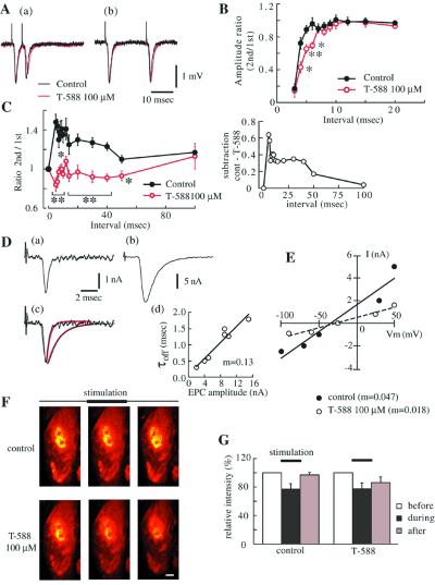
Characteristic presynaptic perineurial currents recorded at levator auris longus muscle in normal saline solution containing 20 μM d-tubocurarine chloride during paired-pulse nerve stimulation at both 50 and 200 Hz are shown in Fig. 3A. Spikes were recorded during paired-pulse stimulation at various intervals (3–20 msec). Fig. 3B shows the relative amplitude (second amplitude/first amplitude) at each interval from five experiments. The second spikes were smaller at less than 5 msec of interval, and in four of five preparations, the second spikes were not detected at 3-msec intervals in control conditions. After 30 min of superfusion with 100 μM T-588, the amplitude of the second spike was reduced significantly at 4-, 5-, and 6-msec intervals (Fig. 3B). There were no significant changes at intervals longer than 6 msec.
Figure 3.
T-588 in mice neuromuscular junction. (A) Presynaptic spike amplitude reduction induced by paired-pulse stimulation at different intervals (a) 5 msec, (b) 20 msec in the presence of 100 μM T-588. Presynaptic perineurial currents are shown as averaged waveforms from 16 successive traces. (B) Presynaptic current amplitude ratio induced by paired-pulse stimulation with various intervals in control and 100 μM T-588. Data are shown as mean with SE from five experiments. Control data were taken after washing out of T-588. *, P < 0.05; **, P < 0.01 paired t test. (C) Synaptic facilitation induced by paired-pulse with various intervals in control and 100 μM T-588. (Left) The ratio of second EPP/first EPP are shown as facilitation indexes at each interval. The subtracted values (T-588 to control) are shown (Right). *, P < 0.05; **, P < 0.01 Student’s t test or Aspin-Welch test subsequent to F-test. (D) Nerve evoked EPCs (average of 32) 50 μM T-588 (a) and washout (45 min) (b). Waves were superimposed after normalization (c). The red lines show first-order exponential curves fitted to the off time course in each current (membrane held at −40 mV) (d). The relationship between decay time constant (off) and maximum value of averaged currents recorded during the washout of T-588. Data obtained at 5, 10, 15, 20, 30, and 45 min washout. (a and b) 0 and 45 minutes of washout, respectively. Experiments were done in 0.8 mM Ca2+/1.5 mM Mg2+ and μ-conotoxin GIIIB (2 μg/ml) to avoid contraction. (E) Current-voltage relationship of the mEPCs recorded at the same motor nerve terminal before (●) and 30-min superfusion of 100 μM T-588 (○). Data are fitted to a linear function. The slope value (m) is shown. (F) Ca2+ uptake during tetanic stimulation. Fura-2 AM loaded from extracellular melieu. Images were captured from brightest plain of the terminal before, during, and after terminate of tetanic stimulation (20 Hz). Fluorescence intensity of Fura-2 decreased with Ca2+ concentration increase. Scale bar represents 2 μm. (G) Fluorescence intensity of unit area measured from captured image with National Institutes of Health image. Data are shown as mean value with SE from five experiments. No significant difference was observed between control and T-588 on relative intensity during stimulation (paired t test).
Synaptic facilitation.
EPPs from levator auris muscle were recorded in 0.9 mM Ca2+/3 mM Mg2+-modified saline solution. Two-pulse protocol was used to study the T-588 effect on synaptic facilitation at this muscle (Fig. 3C; n = 4–32). The level of facilitation was determined by calculating the amplitude ratio of the amplitude of test response (second EPP/first EPP) over the amplitude of the initial EPPs with stimuli delivered at different intervals (5–100 ms). As previously reported (19) facilitation of the order of 20% [129 ± 2% (n = 213)] was observed at intervals of 40 msec and less, in control condition. After 30-min application of 100 μM T-588, paired-pulse facilitation showed a marked reduction through 5- to 50-msec intervals [95 ± 2% (n = 272)] (Fig. 3C).
Presynaptic calcium influx.
The pharmacological effect of T-588 on calcium influx into a synaptic terminal was measured by using Fura-2-AM as described in Materials and Methods. As shown in Fig. 3F, a reduction of fluorescence intensity of the Fura-2 signal was measured for a 3-min tetanic stimulation at 20 Hz, indicating increase in intracellular calcium concentration. This change recovered 5 min after stimulus end for n = 5. In the presence of 100 μM T-588, Fura-2 signal was reduced to the same extent (Fig. 3G) for n = 5. Plot of the relative intensity reduction in the five different experiments demonstrated no significant change in the average calcium concentration in the presence of T-588.
Postsynaptic Evoked EPCs and mEPCs.
To determine the possible effect of T-588 on the mammalian postsynaptic acetylcholine (ACh) receptor, we recorded evoked EPCs by using a two-electrode voltage clamp technique. The amplitude and decay time constant of evoked EPCs recorded at the same cell clamped to −40 mV holding potential in the presence of 50 μM T-588 and after 45 min of washout are shown in Fig. 3D a and b. Note that T-588 reversibly reduced both peak amplitude and decay time constant of EPCs. Fig. 3Dc shows the same signals superimposed to compare their off time courses (Tau values were 0.3 and 1.3 msec for T-588 and washout, respectively). The relationship of amplitude and Tau value was fitted to a linear function (Fig. 3Dd).
mEPCs were recorded at various membrane potentials in 40 mM [NaCl]o-modified saline solution. Data in Fig. 3E were recorded from the same cell. Note how the peak amplitude of mEPCs was decreased by the application of 100 μM T-588, in a nonvoltage-dependent manner. Thus, these results indicate that the reduction in mEPCs is not caused by a T-588 block of the ACh receptor’s pore, as would be expected for a use-dependent ionophore-blocking compound (20).
Synaptic Vesicle Release and Endocytosis.
To assess a possible reduction of transmitter release caused by a reduction of vesicular availability secondary to a reduction in vesicular endocytosis, synaptic vesicule uptake was determined with the dye FM 1–43 (11, 12). Two-photon scanning microscopy was used to image activity-dependent uptake of fluorescent dye FM 1–43 on synaptic vesicle dynamic. Nerve terminals were loaded with FM 1–43 by tetanic stimulation (see Materials and Methods). The results were obtained both in crayfish and mice neuromuscular junction.
Crayfish.
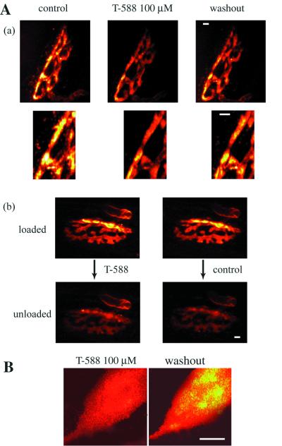
Control images were taken after the loading procedure washing out of T-588. Fluorescence intensity in T-588 was clearly less than that of control. Synaptic facilitation also was suppressed with T-588 in the same preparation. These experiments were repeated six times including the above. We got similar results in every experiment (Fig. 4B).
Figure 4.
Two-photon microscopy imaging of FM 1–43 to determine synaptic vesicle endocytosis in crayfish and mice. (A) (a) The fluorescence image of mice motor nerve terminal-loaded FM 1–43 with tetanic stimulation in the presence of 100 μM T-588. Dye loading was repeated after washing out of T-588. Images were captured every 1-μm step in the Z direction and accumulated. The brightest images in the terminal are shown expanded in the lower column. (b) After full loading of FM 1–43, the preparation was treated with 100 μM T-588 for 30 min and then destained by tetanic stimulation (20 Hz, 10 min) without dye in the bath in the presence of T-588. The same set of experiments was repeated without T-588. (B) Fluorescence image of crayfish motor nerve terminal-loaded FM 1–43 with tetanic stimulation. Dye loading was repeated with 100 μM T-588 and washing out of T-588 after taking under control condition. Scale bar in each image represents 2 μm.
Mice.
Fig. 4Aa shows characteristic images of presynaptic terminals of mice after activity-dependent labeling. Bright regions indicate clusters of labeled vesicles. Fluorescence intensity was clearly reduced by 20-min pretreatment of T-588 followed by a treatment during whole loading period. This uptake block was reversible after washing out of T-588. Similar results were obtained from four more experiments. Presynaptic spikes and postsynaptic responses (recorded without d-tubocurarine chloride) in T-588-treated muscles were present during tetanic stimulation (data not shown). A set of terminals (n = 4) was loaded in the absence of this compound after which T-588 was superfused for 30 min. Under these conditions tetanic stimulation produced excellent unloading of the dye (Fig. 4Ab), demonstrating that vesicular endocytosis, but not vesicular release, is compromised by T-588.
Ultrastructural Findings.
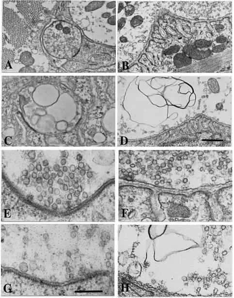
Example views of neuromuscular synapses are shown in Fig. 5. Synaptic vesicles are clustered near the active zone. We compared the number of synaptic vesicles per unit area at docking positions and coated vesicles in the terminals between controls and T-588-treated groups. After the treatment with T-588 (100 μM) for 1 hr, vesicle density in the crayfish claw opener muscle and in the mice levator auris muscle terminals showed significantly lower values than those observed in the control terminals (P = 2.54 × 10⁻¹¹ and P = 1.28 × 10⁻², respectively; Fig. 5, Table 1).
Figure 5.
Electron micrographs of excitatory nerve terminals in crayfish and mice neuromuscular junctions. (A and B) Control terminals show typical numbers of synaptic vesicles in the active zones. In crayfish active zones are restricted to specific areas in the crayfish terminal and are widespread along the endplate area in the mice terminal. (C and D) Terminals exposed to T-588 show synaptic vesicles in very low numbers as compared with the controls. (E and F) As in G and H, compare details of the same terminals at higher magnification. [Scale bars: in D = 1.0 μm for A_–_D; in G = 0.5 μm for E_–_H.]
Table 1.
Number of vesicles at motor nerve terminals
| Crayfish | Mouse | |
|---|---|---|
| Control | ||
| Vesicles/μm2 | 123.00 (±25.00) | 35.54 (±5.36) |
| Avg. cluster area, μm2 | 0.20 | |
| Avg. terminal area, μm2 | 4.25 | |
| T-588 | ||
| Vesicles/μm2 | 70.51 (±15.00) | 24.28 (±5.15) |
| Avg. cluster area, μm2 | 0.21 | |
| Avg. terminal area, μm2 | 4.40 |
Discussion
The present study addresses the mechanism for the pharmacological properties of T-588, a neuroprotective compound (5). The results demonstrate that this compound reduces synaptic facilitation and vesicular endocytosis. In addition, a use-dependent effect on inward sodium currents and a reduction of ACh sensitivity at the mice neuromuscular junction were encountered.
The Issue of Synaptic Facilitation.
Synaptic facilitation during repetitive presynaptic stimulation have been amply demonstrated at both mice and crayfish neuromuscular junctions (3, 4). In the set of experiments described here T-588 markedly reduced synaptic facilitation in a reversible manner. This effect was the result of several mechanisms acting together: (i) the reduction of facilitated transmitter release from the motor nerve terminals, (ii) the use-dependent reduction of sodium and potassium currents that reduce the amplitude of the presynaptic spikes, (iii) the reduction of vesicular endocytosis, and (iv) the reduction in ACh-dependent postsynaptic current.
Although synaptic facilitation has been thought to be driven, almost exclusively, by an increase in intracellular calcium concentration (1), the molecular mechanism underlying this phenomena has not been clearly identified. However, present results with T-588 indicate that facilitation can be powerfully modulated by calcium-independent events. Indeed, as calcium-dependent facilitation is close to linearly related to calcium concentration (19) the expectation would be that any calcium-dependent reduction of facilitation would require an equally large reduction of calcium conductance. The mechanism by which T-588 reduces facilitation can only be partially ascribed by the reduction of vesicular endocytosis, as demonstrated by the reduction of FM1–43 dye uptake that occurs in the presence of this compound. Also the fact that in a preloaded terminal T-588 does not modify FM1–43 release indicates that the reduction of facilitation is not related to an effect on the transmitter release mechanism. This conclusion also is supported by the fact that no appreciable change in the amplitude of single EPPs or EPCs were seen in these experiments. The question does come up regarding the whether this reduction in facilitation may be caused by a decrease in transmitter availability.
The Issues of Vesicular Endocytosis and Use-Dependent Inward Current Reduction.
Concerning the other pharmacological actions of T-588, two aspects deserve further comment. This compound clearly reduces vesicular endocytosis in a reversible manner. When considering that the same compound also modifies voltage-gated and ACh-gated currents at least three possible scenarios may be considered. (a) T-588 may have a local anaesthetic effect as may be suspected by its chemical structure, which does resemble aspects of local anesthetic structures in particular its amino-ethoxy-like chain. This possible anaesthetic effect could explain the use-dependent block of voltage-gated currents and the effect on the ACh receptor. (b) This compound could be envisaged as triggering a molecular signaling sequence having a wide range of effects on synaptic plasticity, to include not only the facilitation and endocytosis effect but also relate to the voltage- and ligand-gated current modulation. (c) Both an anaesthetic and a molecular signaling action may be at work with this compound.
Finally, present results show that T-588 suppressed only accelerated transmitter release without affecting nonfacilitated effect at concentrations similar to the acute neuroprotective levels for this compound. This suppression could be contributed to neuroprotective effect of this compound (5). T-588 thus may become a valuable neuroprotective compound for the treatment of neurodegenerative conditions such as those seen in various diseases in central and peripheral nervous systems.
Acknowledgments
We thank Dolores S. Ferreira and Jose Augusto Maulin for their electron microscopy expertise and Gabriel M. Arisi for statistical analysis. Support was provided by the National Institutes of Health NINDS-13742 (R.L.); Secretaria de Ciencia y Tecnologia-Agencia de Promocion Cientifica y Tecnologica (PICT97/1848) (O.D.U.); and partial support from FAPESP (Fundacão de Amparo à Pesquisa do Estado de São, Brazil) 97/3026-6 and 11097-0 (J.E.M.).
Abbreviations
EPP
endplate potential
EPC
endplate current
mEPC
spontaneous miniature EPC
ACh
acetylcholine
References
- 1.Bittner G D. J Neurobiol. 1989;20:368–408. doi: 10.1002/neu.480200510. [DOI] [PubMed] [Google Scholar]
- 2.Uchitel O D, Protti D A, Sánchez V, Cherksey B D, Sugimori M, Llinás R. Proc Natl Acad Sci USA. 1992;89:3330–3333. doi: 10.1073/pnas.89.8.3330. [DOI] [PMC free article] [PubMed] [Google Scholar]
- 3.Dorlöchter M, Irintchev A, Brinkers M, Werig A. J Physiol. 1991;436:283–292. doi: 10.1113/jphysiol.1991.sp018550. [DOI] [PMC free article] [PubMed] [Google Scholar]
- 4.Zucker R S. Annu Rev Neurosci. 1989;12:13–31. doi: 10.1146/annurev.ne.12.030189.000305. [DOI] [PubMed] [Google Scholar]
- 5.Ono S, Kitamura K, Maekawa M, Hirata K, Ano M, Ukai W, Yamafuji T, Narita H. Jpn J Pharmacol. 1993;62:81–86. doi: 10.1254/jjp.62.81. [DOI] [PubMed] [Google Scholar]
- 6.Dudel J, Kuffler S W. J Physiol. 1961;155:514–529. doi: 10.1113/jphysiol.1961.sp006644. [DOI] [PMC free article] [PubMed] [Google Scholar]
- 7.Van Harreveld A. Proc Soc Exp Biol NY. 1936;34:428–432. [Google Scholar]
- 8.Onodera K, Takeuchi A. J Physiol. 1976;255:669–685. doi: 10.1113/jphysiol.1976.sp011302. [DOI] [PMC free article] [PubMed] [Google Scholar]
- 9.Mallart A. J Physiol. 1985;368:565–575. doi: 10.1113/jphysiol.1985.sp015876. [DOI] [PMC free article] [PubMed] [Google Scholar]
- 10.Denk W, Strickler J H, Webb W W. Science. 1990;248:73–76. doi: 10.1126/science.2321027. [DOI] [PubMed] [Google Scholar]
- 11.Betz W J, Bewick G S. Science. 1992;255:200–203. doi: 10.1126/science.1553547. [DOI] [PubMed] [Google Scholar]
- 12.Betz W J, Mao F, Bewick G S. J Neurosci. 1992;12:363–375. doi: 10.1523/JNEUROSCI.12-02-00363.1992. [DOI] [PMC free article] [PubMed] [Google Scholar]
- 13.Wang C, Zucker R S. Neuron. 1998;21:155–167. doi: 10.1016/s0896-6273(00)80523-1. [DOI] [PubMed] [Google Scholar]
- 14.Quigley P A, Msghina M, Govind C K, Atwood H L. J Neurophysiol. 1999;81:356–370. doi: 10.1152/jn.1999.81.1.356. [DOI] [PubMed] [Google Scholar]
- 15.Reid B, Slater C R, Bewick G S. J Neurosci. 1999;19:2511–2521. doi: 10.1523/JNEUROSCI.19-07-02511.1999. [DOI] [PMC free article] [PubMed] [Google Scholar]
- 16.Kay A R, Alfonso A, Alford S T, Cline H, T, Haas K, Holgado A M, Malinow R, Ryan T A, Sakmann B, Snitsarev V A, et al. Soc Neurosci Abstr. 1999;25:741. [Google Scholar]
- 17.Zucker R S, Delaney K R, Mulkey R, Tank D W. Ann NY Acad Sci. 1991;653:191–207. doi: 10.1111/j.1749-6632.1991.tb36492.x. [DOI] [PubMed] [Google Scholar]
- 18.Wojtowicz J M, Atwood H L. J Neurophysiol. 1984;52:99–113. doi: 10.1152/jn.1984.52.1.99. [DOI] [PubMed] [Google Scholar]
- 19.Zucker R S. Curr Opin Neurobiol. 1999;9:305–313. doi: 10.1016/s0959-4388(99)80045-2. [DOI] [PubMed] [Google Scholar]
- 20.Maleque M A, Souccar C, Cohen J B, Albuquerque E X. Mol Pharmacol. 1982;22:636–647. [PubMed] [Google Scholar]
