Suppression of Bladder Epithelial Cytokine Responses by Uropathogenic Escherichia coli (original) (raw)
Abstract
Urinary tract infections are most commonly caused by uropathogenic strains of Escherichia coli (UPEC), which invade superficial bladder epithelial cells via a type 1 pilus-dependent mechanism. Inside these epithelial cells, UPEC organisms multiply to high numbers to form intracellular bacterial communities, allowing them to avoid immune detection. Bladder epithelial cells produce interleukin-6 (IL-6) and IL-8 in response to laboratory strains of E. coli in vitro. We investigated the ability of UPEC to alter epithelial cytokine signaling by examining the in vitro responses of bladder epithelial cell lines to the cystitis strains UTI89 and NU14. The cystitis strains induced significantly less IL-6 than did the laboratory E. coli strain MG1655 from 5637 and T24 bladder epithelial cells. The cystitis strains also suppressed epithelial cytokine responses to exogenous lipopolysaccharide (LPS) and to laboratory E. coli. We found that insertional mutations in the rfa and rfb operons and in the surA gene all abolished the ability of UTI89 to suppress cytokine induction. The rfa and rfb operons encode LPS biosynthetic genes, while surA encodes a periplasmic cis-trans prolyl isomerase important in the biogenesis of outer membrane proteins. We conclude that, in this in vitro model system, cystitis strains of UPEC have genes encoding factors that suppress proinflammatory cytokine production by bladder epithelial cells.
Urinary tract infections (UTI) represent a significant cause of morbidity and are most frequently caused by uropathogenic Escherichia coli (UPEC). The ability of UPEC to establish infection in the urinary tract is most closely linked to the expression of adhesive organelles called pili that interact with proteins on urinary epithelial cells. P pili are produced by pyelonephritic strains of UPEC and are critical for the establishment of pyelonephritis (34). Isoforms of the P pilus adhesin recognize different globoseries glycolipids on host kidney epithelia, resulting in species specificity of this interaction. Type 1 pili bind to mannose-containing uroplakin molecules on the surfaces of superficial umbrella cells of the bladder epithelium, mediating bacterial entry (26-28). Entry of UPEC into superficial umbrella cells activates the formation and maturation of intracellular bacterial communities (21). The intracellular bacterial community maturation cascade is part of a mechanism that allows UPEC to subvert early innate defenses.
Introduction of UPEC into the bladder in a murine model results in a robust inflammatory response. In part, this innate defense hinges on recognition of lipopolysaccharide (LPS) and other bacterial products by members of the Toll-like receptor (TLR) family expressed on immune cells, such as macrophages, and epithelial cells (reviewed recently in reference 2). TLR4, with its required coreceptors CD14 and MD2, recognizes LPS; mice lacking a functional TLR4 gene fail to produce an inflammatory response upon intravesical challenge with E. coli (13, 17, 32), suggesting that TLR4 is the primary TLR responsible for generation of this response in the bladder. More recently, murine TLR11 was shown to respond specifically to uropathogenic strains of E. coli (43); the ligand is unknown, and it is not clear whether this receptor is expressed in the human urinary tract. Cytoplasmic domains (TIR domains) of the TLRs initiate signaling cascades that culminate in the activation of NF-κB (2). In the nucleus, NF-κB stimulates the transcription of antiapoptotic and proinflammatory genes, such as those encoding interleukin-6 (IL-6) and IL-8, two cytokines found in the urine of patients with UTI (6, 20, 23). IL-6 is secreted by cell lines of urinary tract epithelial origin in response to stimulation with gram-negative bacterial LPS, IL-1, laboratory strains of E. coli, or the pyelonephritic E. coli strain Hu734 (1, 3, 15, 38). In addition, Hu734 evoked secretion of IL-6 and IL-8 from ex vivo human bladder biopsy samples (36). IL-6 is classified as a proinflammatory or immunomodulatory cytokine, though its precise role in many specific infections, including UTI, is unclear. IL-6 may facilitate the transition from a neutrophilic to a predominantly monocytic response during various infectious states (19). In response to IL-8 and other chemotactic stimuli, circulating polymorphonuclear leukocytes infiltrate bladder tissue and engulf UPEC (21).
A number of viral, bacterial, and fungal pathogens possess the ability to interrupt proinflammatory and antiapoptotic NF-κB signaling (41). Among gram-negative bacteria with this property, Yersinia and Salmonella species are the best studied. These pathogens employ a type III contact-dependent secretion system to deliver effector molecules that block host intracellular signaling at various points in the NF-κB pathway (10, 31, 35). It has also been suggested that UPEC may suppress NF-κB signaling, though the exact mechanism must differ from those of Yersinia and Salmonella species because UPEC strains lack a type III secretion system. Klumpp and colleagues found that, in cultured TEU-2 ureteral epithelial cells, the UPEC strain NU14, in contrast to K-12 laboratory strains of E. coli, was able to suppress LPS-induced activation of NF-κB signaling (as measured by a luciferase reporter) at a high multiplicity of infection (MOI) (22). Notably, apoptosis of a significant proportion of the ureteral cells was induced under these infection conditions.
We have previously demonstrated that laboratory strains of E. coli activate bladder epithelial cells through interactions between LPS and host CD14 and TLR4, inducing epithelial cytokines such as IL-6 (38). During murine cystitis, expression of functional TLR4 on epithelial cells as well as stromal cells is critical for the initiation of inflammation in response to UPEC infection (37). Given the unique ability of UPEC strains to establish the pathogenic cascade of murine cystitis despite a robust inflammatory response, we compared the abilities of laboratory (K-12) strains of E. coli and UPEC to induce IL-6 from 5637 and T24 bladder epithelial cells. We found that, unlike laboratory E. coli, the cystitis-derived UPEC strains UTI89 and NU14 suppressed the ability of these bladder cell lines to respond to exogenous LPS. Similarly, UPEC suppressed bladder epithelial cell cytokine responses to laboratory strains of E. coli. Mutations in LPS biosynthetic genes and in the SurA cis-trans prolyl isomerase abolished the ability of the clinical isolate UTI89 to suppress cytokine induction.
MATERIALS AND METHODS
Bacterial strains and plasmids.
Escherichia coli strain MG1655 is a well-characterized laboratory strain (9, 12), while uropathogenic E. coli strains UTI89 and NU14 were obtained from patients with cystitis (18, 29). NU14-1 is a fimH mutant of NU14 (24). The kanamycin resistance marker from MC4100 surA::kan (kind gift of T. Silhavy) was transferred by P1 phage transduction to UTI89, where the _imp_-surA operon is identical in structure to that of MG1655 (C. S. Hung, unpublished data). Plasmid pAER1 (kind gift of T. Silhavy) contains the surA coding sequence under control of the arabinose-inducible PBAD promoter (33).
Transposon mutant construction.
The LPS core polysaccharide (16C1) and O-antigen (11F5) mutants were constructed by transposon-mediated insertion inactivation of rfa and rfb operons, respectively. pUTmini-Tn_5Km2_ (Ampr Kanr), a modified mini-Tn_5_ transposon (11) cloned into a λpir-dependent vector (kind gift of D. Holden), was transformed into E. coli S17-1 λpir. UTI89 was transformed with a temperature-sensitive plasmid, pLSR9.2 (kind gift of L. Robinson), containing a chloramphenicol resistance cassette for counterselection of the exconjugates. The transposon was introduced into UTI89 via conjugal transfer from S17-1 λpir. Briefly, S17-1 λpir carrying pUTmini-Tn_5Km2_ was mixed with UTI89/pLSR9.2 and plated on Luria broth (LB) agar plates. After overnight incubation at 30°C, exconjugates were selected on LB plates containing ampicillin, kanamycin, and chloramphenicol. Approximately 2,000 mutants were individually picked, grown, and frozen at −80°C.
The transposon insertion sites in mutant clones 16C1 and 11F5 were identified by rescue and sequencing of the genomic DNA flanking the transposon. Briefly, pπlessKan, a λpir-dependent vector containing a promoterless kanamycin resistance cassette and a functional ampicillin resistance cassette (kind gift of V. Miller), was transformed into E. coli S17-1 λpir and transferred to the transposon mutants via conjugation. Bacteria with the plasmid inserted in their genome (via a single-crossover event) were selected. Their genomic DNA was isolated and digested with either EcoRV or SacII. After purification of the digested DNA, T4 ligase was added to the genomic DNA fragments to circularize them. The ligated products were electroporated into E. coli S17-1 λpir. The rescued plasmids contain part of pπlessKan and a portion of the genomic DNA either upstream (SacII digested) or downstream (EcoRV digested). These plasmids were sequenced using either P6 (for SacII-digested genomic DNA clones) or P7 (for EcoRV-digested genomic DNA clones) primer (16).
Tissue culture.
5637 (derived from bladder carcinoma; ATCC HTB-9) and T24 (derived from bladder carcinoma; ATCC HTB-4) cells were obtained from the American Type Culture Collection (Manassas, VA). Cell lines were cultured in RPMI 1640 medium (Life Technologies, Carlsbad, CA, or Sigma-Aldrich Co., St. Louis, MO) supplemented with 10% fetal bovine serum (Sigma) at 37°C in a humidified atmosphere of 95% air and 5% CO2.
Cytokine induction assay.
Forty-eight hours prior to assay, bladder epithelial cells were released from flasks with 0.05% trypsin-0.02% EDTA, spun, resuspended, distributed into wells of a 24-well plate, and incubated as above. Bacterial strains were grown overnight statically in 20-ml Luria broth cultures. On the day of assay, confluent bladder cell monolayers were washed once with sterile phosphate-buffered saline (PBS), and fresh medium was applied. Bacteria were centrifuged and resuspended in sterile PBS; dilutions were made in PBS for dose-curve experiments. Expression of type 1 pili was confirmed by mannose-sensitive agglutination of Saccharomyces cerevisiae before each experiment. Ten microliters of bacterial suspension was added to corresponding wells containing 1 ml of fresh medium. Bacterial suspensions were serially diluted and titers were determined with each experiment to verify the number of live bacteria added to the wells. Uninfected cells were used as controls. After inoculation, tissue culture test plates were centrifuged for 3 min at 400 × g and then incubated at 37°C with 5% CO2 for 2 to 3 h. Culture supernatants were collected, centrifuged at 20,000 × g for 3 min to pellet bacteria and cell debris, and stored at −80°C until IL-6 determination. ELISA was performed on Immulon HBX-4 microplates (Thermo Labsystems, Waltham, MA) using anti-human IL-6 capture and detection antibodies from R & D Systems (Minneapolis, MN), streptavidin-horseradish peroxidase from Zymed (South San Francisco, CA), and 3,3′,5,5′-tetramethylbenzidine substrate from Sigma. Absorbances were measured with a VersaMax microplate reader (Molecular Devices, Sunnyvale, CA).
Cytokine suppression assay.
The assay was performed as above, except that fresh cell culture medium included E. coli O55:B5 lipopolysaccharide (Sigma) at a concentration of 5 μg/ml or recombinant human IL-1 (R & D Systems) at 10 ng/ml. Untreated cells and cells treated with LPS or IL-1 only were included as controls.
Library screening.
Approximately 2,000 transposon mutants were screened as follows. 5637 cells were trypsinized, resuspended, and allocated into wells of 96-well tissue culture plates 48 h prior to the screening assay. The day prior to assay, bacterial cultures were prepared by replica plating of frozen stock arrays of transposon mutants into fresh LB in sterile 96-well plates. These were grown statically overnight at 37°C. On the day of assay, 5637 cells were washed once with sterile PBS and overlaid with 195 μl fresh medium. Five microliters of each bacterial culture was then transferred in multichannel fashion to the 5637 cells. Plates were centrifuged and incubated, and supernatants were subjected to human IL-6 ELISA as described above.
Statistical analysis.
Two-tailed Student's t tests were used to compare supernatant IL-6 levels under different infection conditions.
RESULTS
Uropathogenic E. coli strains induce less IL-6 than do E. coli K-12 strains from cultured bladder epithelial cells.
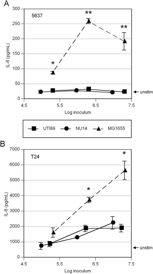
Our previous data demonstrated that 5637 and T24 bladder cells respond in a dose-dependent fashion to laboratory strains of E. coli by producing IL-6 in a TLR4-dependent fashion (38). We evaluated the cytokine responses of these cultured bladder epithelial cell lines to two clinical cystitis strains of UPEC in similar fashion. 5637 or T24 cells were incubated for 2 to 3 h with 105 to 107 CFU/ml of either UTI89 or NU14, and culture supernatants were assayed for IL-6 content by ELISA. In contrast to the type 1 piliated K-12 strain MG1655, cystitis strains UTI89 and NU14 failed to induce IL-6 from 5637 cells that was measurable over baseline (Fig. 1A). Unstimulated T24 cells produce more IL-6 at rest than do 5637 cells, and infection of T24 cells with both UTI89 and NU14 induced a measurable increase in IL-6 production. However, MG1655 induced approximately threefold more IL-6 from T24 cells than did either of the clinical isolates (Fig. 1B). With these infection conditions and over the time period studied, >95% of bladder cells remained viable as measured by exclusion of 0.4% trypan blue in PBS (data not shown). Similarly, after 6 h of infection, epithelial IL-6 induction by the UPEC strains remained significantly less than induction by MG1655. However, up to 40% of bladder cells were nonviable by trypan blue staining after 6 h of infection with the UPEC strains at higher MOI (data not shown).
FIG. 1.
UPEC strains induce less IL-6 from cultured bladder epithelial cells than does E. coli K-12. Confluent bladder cell monolayers were incubated for 2 h with various doses of UPEC strain UTI89 or NU14 or the laboratory E. coli strain MG1655, and IL-6 levels in culture supernatants were measured by sandwich ELISA. (A) IL-6 produced by 5637 cells upon infection with UPEC strain UTI89 or NU14 is equal to unstimulated levels (solid lines), while MG1655 induced significantly more IL-6 at all doses examined (dashed line) (*, P < 0.05; **, P < 0.01). (B) In T24 cells, infection with UPEC induces a modest IL-6 response (solid lines), while stimulation with E. coli K-12 strain MG1655 (dashed line) yields significantly higher IL-6 levels (*, P < 0.03). Experiments were repeated at least three times.
UPEC strains suppress bladder epithelial cell responses to LPS and to K-12 E. coli.
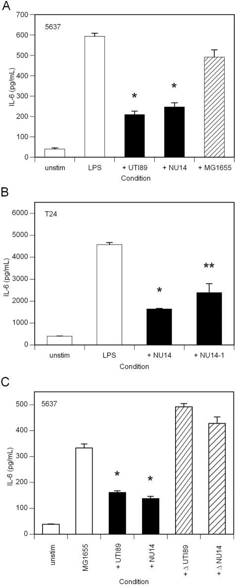
To determine whether UPEC was able to suppress bladder epithelial cell responses to other stimuli that utilize TLR-dependent pathways, we performed coincubation experiments. For these experiments, we used 5637 cells in order to minimize the effect of background IL-6 production in the resting state. Our previous data, using 6-h incubations, demonstrated that 5637 cells respond to E. coli O55:B5 LPS by producing IL-6. These responses were measurable in 5637 cells at an LPS dose of 50 ng/ml and increased in a dose-dependent fashion (38). In the present study, to minimize cytotoxicity to bladder cells during UPEC infection we selected a 2- to 3-h incubation, over which an LPS dose of 5 μg/ml was needed to ensure reliably measurable IL-6 levels. While lower numbers (105 to 106 CFU/ml) of UTI89 or NU14 did not significantly affect IL-6 responses to this dose of exogenous LPS in 5637 cells, 107 CFU/ml of either organism (MOI of 40) suppressed the epithelial IL-6 response to LPS by >50% (Fig. 2A). In contrast, coincubation with 107 CFU/ml of MG1655 did not suppress the IL-6 response to exogenous LPS (Fig. 2A). Both UTI89 and NU14 had a similar cytokine-suppressing effect on T24 cells incubated with exogenous LPS (Fig. 2B and data not shown). The cytokine-suppressing effect did not require the adhesive function of type 1 pili, as an isogenic fimH mutant of NU14 (designated NU14-1) also demonstrated the ability to reduce cytokine responses to exogenous LPS (Fig. 2B).
FIG. 2.
UPEC strains suppress epithelial cytokine responses to exogenously added gram-negative bacterial LPS or E. coli K-12. (A) Confluent monolayers of 5637 cells were left untreated, incubated with 5 μg/ml E. coli O55:B5 LPS, or incubated with LPS plus 107 CFU of the indicated E. coli strains. The UPEC strains UTI89 and NU14 (solid bars) significantly reduced the IL-6 produced in response to exogenous LPS (*, P < 0.01 versus LPS), while MG1655 (striped bar) did not demonstrate such reduction. (B) Confluent monolayers of T24 cells were treated similarly with 500 ng/ml E. coli O55:B5 LPS or with LPSplus 107 CFU of the indicated E. coli strains. The UPEC strain NU14 (*, P < 0.01) and its isogenic fimH mutant NU14-1 (**, P < 0.05) (solid bars) both suppressed T24 cytokine responses to exogenous LPS. (C) Confluent monolayers of 5637 cells were left untreated, treated with 106-CFU/ml MG1655, or treated with MG1655 plus 107 CFU/ml of the indicated UPEC strains. Live UPEC strain UTI89 or NU14 (solid bars) suppressed IL-6 responses to MG1655 (*, P < 0.01 versus MG1655 alone), while heat-killed UTI89 and NU14 (striped bars) have lost this capability. Experiments were repeated at least three times.
We also examined the ability of UTI89 and NU14 to dampen epithelial cytokine responses to K-12 E. coli. 5637 cells were coinfected with 106 CFU/ml of the K-12 strain MG1655 plus 107 CFU/ml of the UPEC strain UTI89 or NU14. As controls, uninfected cells and cells infected only with MG1655 were examined. Coincubation with either UPEC strain downregulated the IL-6 response to MG1655 by >50% (Fig. 2C). The suppressive effect required live bacteria, as heat-killed NU14 or UTI89 did not suppress epithelial cytokine responses to MG1655 (Fig. 2C). In contrast, 107 CFU/ml of UTI89 or NU14 did not suppress 5637 epithelial IL-6 responses to stimulation with IL-1 (data not shown).
Disruption of LPS biosynthetic operons abolishes the cytokine-suppressing activity of UPEC.
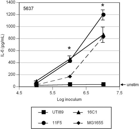
A library of ∼2,000 transposon mutants was created in UTI89 by insertion of a kanamycin resistance marker from the mini-Tn_5Km2_ plasmid (11). Mutants were grown statically in 96-well plates, transferred to epithelial cell monolayers in 96-well tissue culture plates, and incubated for 2 h, and supernatants were examined by human IL-6 ELISA as described above. Two mutant clones, 11F5 and 16C1, were among several identified during primary screening as high inducers of epithelial cytokine activity at 107 CFU/ml. Full dose curves of these mutants compared to those of wild-type UTI89 revealed cytokine induction by the mutants beginning at 106 CFU/ml (Fig. 3). The IL-6 levels induced by the mutants 11F5 and 16C1 were comparable to those induced by the K-12 strain MG1655 at equivalent inocula (Fig. 3). In addition, both 11F5 and 16C1 failed to suppress epithelial cytokine responses to MG1655 (see Fig. 5). The chromosomal DNA sequences flanking the transposon were rescued and sequenced from each of these two clones. The insertion in clone 16C1 was located in the rfa operon upstream of rfaP, while the insertion in clone 11F5 was found in the rfbE gene. Both mutant clones were nonreactive with O18 typing antiserum, in contrast to wild-type UTI89 (data not shown).
FIG. 3.
Disruption of LPS biosynthetic operons in UPEC results in cytokine induction from bladder epithelial cells. Confluent monolayers of 5637 cells were treated with the indicated doses of wild-type UTI89 (squares), the transposon insertion mutant 11F5 (circles) or 16C1 (triangles), or the E. coli K-12 strain MG1655 (diamonds; dashed line). At doses of 106 CFU/ml and above, induction of epithelial IL-6 by the LPS mutants 11F5 and 16C1 was significantly higher than that by UTI89 (*, P < 0.05) and approximated the induction by MG1655. Experiments were repeated at least three times.
FIG. 5.
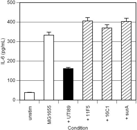
Mutations in LPS biosynthetic genes or in surA abrogate UPEC's ability to suppress cytokine responses to MG1655. Confluent monolayers of 5637 cells were left untreated, treated with 106-CFU/ml MG1655, or treated with MG1655 plus 107 CFU/ml of the indicated UPEC strains. Unlike the UPEC strain UTI89 (solid bar), the LPS biosynthetic mutants 11F5 and 16C1 and the surA mutant (striped bars) fail to suppress epithelial cytokine responses to MG1655. Experiments were repeated at least three times.
Mutation in surA abolishes the cytokine-suppressing activity of UPEC.
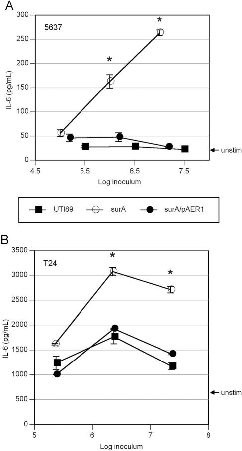
To explore the hypothesis that the UPEC cytokine-suppressing effect may result from expression of a surface-localized or secreted protein, we examined the cytokine profiles of UTI89 mutants deficient in the periplasmic peptidyl-prolyl isomerases. E. coli genes encode at least four of these enzymes (PpiA, PpiD, FkpA, and SurA), which have in vitro cis-trans prolyl isomerase activity. SurA functions as a chaperone in the periplasm (5), and mutation in surA in E. coli K-12 is known to reduce assembly of several major outer membrane proteins (25). Mutation of ppiA, ppiD, or fkpA in UTI89 did not alter cytokine induction from bladder epithelial cells (data not shown). However, mutation in surA led to dramatic changes in the induction of IL-6 from bladder epithelial cells. Dose-response curve assays of 5637 and T24 cells for infection with UTI89 or surA mutant UTI89 were performed as described above. Compared to wild-type UTI89, surA mutant UTI89 induced significantly higher IL-6 production from 5637 and T24 cells (Fig. 4A and B). The wild-type cytokine phenotype was restored by expression of surA from the PBAD promoter on the plasmid pAER1 after induction with 0.1% l-arabinose in overnight static culture (Fig. 4A and B). The surA mutant also failed to suppress epithelial cytokine responses to MG1655 (Fig. 5). The surA mutant, like wild-type UTI89, was reactive with O18 typing antiserum (data not shown).
FIG. 4.
Insertional inactivation of surA results in increased cytokine induction from bladder epithelial cells. Confluent monolayers of 5637 (A) and T24 (B) cells were treated with the indicated doses of wild-type UTI89 (squares), UTI89 surA (open circles), or UTI89 surA/pAER1 (filled circles). The surA mutant displays increased cytokine induction, particularly at doses of 106 CFU/ml and above (*, P < 0.02 versus UTI89), and this phenotype reverts to wild type upon complementation with the plasmid pAER1. Experiments were repeated at least three times.
DISCUSSION
The bladder epithelium is positioned to respond to ascending pathogens, such as uropathogenic Escherichia coli. Bladder epithelial cells expressing TLRs produce proinflammatory cytokines and chemotactic factors that attract neutrophils to the epicenter of infection. On arrival in the bladder tissue, neutrophils engulf available UPEC cells. A critical mechanism used by UPEC to evade this innate defense is to invade superficial umbrella cells and form intracellular bacterial communities (21). This mechanism facilitates the ability of UPEC to establish a foothold in superficial facet cells before the full cadre of neutrophils is able to arrive. We investigated whether UPEC possesses virulence attributes that may dampen or delay the transmission of proinflammatory and chemotactic signals. In this way, the pathogen might prolong its opportunity to establish an intracellular niche within the epithelium.
We employed cultured bladder epithelial cell monolayers infected with uropathogenic and laboratory strains of E. coli. We have previously reported that cell lines of bladder epithelial origin, such as 5637, T24, and J82, respond to LPS and laboratory E. coli by producing IL-6 (38). In contrast, the A498 renal epithelial cell line is not responsive to these stimuli (4, 38). In the present study, infection of bladder epithelial cells with UPEC induced significantly less IL-6 than did MG1655 at all bacterial doses examined. The minimal doses of MG1655 required for a measurable cytokine response were consistent with those we have previously reported (38). 5637 cells, though responsive to significant doses of LPS, did not produce any detectable IL-6 above baseline (uninfected) levels upon infection with the cystitis strain UTI89 or NU14. In T24 cells, where the uninfected baseline is consistently higher, responses to MG1655 were still severalfold higher than those to UPEC. These results argue that UPEC, unlike E. coli K-12, possesses attributes that limit epithelial cytokine responses.
To examine whether UPEC could suppress bladder epithelial responses to known stimuli, we coincubated bladder epithelial cells with UPEC and either exogenous LPS or E. coli K-12. When bladder epithelial cells were coincubated with 107 CFU/ml of UPEC strain UTI89 or NU14 during stimulation with exogenous LPS, cytokine responses to the exogenous LPS were greatly attenuated. The suppressive effect of UPEC on bladder epithelial cytokine responses was not observed at lower UPEC doses; an MOI of approximately 40 was required. The presence of functional type 1 pili on UPEC was not required for cytokine suppression, as NU14-1 (fimH mutant) also was able to suppress LPS responses in bladder epithelial cells. The suppressive capability also required UPEC to be viable, as heat-killed UTI89 and NU14 failed to suppress epithelial responses to E. coli K-12. These requirements for the cytokine-suppressing effect of UPEC in bladder epithelial cells are consistent with those observed in NU14 by Klumpp et al. in their studies with TEU-2 ureteral epithelial cells (22).
These findings suggest at least two hypotheses to account for the mechanism of the suppressive effect of UPEC. One scenario is that LPS of cystitis strains of E. coli is structured or presented to host cells in such a way as to have an inhibitory action on host TLRs. This hypothesis is supported by our observation that UPEC did not suppress 5637 cells' response to IL-1. Stimulation of NF-κB activity by IL-1 utilizes a different surface receptor (IL-1R) than does LPS (TLR4), but both receptors employ a cytoplasmic TIR domain, and the same intracellular machinery accomplishes signal transduction (2).
The stimulatory portion of enterobacterial LPS is generally thought to be lipid A, but there are reports suggesting that some moieties within the polysaccharide portion of LPS may be active in signaling (30). There is also evidence that the acylation state of lipid A may affect the stimulatory properties of gram-negative bacaterial LPS. Most recently, Bäckhed and colleagues demonstrated that penta-acylated lipid A from certain E. coli strains inhibited the production of IL-8 by T24 cells stimulated with hexa-acylated lipid A (3). Other examples of inhibitory LPSs include those of the periodontopathic bacteria Porphyromonas gingivalis and Capnocytophaga ochracea. LPSs from these species fail to stimulate TLR2 and TLR4 in transfected CHO cells and antagonize the stimulatory effects of E. coli K-12 LPS (42).
In our study, initial screening of a transposon mutant library for UPEC clones that augmented epithelial cytokine responses yielded two mutations (presumed to be polar) in operons related to LPS biosynthesis. Clone 16C1 contained an insertion in the rfaP gene, which is part of an operon required for the assembly and modification of the LPS core polysaccharide (40). Clone 11F5 contained an insertion in rfbE. The genes of the rfb operon in enteric gram-negative bacteria encode enzymes for the synthesis and assembly of sugars needed for specific O antigens. Thus, both clones would be predicted to lack O antigen, and in fact they failed to react with O18 typing antiserum. We speculate that the LPS abnormalities resulting from these mutations may lead to the increased availability of lipid A for recognition by host cell surface receptors such as TLR4. These findings are consistent with those of Bäckhed et al., who noted a decrease in IL-8 secretion from T24 epithelial cells when O antigen was present in commercially acquired LPS preparations applied to these cells (4). However, the rfa and rfb mutations would not be predicted to alter the acylation state of lipid A.
A second hypothesis for the UPEC cytokine-suppressing mechanism is that UPEC actively secretes or presents an effector that downregulates host cell NF-κB signaling, as is accomplished by pathogenic Yersinia and Salmonella species. Our observation that heat-killed UPEC fail to suppress cytokine responses to known stimuli supports this notion, though heat killing may also have introduced unrecognized effects on LPS presentation. In addition, insertional inactivation of surA in UTI89 resulted in significantly higher cytokine induction from bladder epithelial cells, a phenotype complemented by addition of the surA gene in trans under the arabinose-inducible PBAD promoter. surA encodes one of at least four periplasmic peptidyl-prolyl isomerases in E. coli. Though surA is not essential for viability in K-12 E. coli, mutation in surA is known to affect proper assembly of outer membrane proteins, reducing levels of OmpA, LamB, and the P pilus usher protein PapC (25; S. S. Justice, submitted for publication). The spectrum of extracytoplasmic proteins acted upon by SurA and the redundancy of function among SurA and the other prolyl isomerases FkpA, PpiA, and PpiD are not precisely known. However, our data suggest that a subset of periplasmic proteins are acted upon uniquely by SurA. Thus far, efforts to delineate the substrates of SurA have revolved around the binding of model peptides to SurA. Bitto and McKay, after solving the structure of SurA (7), reported peptide motifs with the best binding to the N-terminal domain of SurA determined by isothermal titration calorimetry (8). Interestingly, the genome of UTI89 encodes several proteins that contain such motifs and are proposed to act in LPS synthesis (C. S. Hung, unpublished data). Notably, the surA mutant in UTI89 retains reactivity with O18 typing antiserum, indicating that O antigen is likely to be presented intact in this mutant. Additional work is required to determine whether our finding that mutation in surA augments cytokine induction indicates either an active (secreted effector) or passive (LPS-related) mechanism of cytokine suppression by wild-type UPEC.
The in vitro model used in our study differs from ex vivo and in vivo models in that the latter systems include other cell types (e.g., dendritic cells and resident macrophages) present in bladder tissue that participate in the generation of an inflammatory response. Indeed, it is well established that inflammation is a primary feature of bacterial cystitis and that innate responses to LPS and other bacterial products lead to the secretion of inflammatory cytokines from various cell types (2, 14). In the murine model of cystitis, bladder inflammation is absent in C3H/HeJ mice, which express a defective TLR4 (13, 39). In addition, bone marrow transfer experiments between TLR4 wild-type and TLR4 mutant mice demonstrated that TLR4 expression on epithelial cells is critical in initiation of the inflammatory response during murine UTI (37). Our discovery that clinical cystitis strains have a cytokine-suppressing mechanism, as measured in an in vitro epithelial system, is meant to be interpreted in the context of the more complex in vivo situation. Specifically, the ability of the cystitis strains to dampen the cytokine response of the epithelium may give the bacteria an advantage in invading the epithelium and escaping early innate defenses.
The early events of murine cystitis, and presumably those in humans, offer an array of opportunities for uropathogenic E. coli to interact with host epithelia, soluble factors, and immune effector cells. UPEC is able to subvert host defenses by invasion into facet cells and by eventual formation of a quiescent reservoir, critical steps that may be aided by its modulation of host innate responses. The present study demonstrates the ability of UPEC to downregulate epithelial cytokine responses in vitro and offers clues to the mechanism of this effect. Further studies will determine whether a similar pathogenic strategy can be demonstrated during specific host-pathogen interactions in cystitis.
Acknowledgments
This work was supported by National Institutes of Health grants K08-DK067894 (D.A.H.) and P50-DK64540 and R01-DK51406 (S.J.H.). D.A.H. is an NICHD Fellow of the Pediatric Scientist Development Program (K12-HD00850).
We thank C. DebRoy for O-antigen serotyping and T. Silhavy for helpful discussions.
REFERENCES
- 1.Agace, W., S. Hedges, U. Andersson, Y. Andersson, M. Ceska, and C. Svanborg. 1993. Selective cytokine production by epithelial cells following exposure to Escherichia coli. Infect. Immun. 61**:**602-609. [DOI] [PMC free article] [PubMed] [Google Scholar]
- 2.Akira, S., and K. Takeda. 2004. Toll-like receptor signalling. Nat. Rev. Immunol. 4**:**499-511. [DOI] [PubMed] [Google Scholar]
- 3.Bäckhed, F., S. Normark, E. K. Schweda, S. Oscarson, and A. Richter-Dahlfors. 2003. Structural requirements for TLR4-mediated LPS signalling: a biological role for LPS modifications. Microbes Infect. 5**:**1057-1063. [DOI] [PubMed] [Google Scholar]
- 4.Bäckhed, F., M. Soderhall, P. Ekman, S. Normark, and A. Richter-Dahlfors. 2001. Induction of innate immune responses by Escherichia coli and purified lipopolysaccharide correlate with organ- and cell-specific expression of Toll-like receptors within the human urinary tract. Cell. Microbiol. 3**:**153-158. [DOI] [PubMed] [Google Scholar]
- 5.Behrens, S., R. Maier, H. de Cock, F. X. Schmid, and C. A. Gross. 2001. The SurA periplasmic PPIase lacking its parvulin domains functions in vivo and has chaperone activity. EMBO J. 20**:**285-294. [DOI] [PMC free article] [PubMed] [Google Scholar]
- 6.Benson, M., U. Jodal, A. Andreasson, A. Karlsson, J. Rydberg, and C. Svanborg. 1994. Interleukin-6 response to urinary tract infection in childhood. Pediatr. Infect. Dis. J. 13**:**612-616. [DOI] [PubMed] [Google Scholar]
- 7.Bitto, E., and D. B. McKay. 2002. Crystallographic structure of SurA, a molecular chaperone that facilitates folding of outer membrane porins. Structure (Cambridge) 10**:**1489-1498. [DOI] [PubMed] [Google Scholar]
- 8.Bitto, E., and D. B. McKay. 2003. The periplasmic molecular chaperone protein SurA binds a peptide motif that is characteristic of integral outer membrane proteins. J. Biol. Chem. 278**:**49316-49322. [DOI] [PubMed] [Google Scholar]
- 9.Blattner, F. R., G. Plunkett, C. A. Bloch, N. T. Perna, V. Burland, M. Riley, J. Collado-Vides, J. D. Glasner, C. K. Rode, G. F. Mayhew, J. Gregor, N. W. Davis, H. A. Kirkpatrick, M. A. Goeden, D. J. Rose, B. Mau, and Y. Shao. 1997. The complete genome sequence of Escherichia coli K-12. Science 277**:**1453-1474. [DOI] [PubMed] [Google Scholar]
- 10.Collier-Hyams, L. S., H. Zeng, J. Sun, A. D. Tomlinson, Z. Q. Bao, H. Chen, J. L. Madara, K. Orth, and A. S. Neish. 2002. Cutting edge: Salmonella AvrA effector inhibits the key proinflammatory, anti-apoptotic NF-κB pathway. J. Immunol. 169**:**2846-2850. [DOI] [PubMed] [Google Scholar]
- 11.de Lorenzo, V., M. Herrero, U. Jakubzik, and K. N. Timmis. 1990. Mini-Tn_5_ transposon derivatives for insertion mutagenesis, promoter probing, and chromosomal insertion of cloned DNA in gram-negative eubacteria. J. Bacteriol. 172**:**6568-6572. [DOI] [PMC free article] [PubMed] [Google Scholar]
- 12.Guyer, M. S., R. R. Reed, J. A. Steitz, and K. B. Low. 1981. Identification of a sex-factor-affinity site in E. coli as gamma delta. Cold Spring Harbor Symp. Quant. Biol. 45**:**135-140. [DOI] [PubMed] [Google Scholar]
- 13.Hagberg, L., R. Hull, S. Hull, J. R. McGhee, S. M. Michalek, and C. Svanborg Eden. 1984. Difference in susceptibility to gram-negative urinary tract infection between C3H/HeJ and C3H/HeN mice. Infect. Immun. 46**:**839-844. [DOI] [PMC free article] [PubMed] [Google Scholar]
- 14.Hedges, S., P. Anderson, G. Lidin-Janson, P. de Man, and C. Svanborg. 1991. Interleukin-6 response to deliberate colonization of the human urinary tract with gram-negative bacteria. Infect. Immun. 59**:**421-427. [DOI] [PMC free article] [PubMed] [Google Scholar]
- 15.Hedges, S., M. Svensson, and C. Svanborg. 1992. Interleukin-6 response of epithelial cell lines to bacterial stimulation in vitro. Infect. Immun. 60**:**1295-1301. [DOI] [PMC free article] [PubMed] [Google Scholar]
- 16.Hensel, M., J. E. Shea, C. Gleeson, M. D. Jones, E. Dalton, and D. W. Holden. 1995. Simultaneous identification of bacterial virulence genes by negative selection. Science 269**:**400-403. [DOI] [PubMed] [Google Scholar]
- 17.Hoshino, K., O. Takeuchi, T. Kawai, H. Sanjo, T. Ogawa, Y. Takeda, K. Takeda, and S. Akira. 1999. Cutting edge: Toll-like receptor 4 (TLR4)-deficient mice are hyporesponsive to lipopolysaccharide: evidence for TLR4 as the Lps gene product. J. Immunol. 162**:**3749-3752. [PubMed] [Google Scholar]
- 18.Hultgren, S. J., W. R. Schwan, A. J. Schaeffer, and J. L. Duncan. 1986. Regulation of production of type 1 pili among urinary tract isolates of Escherichia coli. Infect. Immun. 54**:**613-620. [DOI] [PMC free article] [PubMed] [Google Scholar]
- 19.Hurst, S. M., T. S. Wilkinson, R. M. McLoughlin, S. Jones, S. Horiuchi, N. Yamamoto, S. Rose-John, G. M. Fuller, N. Topley, and S. A. Jones. 2001. IL-6 and its soluble receptor orchestrate a temporal switch in the pattern of leukocyte recruitment seen during acute inflammation. Immunity 14**:**705-714. [DOI] [PubMed] [Google Scholar]
- 20.Jantausch, B. A., R. O'Donnell, and B. L. Wiedermann. 2000. Urinary interleukin-6 and interleukin-8 in children with urinary tract infection. Pediatr. Nephrol. 15**:**236-240. [DOI] [PubMed] [Google Scholar]
- 21.Justice, S. S., C. Hung, J. A. Theriot, D. A. Fletcher, G. G. Anderson, M. J. Footer, and S. J. Hultgren. 2004. Differentiation and developmental pathways of uropathogenic Escherichia coli in urinary tract pathogenesis. Proc. Natl. Acad. Sci. USA 101**:**1333-1338. [DOI] [PMC free article] [PubMed] [Google Scholar]
- 22.Klumpp, D. J., A. C. Weiser, S. Sengupta, S. G. Forrestal, R. A. Batler, and A. J. Schaeffer. 2001. Uropathogenic Escherichia coli potentiates type 1 pilus-induced apoptosis by suppressing NF-κB. Infect. Immun. 69**:**6689-6695. [DOI] [PMC free article] [PubMed] [Google Scholar]
- 23.Ko, Y. C., N. Mukaida, S. Ishiyama, A. Tokue, T. Kawai, K. Matsushima, and T. Kasahara. 1993. Elevated interleukin-8 levels in the urine of patients with urinary tract infections. Infect. Immun. 61**:**1307-1314. [DOI] [PMC free article] [PubMed] [Google Scholar]
- 24.Langermann, S., S. Palaszynski, M. Barnhart, G. Auguste, J. S. Pinkner, J. Burlein, P. Barren, S. Koenig, S. Leath, C. H. Jones, and S. J. Hultgren. 1997. Prevention of mucosal Escherichia coli infection by FimH-adhesin-based systemic vaccination. Science 276**:**607-611. [DOI] [PubMed] [Google Scholar]
- 25.Lazar, S. W., and R. Kolter. 1996. SurA assists the folding of Escherichia coli outer membrane proteins. J. Bacteriol. 178**:**1770-1773. [DOI] [PMC free article] [PubMed] [Google Scholar]
- 26.Martinez, J. J., M. A. Mulvey, J. D. Schilling, J. S. Pinkner, and S. J. Hultgren. 2000. Type 1 pilus-mediated bacterial invasion of bladder epithelial cells. EMBO J. 19**:**2803-2812. [DOI] [PMC free article] [PubMed] [Google Scholar]
- 27.Min, G., M. Stolz, G. Zhou, F. Liang, P. Sebbel, D. Stoffler, R. Glockshuber, T. T. Sun, U. Aebi, and X. P. Kong. 2002. Localization of uroplakin Ia, the urothelial receptor for bacterial adhesin FimH, on the six inner domains of the 16 nm urothelial plaque particle. J. Mol. Biol. 317**:**697-706. [DOI] [PubMed] [Google Scholar]
- 28.Mulvey, M. A., Y. S. Lopez-Boado, C. L. Wilson, R. Roth, W. C. Parks, J. Heuser, and S. J. Hultgren. 1998. Induction and evasion of host defenses by type 1 piliated uropathogenic Escherichia coli. Science 282**:**1494-1497. [DOI] [PubMed] [Google Scholar]
- 29.Mulvey, M. A., J. D. Schilling, and S. J. Hultgren. 2001. Establishment of a persistent Escherichia coli reservoir during the acute phase of a bladder infection. Infect. Immun. 69**:**4572-4579. [DOI] [PMC free article] [PubMed] [Google Scholar]
- 30.Muroi, M., and K. Tanamoto. 2002. The polysaccharide portion plays an indispensable role in Salmonella lipopolysaccharide-induced activation of NF-κB through human Toll-like receptor 4. Infect. Immun. 70**:**6043-6047. [DOI] [PMC free article] [PubMed] [Google Scholar]
- 31.Orth, K., Z. Xu, M. B. Mudgett, Z. Q. Bao, L. E. Palmer, J. B. Bliska, W. F. Mangel, B. Staskawicz, and J. E. Dixon. 2000. Disruption of signaling by Yersinia effector YopJ, a ubiquitin-like protein protease. Science 290**:**1594-1597. [DOI] [PubMed] [Google Scholar]
- 32.Poltorak, A., X. He, I. Smirnova, M. Y. Liu, C. V. Huffel, X. Du, D. Birdwell, E. Alejos, M. Silva, C. Galanos, M. Freudenberg, P. Ricciardi-Castagnoli, B. Layton, and B. Beutler. 1998. Defective LPS signaling in C3H/HeJ and C57BL/10ScCr mice: mutations in Tlr4 gene. Science 282**:**2085-2088. [DOI] [PubMed] [Google Scholar]
- 33.Rizzitello, A. E., J. R. Harper, and T. J. Silhavy. 2001. Genetic evidence for parallel pathways of chaperone activity in the periplasm of Escherichia coli. J. Bacteriol. 183**:**6794-6800. [DOI] [PMC free article] [PubMed] [Google Scholar]
- 34.Roberts, J. A., B. I. Marklund, D. Ilver, D. Haslam, M. B. Kaack, G. Baskin, M. Louis, R. Mollby, J. Winberg, and S. Normark. 1994. The Gal(α1-4)Gal-specific tip adhesin of Escherichia coli P-fimbriae is needed for pyelonephritis to occur in the normal urinary tract. Proc. Natl. Acad. Sci. USA 91**:**11889-11893. [DOI] [PMC free article] [PubMed] [Google Scholar]
- 35.Ruckdeschel, K., O. Mannel, K. Richter, C. A. Jacobi, K. Trulzsch, B. Rouot, and J. Heesemann. 2001. Yersinia outer protein P of Yersinia enterocolitica simultaneously blocks the nuclear factor-kappa B pathway and exploits lipopolysaccharide signaling to trigger apoptosis in macrophages. J. Immunol. 166**:**1823-1831. [DOI] [PubMed] [Google Scholar]
- 36.Samuelsson, P., L. Hang, B. Wullt, H. Irjala, and C. Svanborg. 2004. Toll-like receptor 4 expression and cytokine responses in the human urinary tract mucosa. Infect. Immun. 72**:**3179-3186. [DOI] [PMC free article] [PubMed] [Google Scholar]
- 37.Schilling, J. D., S. M. Martin, C. S. Hung, R. G. Lorenz, and S. J. Hultgren. 2003. Toll-like receptor 4 on stromal and hematopoietic cells mediates innate resistance to uropathogenic Escherichia coli. Proc. Natl. Acad. Sci. USA 100**:**4203-4208. [DOI] [PMC free article] [PubMed] [Google Scholar]
- 38.Schilling, J. D., S. M. Martin, D. A. Hunstad, K. P. Patel, M. A. Mulvey, S. S. Justice, R. G. Lorenz, and S. J. Hultgren. 2003. CD14- and Toll-like receptor-dependent activation of bladder epithelial cells by lipopolysaccharide and type 1 piliated Escherichia coli. Infect. Immun. 71**:**1470-1480. [DOI] [PMC free article] [PubMed] [Google Scholar]
- 39.Schilling, J. D., M. A. Mulvey, C. D. Vincent, R. G. Lorenz, and S. J. Hultgren. 2001. Bacterial invasion augments epithelial cytokine responses to Escherichia coli through a lipopolysaccharide-dependent mechanism. J. Immunol. 166**:**1148-1155. [DOI] [PubMed] [Google Scholar]
- 40.Schnaitman, C. A. 2001. The genetics and biosynthesis of lipopolysaccha-rides, p. 93-136. In M. Sussman (ed.), Molecular medical microbiology, vol. 1. Academic Press, San Diego, Calif.
- 41.Tato, C. M., and C. A. Hunter. 2002. Host-pathogen interactions: subversion and utilization of the NF-κB pathway during infection. Infect. Immun. 70**:**3311-3317. [DOI] [PMC free article] [PubMed] [Google Scholar]
- 42.Yoshimura, A., T. Kaneko, Y. Kato, D. T. Golenbock, and Y. Hara. 2002. Lipopolysaccharides from periodontopathic bacteria Porphyromonas gingivalis and Capnocytophaga ochracea are antagonists for human Toll-like receptor 4. Infect. Immun. 70**:**218-225. [DOI] [PMC free article] [PubMed] [Google Scholar]
- 43.Zhang, D., G. Zhang, M. S. Hayden, M. B. Greenblatt, C. Bussey, R. A. Flavell, and S. Ghosh. 2004. A Toll-like receptor that prevents infection by uropathogenic bacteria. Science 303**:**1522-1526. [DOI] [PubMed] [Google Scholar]
