TERMINAL FLOWER1 Is a Mobile Signal Controlling Arabidopsis Architecture (original) (raw)
Abstract
Shoot meristems harbor stem cells that provide key growing points in plants, maintaining themselves and generating all above-ground tissues. Cell-to-cell signaling networks maintain this population, but how are meristem and organ identities controlled? TERMINAL FLOWER1 (TFL1) controls shoot meristem identity throughout the plant life cycle, affecting the number and identity of all above-ground organs generated; tfl1 mutant shoot meristems make fewer leaves, shoots, and flowers and change identity to flowers. We find that TFL1 mRNA is broadly distributed in young axillary shoot meristems but later becomes limited to central regions, yet affects cell fates at a distance. How is this achieved? We reveal that the TFL1 protein is a mobile signal that becomes evenly distributed across the meristem. TFL1 does not enter cells arising from the flanks of the meristem, thus allowing primordia to establish their identity. Surprisingly, TFL1 movement does not appear to occur in mature shoots of leafy (lfy) mutants, which eventually stop proliferating and convert to carpel/floral-like structures. We propose that signals from LFY in floral meristems may feed back to promote TFL1 protein movement in the shoot meristem. This novel feedback signaling mechanism would ensure that shoot meristem identity is maintained and the appropriate inflorescence architecture develops.
INTRODUCTION
Growth and development are coordinated through diverse signaling networks. Cell-to-cell signals communicate positional information, induce cellular and tissue identities, and ultimately lead to complex developmental patterns of differentiation. Animals and plants have evolved diverse modes of signaling to establish cell and organ identities. Diffusible protein morphogens, secreted peptides, and small hormones promote differential tissue development and growth (Crickmore and Mann, 2006; Xu et al., 2006). An important aim is to identify the range of such signals, how they are regulated, and how they coordinately act upon their targets.
Plant meristems integrate a diverse array of signals. Meristems are key growth points of plants, adding new organs appropriate to each developmental phase. They are composed of central cells, which maintain the population, and peripheral cells that are programmed to form the different types of organ primordia on the flanks. Various signaling networks maintain this pattern and regulate the size and form of the meristem (Clark et al., 1993; Long et al., 1996; Mayer et al., 1998; Fletcher et al., 1999; Wu et al., 2005). Overlaying this pattern are gene activities and signals that determine where and when primordia are made, what will be their identity, and, thus, what form of overall plant architecture will arise.
Shoot meristems give rise to above-ground organs of the plant. Organ identity is tightly controlled, reflecting the ability of the shoot meristem to precisely interpret a diverse array of signals that affect the growth and identity of primordia. In Arabidopsis thaliana, upon germination, the shoot meristem passes though a vegetative phase, making leaf primordia in a compact rosette, with the first leaves showing juvenile traits and later leaves adult traits (Poethig, 2003; Baurle and Dean, 2006). Later, in response to developmental and environmental signals, the shoot meristem changes its growth pattern: the stem elongates, makes modified leaves, and then switches to making flowers to give the inflorescence. Secondary shoots (branches) arise from the axils of leaves and have a similar growth pattern to the main shoot. However, which axillary meristems grow out, when this occurs, and to which degree are controlled by many axillary factors and branching signals (Beveridge, 2006). Shoot meristems therefore integrate many different signals, but what are these and how do they act?
Specific signals act upon the different growth phases. Various signaling pathways define the vegetative phase, its identity, and its length. Many of these signals are components of RNA interference pathways, affecting the expression of genes that influence the identity of leaves (Hunter et al., 2006). The number of leaves is also tightly controlled, with many pathways promoting a longer vegetative phase and others shortening it (Baurle and Dean, 2006; Corbesier and Coupland, 2006). The switch to flowering involves an integration of these signaling pathways, resulting in the upregulation of the flowering genes. Activation of key integrators, FLOWERING LOCUS T (FT) and SUPPRESSOR OF CONSTANS1, results in a switch in shoot meristem identity and the induction of flowering (Kardailsky et al., 1999; Kobayashi et al., 1999; Samach et al., 2000; Abe et al., 2005; Wigge et al., 2005). FT is expressed in leaves, but its mRNA appears to be a mobile signal that moves to activate APETALA1 (AP1) and LEAFY (LFY) on the flanks of the shoot meristem where they promote floral meristem development (Mandel et al., 1992; Weigel et al., 1992; Hempel et al., 1997; Takada and Goto, 2003; An et al., 2004; Huang et al., 2005).
TERMINAL FLOWER1 (TFL1) is a key signaling protein that controls shoot meristem identity during all of the plant's life cycle. TFL1 acts throughout development to influence each phase of growth; tfl1 mutants have a shorter vegetative phase, making less leaves, branches, and flowers than the wild type (Shannon and Meeks-Wagner, 1991; Alvarez et al., 1992; Schultz and Haughn, 1993). Also, the shoot meristem converts to a terminal flower in tfl1 mutants, unlike the wild type, where it grows indefinitely, never differentiating and only finally senescing. TFL1 is a temporal and spatial repressor of flowering genes and acts opposite to FT, which promotes flowering genes. Interestingly, these two proteins are also homologs (Kardailsky et al., 1999; Kobayashi et al., 1999; Bradley et al., 1997; Ohshima et al., 1997).
Expression of TFL1 is restricted to the inner cells of mature shoot meristems. TFL1 mRNA is very low during the vegetative phase, but its levels are strongly upregulated at the switch to flowering (Simon et al., 1996; Bradley et al., 1997; Ratcliffe et al., 1999). TFL1 expression does not extend into the outer cell layers, the epidermis of the shoot meristem, or into primordia, yet it controls the identity of all of these cells. First, TFL1 determines the identity of primordia made. Second, in tfl1 mutants, all cells, even the epidermis, ectopically express flowering genes (Weigel et al., 1992; Bowman et al., 1993; Gustafson-Brown et al., 1994; Bradley et al., 1997; Liljegren et al., 1999). Thus, TFL1 acts beyond its expression domain, but how does TFL1 achieve this? Where does the protein act to repress flowering genes?
We find that the TFL1 protein acts as a mobile signal to coordinate shoot meristem identity. TFL1 moves from inner cells to outer cells but is restricted from lateral primordia. Its pattern does not show a gradient, suggesting that it may be actively transported to coordinate shoot meristem identity and reach its targets. Strikingly, we found that LFY may control TFL1 movement, suggesting that floral meristems signal back to influence shoot development. This feedback may provide a general mechanism to keep floral meristem identity genes expressed on the flanks and thus generate an indeterminate growing inflorescence bearing many flowers. As lfy mutants terminate in floral structures, our observations suggest that TFL1 movement may also be necessary. Finally, we demonstrate that TFL1 expression is regulated differently in the mature shoot meristem compared with young axillary meristems, both in terms of RNA expression and protein patterns.
RESULTS
TFL1 Is a Cytoplasmic, Unmodified 20-kD Protein of Shoot Meristems
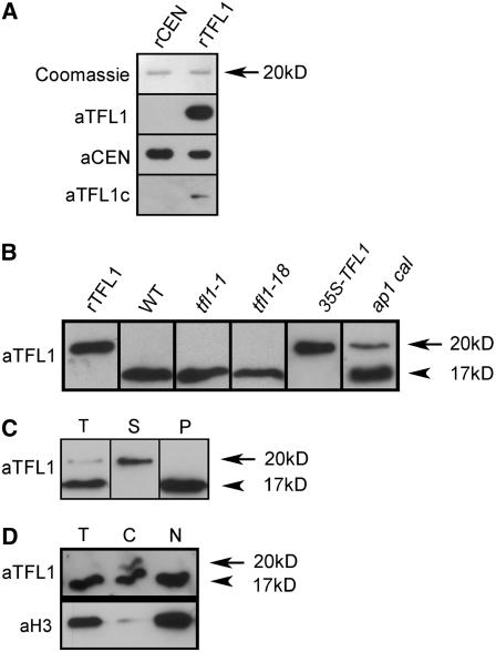
To analyze the amount, size, and distribution of the TFL1 protein, we raised antibodies against TFL1. This anti-TFL1 sera detected _Escherichia coli_–expressed TFL1, but not its Antirrhinum majus functional homolog CENTRORADIALIS (CEN), which is 69% identical (Figure 1A) (Bradley et al., 1996; Cremer et al., 2001). A commercial anti-TFL1 serum gave only a weak signal.
Figure 1.
TFL1 Is a Cytoplasmic 20-kD Protein of Inflorescences.
(A) Recombinant (r) CEN and TFL1 proteins (100 ng) were size-fractioned by SDS-PAGE and visualized with Coomassie staining to reveal the predicted size of 20 kD. Equivalent gels were blotted and probed with anti-TFL1 sera (aTFL1), anti-CEN sera (aCEN), or commercial anti-TFL1 sera (aTFL1c).
(B) Anti-TFL1 sera were used to probe blots of rTFL1 compared with plant extracts. Total protein extracts were analyzed from inflorescences (1-cm-long shoots) of 16-d-old wild-type, tfl1-1, and tfl1-18 plants. Extracts of 12-d-old vegetative 35S-TFL1 seedlings and 26-d-old ap1 cal shoot meristem tissues were also analyzed. The arrow indicates the 20-kD TFL1 protein. The arrowhead indicates a nonspecific protein of 17 kD detected by the sera.
(C) Total (T) protein extracts derived from ap1 cal meristem tissues were fractionated into a high-speed supernatant (S) fraction and membrane pellet (P). Proteins were blotted and probed with anti-TFL1 sera.
(D) Total (T) protein extracts derived from ap1 cal meristem tissues were fractionated into a cytosolic (C) and nuclear fraction (N). Equal amounts of proteins were analyzed as in (C) and probed with anti-TFL1 sera (aTFL1). An equivalent blot was probed with anti-histone3 (aH3) sera.
We tested whether our anti-TFL1 sera detected TFL1 in plant extracts. Initial immunoblots with extracts of 35S-TFL1 control plants (Ratcliffe et al., 1998) showed that only two bands were detected: one of the predicted size of 20 kD and one of 30 kD. This 30-kD band was nonspecific, as it was detected by all antisera and controls and was found in tfl1 null mutant plants and only in leaf tissue. Therefore, to characterize the 20-kD band in more detail, we analyzed protein extracts of young wild-type Arabidopsis and two tfl1 mutant alleles (tfl1-1 and tfl1-18). The tfl1-18 mutant allele is a newly described insertion allele (available in the GABI-Kat collection; Y. Hanzawa and D. Bradley, unpublished data) that should delete most of the protein, and no significant mRNA was detected in long days (LD) by in situ analysis. Therefore, tfl1-18 was probably a very strong or null allele, and TFL1 protein was expected to be absent or much reduced in tfl1-18 compared with the wild type.
High-resolution gels showed that the initial 20 kD band detected could be resolved into two proteins of 20 and 17 kD. The 20-kD protein was present in E. coli TFL1 expression extracts, but wild-type plant extracts only had the 17-kD protein, and this was also present in tfl1-1 and tfl1-18 mutants, suggesting that it was a nonspecific cross-reaction of the anti-TFL1 sera (Figure1B). The 17-kD protein was enriched in extracts of young flowers and was not found in vegetative seedlings (e.g., Figure 1B, 35S-TFL1). The 20-kD TFL1 protein was detected in transgenic lines overexpressing TFL1.
To detect TFL1 in extracts, we used plant material enriched in meristems expressing TFL1, namely, the ap1 cal double mutant. This produces large cauliflower-like apices of proliferating meristems that have high levels of functional TFL1 mRNA (Bowman et al., 1993; Ratcliffe et al., 1999; Ferrandiz et al., 2000). The 20-kD TFL1 protein was clearly detected in these extracts, along with the 17-kD protein (Figure 1B). This reflected the dual nature of ap1 cal meristems being part inflorescence and part floral. Therefore, our anti-TFL1 sera could successfully detect the 20-kD TFL1 protein, which was stable in tissue extracts. TFL1 was apparently of the predicted size; repeated measurements gave a size of 19.5 ± 1.9 kD compared with the predicted 20.2 kD.
We checked whether there were any modifications in TFL1 not affecting apparent gel size. We affinity-purified TFL1 from transgenic plants expressing a constitutive TAPtagTFL1 fusion protein and subjected TFL1 to peptide fingerprinting/mass spectrometry (matrix-assisted laser-desorption ionization time of flight) (see Supplemental Figures 1 and 2 online). Repeat analyses provided extensive coverage (86%) of this version of TFL1 and spanned from V8 to R170. This revealed no novel peptide fragments, suggesting that no significant covalent modifications or processing of TFL1 occurs.
To determine where TFL1 was located inside cells, we fractionated native TFL1 from ap1 cal tissues. The final steps involved ultracentrifugation to separate the total mixture into soluble components and pelleted membranes/membrane vesicles (Figure 1C). This showed that TFL1 was a soluble 20-kD protein, while the nonspecific 17-kD protein was membrane-bound and thus separable from TFL1.
As the TFL1 homolog FT is found in the nucleus (from FT–green fluorescent protein [GFP] studies) and FT binds bZIP transcription factors, we asked whether TFL1 was also nuclear (Pnueli et al., 2001; Abe et al., 2005; Wigge et al., 2005). Fractionation of ap1 cal tissues yielded a nuclear-enriched fraction (as confirmed by histone detection) and a supernatant fraction, containing the cytosol and low-density membranes and organelles (Figure 1D). TFL1 was only found in the supernatant, suggesting that the majority of TFL1 is not in the nucleus (Figure 1D). To resolve exactly where TFL1 protein accumulated, in which specific tissues, and which domains of the inflorescence, we probed sections of plant tissue.
TFL1 Moves beyond Its mRNA Expression Domain in Inflorescence Meristems
Wild-type and control tfl1 mutant plants were grown in LDs and harvested at various time points for both protein (immunolocalization) and mRNA in situ analyses.
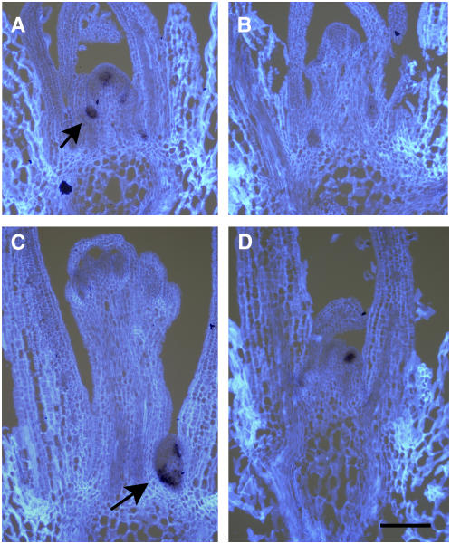
In wild-type plants, TFL1 mRNA was very low during the vegetative phase but was strongly upregulated in the shoot meristem when it switched to an inflorescence identity at about day 12 (Figure 2A). TFL1 mRNA was strong in both the main and lateral shoot meristems at this stage and at later time points (Figure 2A, arrow). No tfl1 mRNA was detected in the tfl1-18 mutant plants at any time point (Figure 2B). Unlike tfl1-18 mutant plants, both tfl1-1 and tfl1-13 had tfl1 mRNA (Figures 2C and 2D). In these cases, their tfl1 mRNA was absent from the main shoot by day 12, as it had already converted into a floral meristem (as shown by AP1 expression). However, tfl1 mRNA was present in secondary shoots arising from the axils of rosette leaves, and this acted as a potential marker for inflorescence shoot meristem identity (Figure 2C). Therefore, as previously reported, TFL1 and tfl1-1 mRNA accumulated in the central, internal regions of inflorescence meristems. However, tfl1-18 mutant plants had no detectable tfl1 mRNA.
Figure 2.
TFL1 mRNA Patterns in Shoot Inflorescence Meristems.
(A) TFL1 mRNA pattern (purple stain) in sections of wild-type plants harvested after 12 LD. Strong TFL1 mRNA was detected in the main shoot and young axillary meristems (arrow). The tissue was counterstained to show cells (white).
(B) No TFL1 mRNA was seen in any tfl1-18 meristems (10 LD is shown).
(C) and (D) TFL1 expression was seen in 12-d-old tfl1-1 and tfl1-13 mutant plants in axillary meristems (arrow). Bar = 100 μm.
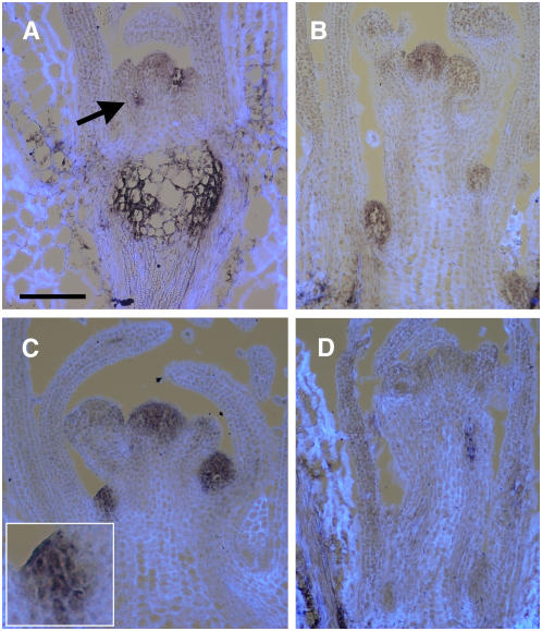
In wild-type Arabidopsis, Columbia (Col) or Landsberg erecta (L_er_) backgrounds, TFL1 protein was not observed at early time points of 8 or 10 LD. Strong background, nonspecific signal was often observed but was confined to the pith region (the central body) of the rosette stem and more variably in older stems or leaves (Figure 3A). This was confirmed as background by probing tfl1-18 mutants that had no TFL1 mRNA or protein. In the wild type, the TFL1 protein was detected only clearly at the switch to inflorescence identity at about day 12 onwards; TFL1 was then strong and found throughout upper axillary shoot meristems (Figures 3A to 3C). TFL1 protein was detected in the main shoot meristem only later, after it was seen in the secondary axillary meristems, where it again appeared throughout the meristem (Figures 3B and 3C). Therefore, independent of their position, wild-type inflorescences had TFL1 protein distributed throughout their entire meristems, unlike its mRNA, which was restricted to the central region.
Figure 3.
TFL1 Protein Moves beyond Its mRNA Domain.
(A) and (B) TFL1 protein in wild-type (Col) plants harvested after 12 or 16 LD, respectively. TFL1 protein (purple stain) was detected at 12 LD in young axillary shoot meristems (arrow). At 16 LD, TFL1 protein was detected also in the main shoot meristem, its upper axillary meristems, and axillary shoot meristems of rosette leaves. The tissue was counterstained to show cells (white). Bar = 100 μm.
(C) TFL1 protein was also detected in wild-type L_er_ plants harvested after 14 LD. Inset shows higher magnification (×3) of a meristem, highlighting TFL1 in epidermis/L1.
(D) TFL1 protein was not detected in tfl1-18 mutant plants harvested after any time point (12 LD is shown).
Probing late wild-type time points showed that TFL1 protein signal was variable but present up to 28 LDs. TFL1 protein did not appear in floral meristems, despite being throughout the inflorescence meristem dome (Figures 3B and 3C). The floral meristems did have some weak background staining, but this was also found in tfl1-18 mutants and the wild type probed with preimmune sera (Figure 3D; see Supplemental Figure 3 online).
TFL1 protein did not appear to extend far below the mRNA expression domain (Figure 3C). This suggested that TFL1 moved toward the apex and to the outer layers (epidermis) of the meristem. If TFL1 did move inward, it did not accumulate, either because it was degraded or it moved rapidly away to distant tissues. We probed 35S-TFL1 plants and showed that TFL1 protein was present and stable in all tissues observed (see Supplemental Figure 3 online). This indicated that TFL1 was not rapidly degraded if present outside the inflorescence meristem.
We analyzed the effect of tfl1 mutations on protein levels and its distribution. The tfl1-1 and tfl1-13 alleles were chosen because they derive from single amino acid substitutions that map to two different sites of the TFL1 protein crystal structure, suggesting that they might affect two different domains of TFL1 function (Ahn et al., 2006). No tfl1-1 or tfl1-13 mutant protein was observed at any time point in mutant plants. This suggested that tfl1 mutant protein was perhaps not made or unstable. However, as tfl1 mutants are more advanced in their growth phase compared with the wild type and shoots converted to flowers, it was difficult to directly compare meristem stages. Therefore, to overcome these problems, we looked at short day (SD) grown plants, where tfl1 mutants have extensive vegetative and inflorescence phases.
TFL1 Protein Also Moves in Vegetative Meristems, but tfl1 Mutant Proteins Are Absent
Arabidopsis plants grown in SD have an extensive vegetative phase, during which time the main shoot meristem generates many leaves with axillary shoot meristems. These axillary meristems develop in a general gradient, being more developed in the older leaves (these axillary meristems make a few leaf primordia) and least developed nearer the main shoot meristem (the apex) (Grbic and Bleeker, 2000; Long and Barton, 2000). These meristems remain small and vegetative, until the main shoot has switched to inflorescence identity, at which point many grow out and switch to inflorescence identity. This switch can be simply controlled by shifting plants to LD.
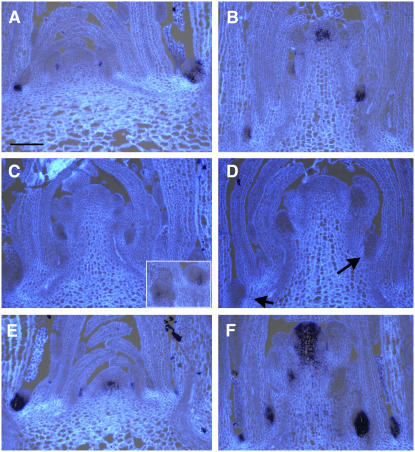
We grew plants for 30 SDs before shifting them to LD to induce flowering and harvest at various time points for mRNA and protein in situ. This showed that TFL1 mRNA was strong in wild-type vegetative meristems before LD induction (Figure 4A). However, this expression was only observed in axillary meristems, not the main shoot meristem. All axillary shoot meristems observed in sections appeared to have TFL1 mRNA, from young to old (Figure 4A). At the very youngest stages, TFL1 mRNA appeared to occupy all cell layers, but at later meristem stages, this resolved into TFL1 being restricted and only occupying the inner cells, at the base of the meristem, next to the leaf. After LD induction, TFL1 mRNA increased first in the upper developing secondary shoots of the elongating main inflorescence. TFL1 was still low in the main shoot meristem between +1 to +3 LD stages but generally increased. Eventually, by approximately +4 LD, TFL1 mRNA was strong in the main shoot in the central region, at the time when floral meristems developed as the uppermost lateral meristems, as shown by AP1 expression (Figure 4B; see Supplemental Figure 4 online) This suggested that the main shoot only expressed TFL1 strongly when it was in the inflorescence stage, but axillary meristems expressed TFL1 strongly in the vegetative phase.
Figure 4.
TFL1 mRNA in SD Vegetative Axillary Meristems.
(A) and (B) TFL1 mRNA expression patterns in sections of wild-type plants grown for 30 SD (A) and then transferred to inductive LD and harvested after 4 LD (B). Bar = 100 μm.
(C) and (D) tfl1-18 mRNA expression patterns in tfl1-18 mutant plants after 30 SD ([C]; inset shows that axillary meristems can have weak tfl1-18 mRNA at low frequency) and then after 2 LD induction (D). A section from 2 LD probed for AP1 expression (see Supplemental Figure 4 online) showed that many shoot meristems were present (arrows in [D]).
(E) and (F) tfl1-1 mRNA expression pattern in tfl1-1 mutant plants after 30 SD (E) or after 4 LD induction (F).
In sections of tfl1-18 mutant plants, weak tfl1-18 mRNA was very occasionally observed in a few meristems (Figure 4C). After LD induction, no tfl1-18 mRNA was seen (Figure 4D). This was not due to conversion of all meristems to flowers directly, as both the main shoot meristem and many lateral meristems were vegetative or inflorescences, as shown by lack of AP1 expression (Figure 4D, arrows; see Supplemental Figure 4 online).
The tfl1-1 and tfl1-13 mutant plants had strong levels of tfl1 mRNA in SD, similar to the wild type (Figure 4E). After LD induction, both point mutation alleles again appeared like the wild type, with strong tfl1 mRNA accumulating in secondary meristems and later in the main shoot meristem (Figure 4F). Also, in some sections, the position of tfl1 mutant mRNA appeared altered compared with the wild type, occupying nearly the whole meristem. AP1 expression analyses also confirmed that many shoots remained as vegetative or inflorescence meristems throughout these time points for both tfl1 alleles, including the main shoot meristem (see Supplemental Figure 4C online).
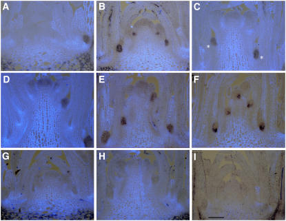
Protein in situ showed high levels of TFL1 protein in axillary meristems in SD where TFL1 mRNA was also strong and not the main shoot meristem where mRNA was low (Figure 5A). TFL1 protein was present throughout these SD vegetative meristems, in contrast with TFL1 mRNA, which was generally restricted to central/basal cells. After LD induction, the TFL1 protein appeared strong in the axillary shoot meristems of the elongating main shoot (Figures 5B and 5C). TFL1 protein was consistently not clearly detected in the main shoot meristem till about day 6 (Figure 5D). Again, the pattern of TFL1 protein did not match the mRNA pattern; the TFL1 protein was distributed throughout the meristems, vegetative or inflorescence, and so extended beyond the mRNA domain.
Figure 5.
TFL1 Protein Moves in Vegetative Meristems.
(A) to (D) TFL1 protein in wild-type plants grown for 30 SD (A) or after 3, 5, or 6 LD induction, respectively ([B] to [D]). Axillary meristems with strong TFL1 protein are marked in (C) with asterisks.
(E) and (F) TFL1 protein pattern (E) was directly compared with its TFL1 mRNA pattern (F) in sections of the same material harvested after 30 SD + 5 LD. Again, TFL1 mRNA was restricted, while TFL1 protein was throughout the shoot meristems.
(G) and (H) No mutant protein was detected in tfl1-18 mutant plants after 30 SD (G), after 1 LD induction (H), or any other times. Dark spots are dirt on tissue/slide.
(I) Similarly, no mutant protein was detected in tfl1-1 mutant plants at any time point (30 SD + 2 LD is shown) despite abundant mRNA. Bar = 100 μm.
No tfl1-18 protein could be detected in mutant plants at any stage in SD or after LD induction, despite the presence of vegetative shoot meristems and inflorescences (Figures 5G and 5H). Also, no tfl1 mutant protein was seen in either tfl1-1 or tfl1-13 mutant plants at any time point, in any stage of meristem development (Figure 5I). Thus, despite significant levels of mRNA for these two different point mutation alleles, their proteins did not accumulate.
TFL1 protein had a different distribution compared with the wild-type mRNA. This difference might be explained by one possible artifact, namely, different fixation/embedding methods for protein versus mRNA. We tested this by probing consecutive sections of the same material for both protein and mRNA distribution. This showed that indeed the protein had a broader expression domain compared with the mRNA (Figures 5E and 5F). Thus, protein movement is most likely to account for TFL1 protein distribution inside shoot meristems. No obvious signal peptide could be found in the TFL1 protein sequence that suggested it might be secreted, moved, and taken up by target cells. This suggests that movement occurs cell-to-cell through plasmodesmata.
Floral Meristem Identity Genes Regulate the TFL1 Protein Pattern
TFL1 is antagonistic to the floral meristem identity genes. TFL1 mRNA is found in a separate domain from the floral meristem identity genes LFY, AP1, and CAL. This also appears true for the TFL1 protein in wild-type plants because although TFL1 protein moves beyond its mRNA domain, it does not move into floral meristems. To see how this pattern of protein movement might be regulated, we looked at the TFL1 protein in lfy and ap1 cal mutants.
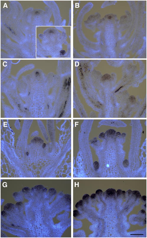
In lfy mutants, shoots replace flowers and TFL1 mRNA is present in a similar pattern to wild-type shoot meristems, being strong in the central region (Huala and Sussex, 1992; Weigel et al., 1992; Liljegren et al., 1999; Ratcliffe et al., 1999). The shoot meristems eventually convert to terminal carpel/flower-like structures and TFL1 mRNA decreases. Samples from independent lfy alleles were harvested just as they began to elongate (bolt) at ∼23 LD. TFL1 protein was found to be strong in young, axillary meristems, similar to the wild type (Figure 6A, inset). Thus, the distribution and stability of TFL1 protein in young axillary shoot meristems was not affected in lfy mutants. However, in the elongated main, secondary, or tertiary shoots, the TFL1 protein was often weak or absent or in a novel pattern of a restricted domain in the center of the meristem (Figures 6A to 6C). Control wild-type plant segregants at the same time point showed TFL1 protein distribution throughout all shoot meristems, indicating that this restriction of TFL1 movement in lfy plants did not depend on the age of the meristem (Figure 6D).
Figure 6.
TFL1 Protein Patterns in lfy and ap1 cal Mutants.
(A) to (D) Analysis of TFL1 protein in lfy mutants ([A] to [C]) and a wild-type segregant (D). The lfy-6 allele was analyzed after 23 LD ([A], main shoot; inset shows secondary shoot with young axillary shoot meristem) or after 27 LD (B). The lfy-14 allele was analyzed after 27 LD (C) and compared with the wild type (D).
(E) to (H) TFL1 protein was detected in ap1 cal mutant shoot meristems after 12, 14, 16, and 20 LD, respectively. Bar = 100 μm.
This restriction of TFL1 protein to the center of the meristem was seen in more than four independent experiments, for two different alleles (lfy-6 [L_er_] and lfy-14 [Col]), and for multiple meristems from any sample. However, it was only seen in 23 or 27 d samples, being absent from the four 36 d samples analyzed. This localized pattern was not seen in wild-type segregants (or other wild types analyzed in our experiments), and a possibly similar signal was only rarely found in tfl1 mutants grown in SD. Such a signal could arise if the antisera cross-reacted with pith/vascular tissue developing below the apex. However, such artifacts might be expected to be evident also at later time points or in the wild type, but this was not observed. Rather, given the high frequency of this novel TFL1 pattern in lfy mutants, it appears to show an inability for TFL1 protein to move in lfy shoots.
AP1 and CAL are also required restriction of TFL1 mRNA to the shoot. AP1 and CAL are required for floral meristem identity and are themselves activated by LFY. In ap1 cal mutants, TFL1 mRNA is present throughout the reiterated meristems of the cauliflower phenotype, in the same meristems as LFY, though the youngest meristems lack LFY (Weigel et al., 1992; Liljegren et al., 1999; Ratcliffe et al., 1999). Therefore, we looked at TFL1 protein distribution in ap1 cal. At day 12, ap1 cal mutant shoots looked similar to the wild type, and TFL1 protein was present at high levels in axillary shoot meristems similar to the wild type, while the main shoot meristem had moderate levels of TFL1 protein (Figure 6E). By day 14, the morphology of the main shoot had changed compared with the wild type, and the meristems arising from the flanks of the ap1 cal main shoot also had high levels of TFL1 protein (Figure 6F). By contrast, these meristems were floral in the wild type and had no TFL1 protein. In ap1 cal mutant plants, the lateral meristems proliferated to form more meristems on each of their flanks. Therefore, by days 16 or 20, a proliferating series of meristems was generated and these all had TFL1 protein (Figures 6G and 6H). In all meristems, at all time points, TFL1 protein appeared throughout the meristem, though levels were variable. Because LFY is not upregulated in the youngest stages of ap1 cal meristems, these data again show that early TFL1 protein movement is independent of all these floral genes. Thus, the ap1 cal mutations affected meristem identity but did not affect TFL1 movement.
DISCUSSION
We analyzed the biochemistry and tissue patterns of TFL1 to see how this signaling protein controls shoot meristem identity. Intriguingly, TFL1 mRNA is restricted to the inner cells of the meristem, but TFL1 protein is small and mobile and may thus coordinate meristem functions by moving to interact with its targets. Our data suggest a new model for how floral and shoot meristems may establish a feedback loop of expression and protein movement to control inflorescence architecture.
TFL1 Is a Small Cytoplasmic Protein of Shoot Meristems
The TFL1 protein accumulates in shoot meristems and is mostly cytoplasmic, not being readily detected in nuclear extracts. Consistent with this pattern, in situ studies showed that the TFL1 protein is found throughout the cell and not in a discrete nuclear-localized pattern similar to transcription factors, such as LFY (Levin and Meyerowitz, 1995; Parcy et al., 1998; Sessions et al., 2000). TFL1 affinity purification for peptide analysis indicated that extractable TFL1 is largely unmodified in plants. Therefore, to signal and regulate meristem identity genes, TFL1 may usually reside in the cytoplasm, where it may transiently associate with its interactors. If TFL1 moves to the membrane, nucleus, or any other organelle, then this may affect only a small amount of the TFL1 pool.
Many TFL1 plant homologs are known, and their functions, mRNA expression patterns, three-dimensional structures, and interactions are all very well described, yet only a few aspects of protein action have been described (Banfield and Brady, 2000; Pnueli et al., 2001; Abe et al., 2005; Hanzawa et al., 2005; Wigge et al., 2005; Ahn et al., 2006). Use of fusions to marker proteins showed that one protein homolog, FT, resides in both the cytoplasm and nucleus and interacts with a bZIP transcription factor to regulate its flowering target, AP1 (Abe et al., 2005; Wigge et al., 2005). Similarly, TFL1 can interact with bZIPs. Animal homologs have been shown to weakly associate with membranes but appear to be mostly cytoplasmic, interacting with a range of proteins, such as kinases and proteases (Hengst et al., 2001; Yeung et al., 2001). Therefore, one general model for this family of proteins may be that they reside in the cytoplasm and interact with potentially many partners, and this regulates their distribution and action throughout the cell. Since TFL1 moves from cell to cell, some of these interactions may guide TFL1 on its journey.
TFL1 Moves from Inner to Outer Meristem Cells
We have addressed a key question on how TFL1 regulates shoot meristems. Gene expression data show that TFL1 mRNA is restricted to the inner cells of the shoot meristem, yet TFL1 must prevent flowering genes from expressing in all layers and cells (Weigel et al., 1992; Bowman et al., 1993; Gustafson-Brown et al., 1994; Bradley et al., 1997; Liljegren et al., 1999). To achieve this, the TFL1 protein apparently moves beyond its gene expression domain. TFL1 moves from inner cells to outer cells. Therefore, TFL1 itself is likely a signal that relays and coordinates shoot identity. Subsequently, TFL1 may find its interactors to act upon its targets to regulate cell identity (Abe et al., 2005; Wigge et al., 2005). A future goal will be to find the distribution of these interactors and how they overlap with TFL1.
TFL1 movement may allow it to act, not only in central cells, but also in peripheral cells of the shoot meristem that proliferate to form floral meristems. LFY is expressed in this peripheral region, in anlagen of future primordia and floral meristems (Weigel et al., 1992; Hempel et al., 1997). Thus, TFL1 protein may overlap with LFY mRNA at the periphery, though it is not clear if LFY protein also accumulates to significant levels in these cells (Levin and Meyerowitz, 1995; Parcy et al., 1998). FD is also expressed in anlagen and is involved in AP1 activation (Abe et al., 2005; Wigge et al., 2005). By contrast, AP1 mRNA is not upregulated until cells emerge from the shoot meristem to give rise to a stage 1 floral meristem (Mandel et al., 1992; Hempel et al., 1997). In tfl1 mutants, AP1 becomes expressed in the shoot meristem. Thus, TFL1 movement may be critically involved in ensuring that LFY and AP1 are not expressed in the outer cells of shoot meristems, as even their epidermal expression is sufficient to change shoot identity (Sessions et al., 2000).
Other proteins can move in the shoot meristem. KN1 moves from inner to outer cells of the epidermis (Jackson et al., 1994). CLV3-GFP can move from outer cells to inner cells, to a limited extent (Rojo et al., 2002; Lenhard and Laux, 2003). Transgenic methods involving chimeras or cell-specific promoters show that LFY or GFP can move within shoot or floral meristems (Sessions et al., 2000; Wu et al., 2003; Kim et al., 2005). By contrast, some factors have more restricted movement, such as DEF in floral meristems, that indicate regulation of movement (Perbal et al., 1996). SHR moves within root tissues in functionally important and very precise patterns (Nakajima et al., 2001). Thus, TFL1 is not unique in moving but appears to have its own specific pattern, regulation, and function in generating a particular architecture.
Mechanism of TFL1 Movement
Two general modes of protein movement in meristems have been suggested. The first requires export of the protein from the expressing cell, extracellular diffusion to the target cells, and interaction with receptors. Examples, such as CLV3, reveal signal sequences that target such export (Fletcher et al., 1999; Rojo et al., 2002; Lenhard and Laux, 2003). TFL1, however, has no clear signal sequence.
The second mode involves proteins moving through plasmodesmata that directly connect cells (Kim and Zambryski, 2005; Lough and Lucas, 2006). This movement may be passive by simple diffusion or controlled and targeted. Studies suggest that LFY moves passively, a default situation of not being retained (Sessions et al., 2000; Wu et al., 2003). Similarly, the small protein GFP diffuses. Both show a gradient in protein levels across the tissue from their source. By contrast, KN1 has specific sequences that are sufficient and necessary for movement (Kim et al., 2005). Similarly, viruses make specific proteins to interact with plasmodesmata to change size of their pores and promote virus movement (Lough and Lucas, 2006). Such modes result in a more even distribution across tissues. The TFL1 pattern appears to be quite uniform across the shoot meristem, suggesting it is also actively transported, but this is not proof. If specificity is involved in TFL1 transport, protein structure motifs might be found through functional bioassays (Kim et al., 2005). However, it could still be that size and TFL1 properties (small, stable, and cytoplasmic) might allow it to passively diffuse and accumulate to equivalent levels across many cell distances in a tissue. It could possibly move in both directions to reach an even distribution at equilibrium. It may prove difficult to address whether size limits TFL1 movement, as our initial fusions of TFL1 to other proteins showed that activity was lost if domains as small as 20 kD were fused to either end (our unpublished data).
Our data suggest that TFL1 is readily moving at all stages of development and thoughout the shoot meristems. One key aspect for Arabidopsis and TFL1 may be limiting its movement so that it does not enter floral meristems. Such restriction of movement between floral and shoot meristems has been described for different proteins and markers (Gisel et al., 1999; Sessions et al., 2000; Wu et al., 2003).
TFL1 Is Regulated Differently in the Main versus Axillary Meristems
TFL1 mRNA is already strongly expressed in axillary meristems in SD during vegetative growth but is weak or absent from the main shoot. Why is TFL1 strong in axillary meristems when not induced to flower? Perhaps axillary meristems need stronger protection from florigen signals, as they are closely associated with leaves, the source of florigens, while the main shoot is perhaps separate and more isolated (Grbic and Bleeker, 2000; Long and Barton, 2000). LFY itself promotes flowering and is expressed weakly in leaves (Weigel and Nilsson, 1995; Blazquez et al., 1997). High levels of initial TFL1 protein may therefore be needed to inhibit LFY effects. Interestingly, tfl1 mutants make extra axillary meristems compared with wild-type plants (Grbic and Bleeker, 2000). Perhaps TFL1 plays additional roles compared with the main shoot and this high level of protein is needed for this function. Lower levels of TFL1 in the mature shoot may be sufficient to prevent florigen action, as the mature shoot may be less competent to respond to such signals.
Previously it was suggested that high levels of TFL1 mRNA could be a marker for a switch to bolting and inflorescence stages (Bradley et al., 1997; Ratcliffe et al., 1999). This now appears only true for the main shoot meristem, not axillary meristems. In SD, axillary meristems have high TFL1 levels, yet are not flowering. Could they be at the stage of induced to bolt but are simply not growing out? Or are they truly in a nonbolting, vegetative rosette-like state? Elegant and detailed analyses of traits have been extensively made on the main shoot to define its phases (Telfer et al., 1997; Poethig, 2003). However, less is known about the phase changes in axillary meristems in SD despite detailed analysis of their early development. It may be possible to find markers, perhaps STP, that are in vegetative, but not inflorescence meristems, and so follow the changes in axillary meristem identity (Wu et al., 2005).
TFL1 mRNA was present through all cells of young axillary meristems. Only as these meristems developed did TFL1 mRNA become restricted to inner cells like the main shoot meristem. How is early and late expression controlled? Many factors play specific roles in control of axillary meristem development. Genes expressed before TFL1 regulate axillary meristem initiation and growth and are specifically expressed there, while hormone targets influence axillary meristem growth (Schmitz and Theres, 2005; Beveridge, 2006; Keller et al., 2006; Muller et al., 2006). These are candidates for early regulators of TFL1 expression or protein levels in axillary meristems. Later, the TFL1 pattern is limited to central cells similar to the pattern of WUS or CLV1. How do these or regulators of this pattern affect TFL1 mRNA? Dissection of the TFL1 promoter will give insights as to how these and flowering signals are integrated to control shoot meristem identity.
Spatial Model for TFL1 Action
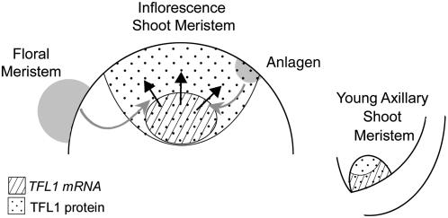
Our studies suggest a model for how spatial regulation of TFL1 is linked to the control of plant architecture (Figure 7). In young axillary meristems, TFL1 mRNA is first seen in all meristem cells and suggests early regulation of TFL1 expression by axillary factors. This pattern changes as the meristem develops, with the most apical cells no longer expressing TFL1. However, TFL1 protein moves into these cells, and this pattern further resolves into that seen in mature shoots.
Figure 7.
Model for TFL1 Expression, Movement, Regulation, and Action.
TFL1 mRNA is first found throughout all cells of young axillary meristems in the axils of leaves. As the meristem develops, TFL1 mRNA is excluded from the tip, but TFL1 protein moves into this region to coordinate cell identity. In the mature shoot meristem, TFL1 mRNA becomes limited to central cells, while TFL1 protein continues to move to the outer cells (black arrows). In the inflorescence, LFY is expressed in peripheral cells (anlagen) and floral meristems, and signals (gray arrows) from LFY promote TFL1 protein movement in the shoot meristem and thus restrict LFY and AP1 expression.
In a mature inflorescence shoot, TFL1 mRNA is restricted to cells in the center of the meristem, in a domain similar to WUS or CLV1 (Castellano and Sablowski, 2005; Williams and Fletcher, 2005). TFL1 protein moves outward to the epidermal tissues but does not appear to enter primordia, suggesting barriers to movement. This barrier is necessary, as expression of TFL1 in primordia may change their identity. We propose that TFL1 movement is required to inhibit LFY and AP1 expression in the inflorescence meristem because expression of these genes, even when restricted to the shoot epidermis, is sufficient to change meristem identity (Sessions et al., 2000). TFL1 restricts AP1 expression to developing floral meristems outside of the shoot meristem. TFL1 cannot prevent LFY expression in some cells, in future primordia on the periphery of the meristem (anlagen), suggesting that activating factors are expressed in this region and these are independent of TFL1. It is not clear if LFY protein accumulates in anlagen, but only in floral meristems, where it also moves (Levin and Meyerowitz, 1995; Parcy et al., 1998). In young axillary meristems of lfy mutants, TFL1 was found throughout the meristem, showing that initial movement was independent of LFY. However, TFL1 protein was limited to central regions in mature lfy mutant shoots. This might reflect a change in protein stability in outer cells, but TFL1 appears stable in young lfy mutants and in extracts from diverse genotypes. Rather, it suggests that movement of TFL1 in the inflorescence meristem is regulated by LFY. In lfy mutants, TFL1 becomes restricted to central cells and the shoot meristem changes identity, differentiating into carpel/floral-like structures. We therefore propose that a LFY-TFL1 feedback loop may operate; LFY promotes TFL1 movement (to overcome its limited expression domain), and this ensures that LFY and AP1 are limited to lateral meristems, while shoot meristems proliferate. We expect these interactions to be indirect, as LFY and TFL1 proteins are apparently in separate domains and no reports of molecular association have been reported.
METHODS
Plant Materials and Growth Conditions
We used wild-type Arabidopsis thaliana plants, ecotype Col or L_er_, mutant lines tfl1-1, tfl1-13 (Shannon and Meeks-Wagner, 1991), ap1-1 cal-1 (Bowman et al., 1993), lfy-6, and lfy-14 (Huala and Sussex, 1992; Weigel et al., 1992), and transgenic line 35S-TFL1 (Ratcliffe et al., 1998). The tfl1-18 mutant is a T-DNA insertion allele (GABI-Kat ID 228A07; Rosso et al., 2003). Plants were grown as described (Ratcliffe et al., 1998). For LD experiments, plants were grown under a 16-h-light/8-h-dark photoperiod in a controlled environment at a temperature of 20 to 22°C. Light was cool light fluorescent tubes at an intensity of 90 to 120 μmol m−2 s−1.
For the SD to LD shift experiments, plants were grown in SDs (8 h light/16 h dark) in a controlled environment at 20 to 22°C, with a light fluency of 120 to 170 μmol m−2 s−1. Plants were transferred to a LD cabinet and grown under 90 μmol m−2 s−1 white cool white lamps at a temperature of 20 to 22°C. Five independent shift experiments were performed, with transfers to LD after a range of 20 to 30 SD.
Antisera and Recombinant Proteins
Anti-TFL1 sera were raised in rabbits by repeated injections of 100 μg of His-TFL1 protein. Recombinant proteins were produced in BL21 (DE3) pLysS Escherichia coli cells (Novagen). His-TFL1 was found in inclusion bodies but could be solubilized and purified with a nickel column kit according to the manufacturer's instructions (Novagen). Untagged recombinant TFL1 protein was produced from a CHITIN BINDING DOMAIN-INTEIN-TFL1 fusion (CBD-INTEIN-TFL1) via intein-mediated cleavage according to the manufacturer's instructions (New England Biolabs). CEN protein was purified as previously detailed (Banfield and Brady, 2000). Anti-CEN sera were raised in rabbits (S. Rothstein, R. Stahl, and D. Bradley, unpublished data).
For protein in situ analysis, anti-TFL1 sera (final bleed) were affinity-purified against a glutathione _S_-transferase-TFL1 (GST-TFL1) column. Glutathione affinity-purified GST-TFL1 (150 μg) was cross-linked to a Thiogel (Severn Biotech). Sera (300 μL) were applied to the column and treated according to instructions, except that column washes involved PBS containing 0.1% (v/v) Tween 20. High-affinity antibodies were eluted, identified by immunoblot, pooled, dialyzed against PBS, and concentrated to ∼400 μg/mL. Affinity-purified antibodies were mixed with an equal volume of 10% (w/v) BSA in PBS and frozen.
Plasmid Construction
To produce different recombinant TFL1-tag proteins, the TFL1 open reading frame (ORF) was amplified from the cDNA with different sets of oligonucleotides and cloned into pGEM-T vector (Stratagene). For poly-histidine-TFL1 (His-TFL1), the TFL1 ORF was amplified from the cDNA with oligos s169 (5′-CATATGGAGAATATGGGAACTAGAGT-3′) and s170 (5′-CTGCAGCTAGCGTTTGCGTGCAGCGGT-3′) and cloned into pGEM-T to give plasmid pLC3. pLC3 was digested with _Nde_I and the TFL1 insert isolated and cloned into the _Nde_I site of pET-19b (Novagen) to give plasmid pLC7.
For GST-TFL1, the TFL1 ORF was amplified with oligos s136 (5′-AACTAGCGTTTGCGTGCAGCGG-3′) and s145 (5′-CCTCCCGGGGAATTCCATGGAGAATATGGGAAC-3′) and cloned into pGEM-T vector to give plasmid pLC23. The _Eco_RI/_Sal_I fragment from pLC23 was ligated into the _Eco_RI/_Sal_I sites of pGEX5X-3 (Pharmacia) to give the GST-TFL1 fusion protein.
To produce CBD-INTEIN-TFL1, the TFL1 ORF was amplified with oligos s170 (5′-CTGCAGCTAGCGTTTGCGTGCAGCGGT-3′) and s171 (5′-GCTCTTCTAACATGGAGAATATGGGAACT-3′) and cloned into pGEM-T to give pLC1. pLC1 was digested with _Sap_I and a vector-derived _Pst_I site and ligated into pTWIN2 (New England Biolabs) previously cut with _Sap_I/_Pst_I to give plasmid pLC6, resulting in the CBD-INTEIN-TFL1 fusion.
To produce 35S-TAPtag-TFL1, the TFL1 ORF was amplified by PCR from the TFL1 cDNA with oligos s174 (5′-TAAGCTTATGGAGAAATATGGGAACT-3′) and s175 (5′-TAAGCTTGGATCCTCTAGACTAGCGTTTGCGTGCA-3′) and cloned into pGEM-T to give plasmid pLC10. The TAPtag ORF was amplified by PCR from pBS1761 (Puig et al., 2001) with oligos s176 (5′-AGGATCCATGGCAGGCCCCTTGCG-3′) and s177 (5′-AAAGCTTATCGTCATCATC-3′) and cloned in pGEM-T to give plasmid pLC11. pLC10 was digested with _Hin_dIII and ligated into pLC11 to give pLC14. This was digested with _Bam_HI, and the TAPtag-TFL1 fragment was cloned into the _Bam_HI site of pJIT62 to give plasmid pLC15 (CaMV35S-TAPtag-TFL1-CaMVTerm). pLC15 was cut with _Kpn_I to release the overexpression cassette, and this was cloned into the _Kpn_I site of the pGREEN 0229 Bar binary vector to give pLC37 (Hellens et al., 2000).
SDS-PAGE and Immunoblots
Proteins were size-fractionated on 15% SDS-PAGE gels and gels stained with Coomassie Brilliant Blue, or proteins were transferred to nitrocellulose filters for immunoblots (Sammy Dry; Schleicher and Schuell). High-resolution gels used modified conditions as described (Schagger and von Jagow, 1987).
Filters were incubated in TTBS (5% [w/v] dry nonfat milk, 25 mM Tris-HCl, pH 8, 150 mM NaCl, and 0.05% [v/v] Tween 20) before incubation with different primary antibodies: rabbit anti-TFL1 (diluted 1:1000), rabbit anti-CEN (diluted 1:1000), rabbit anti-histone H3 acetyl K9 (Abcam) (diluted 1:500), or goat anti-TFL1 antibody (Santa Cruz Biotechology) (diluted 1:200). Filters were washed and incubated with the secondary antibody (anti-rabbit or anti-goat horseradish peroxidase–conjugated [Pierce]), diluted 1:20,000 in TTBS. Filters were washed in TTBS and incubated with the peroxidase substrate (Super Signal West Pico; Pierce) before exposure to film (Hyperfilm ECL; Amersham Pharmacia Biotech).
Arabidopsis Protein Extractions
Arabidopsis proteins were extracted as described (Martinez-Garcia et al., 1999). Cell membranes were isolated from ap1 cal meristem tissues (2 g) as described (Kjellbom and Larsson, 1984). Total homogenates were clarified by centrifugation at 7800_g_, and the resulting solution referred to as the total (T) protein extract. This was spun at 140,000_g_ to produce a supernatant soluble fraction (S) and a high-speed pellet. This pellet was washed and respun to give the final pelleted membrane fraction (P).
Nuclei-enriched fractions were prepared from ap1 cal meristem tissues (2 g) as described (Xia et al., 1997). Fresh tissues were ground in a chilled mortar with a pestle and clarified by filtration through Miracloth and nylon mesh (50-μM pore) to give the total protein extract (T). Nuclei were pelleted by low-speed centrifugation (1500_g_), and the resulting supernatant was referred to as the cytosolic fraction (C). The nuclear fraction was washed and repelleted to give the nuclei-enriched fraction (N).
Proteins were quantified with the Bradford assay kit (Sigma-Aldrich) using BSA as a standard. Forty micrograms of protein extracts were mixed with an equal volume of 2× Laemmli protein loading buffer, heated to 95°C for 5 min, and loaded onto gels.
Protein in Situ
Arabidopsis plants were harvested, and all visible leaves (>2 mm) were removed with a sharp razor blade. Samples were fixed in formaldehyde-acetic acid solution and embedded in Paramat extra (BDH Laboratory Supplies) as previously detailed (Sessions et al., 2000).
Sections (8 μm) were dewaxed and hydrated through a decreasing ethanol series. Slides were boiled in an antigen retrieval solution of 10 mM citrate (pH 6) for 10 min and then left to cool for ∼30 min at room temperature (Shi et al., 1997). Sections were rinsed in deionized water and incubated with BB (5% [w/v] nonfat dry milk, 0.9% [w/v] NaCl, and 10 mM Na phosphate, pH 7.4) overnight at 4°C in a humidified chamber. Sections were incubated with affinity-purified anti-TFL1 antibodies diluted 1:200 in BB for 6 h at 4°C. Slides were washed three times at room temperature with PBSX (PBS and 0.3% [v/v] Triton X-100) with gentle rocking. Secondary antibody (alkaline phosphatase-conjugated AffiPure goat anti-rabbit IgG, Fc fragment specific; Jackson Immunoresearch) was diluted in BB to 1:2000 and added to slides that were incubated again overnight at 4°C in a humidified chamber. The next day, slides were washed three times in PBSX, followed by 10 min in 100 mM Tris-HCl, pH 7.5, and 150 mM NaCl, followed by a 10-min wash in buffer 5 (100 mM Tris-HCl, pH 9.5, 100 mM NaCl, and 50 mM MgCl2). Slides were then incubated in the presence of the substrate solution with 5-bromo-4-chloro-3-indolyl phosphate (BCIP) and nitroblue tetrazolium (NBT) diluted in buffer 5 (0.33 mg/mL of BCIP and 0.165 mg/mL of NBT) and left in the dark to develop signal (∼2 to 6 h). The color reaction was stopped by several rinses in deionized water. Slides were then washed through an ethanol series, counterstained with 0.1% (w/v) calcofluor, and dried before mounting with Entellan (Merck) and a cover slip.
RNA in Situ Experiments
The RNA in situ experiments were performed as described (Ratcliffe et al., 1999).
Accession Numbers
Sequence data from this article can be found in the GenBank/EMBL data libraries under accession numbers U77674 (TFL1), M91208 (LFY), and Z16421 (AP1).
Supplemental Data
The following materials are available in the online version of this article.
- Supplemental Figure 1. Purification of TAPtagTFL1.
- Supplemental Figure 2. MALDI-TOF Analysis of Purified TAPtagTFL1.
- Supplemental Figure 3. TFL1 Protein in Situ Controls.
- Supplemental Figure 4. AP1 Is Found Only in Floral Meristems.
Supplementary Material
[Supplemental Data]
Acknowledgments
We thank Kim Baumann, Enrico Coen, and Yoshie Hanzawa for critical reading of the manuscript and stimulating discussions.
The author responsible for distribution of materials integral to the findings presented in this article in accordance with the policy described in the Instructions for Authors (www.plantcell.org) is: Desmond Bradley (desmond.bradley@bbsrc.ac.uk).
[W]
Online version contains Web-only data.
References
- Abe, M., Kobayashi, Y., Yamamoto, S., Daimon, Y., Yamaguchi, A., Ikeda, Y., Ichinoki, H., Notaguchi, M., Goto, K., and Araki, T. (2005). FD, a bZIP protein mediating signals from the floral pathway integrator FT at the shoot apex. Science 309 1052–1056. [DOI] [PubMed] [Google Scholar]
- Ahn, J.H., Miller, D., Winter, V.J., Banfield, M.J., Lee, J.H., Yoo, S.Y., Henz, S.R., Brady, R.L., and Weigel, D. (2006). A divergent external loop confers antagonistic activity on floral regulators FT and TFL1. EMBO J. 25 605–614. [DOI] [PMC free article] [PubMed] [Google Scholar]
- Alvarez, J., Guli, C.L., Yu, X.-H., and Smyth, D.R. (1992). terminal flower: A gene affecting inflorescence development in Arabidopsis thaliana. Plant J. 2 103–116. [Google Scholar]
- An, H., Roussot, C., Suarez-Lopez, P., Corbesier, L., Vincent, C., Pineiro, M., Hepworth, S., Mouradov, A., Justin, S., Turnbull, C., and Coupland, G. (2004). CONSTANS acts in the phloem to regulate a systemic signal that induces photoperiodic flowering of Arabidopsis. Development 131 3615–3626. [DOI] [PubMed] [Google Scholar]
- Banfield, M.J., and Brady, R.L. (2000). The structure of Antirrhinum centroradialis protein (CEN) suggests a role as a kinase regulator. J. Mol. Biol. 297 1159–1170. [DOI] [PubMed] [Google Scholar]
- Baurle, I., and Dean, C. (2006). The timing of developmental transitions in plants. Cell 125 655–664. [DOI] [PubMed] [Google Scholar]
- Beveridge, C.A. (2006). Axillary bud outgrowth: Sending a message. Curr. Opin. Plant Biol. 9 35–40. [DOI] [PubMed] [Google Scholar]
- Blazquez, M.A., Soowal, L.N., Lee, I., and Weigel, D. (1997). LEAFY expression and flower initiation in Arabidopsis. Development 124 3835–3844. [DOI] [PubMed] [Google Scholar]
- Bowman, J.L., Alvarez, J., Weigel, D., Meyerowitz, E.M., and Smyth, D.R. (1993). Control of flower development in Arabidopsis thaliana by APETALA1 and interacting genes. Development 119 721–743. [Google Scholar]
- Bradley, D., Carpenter, R., Copsey, L., Vincent, C., Rothstein, S., and Coen, E. (1996). Control of inflorescence architecture in Antirrhinum. Nature 379 791–797. [DOI] [PubMed] [Google Scholar]
- Bradley, D., Ratcliffe, O., Vincent, C., Carpenter, R., and Coen, E. (1997). Inflorescence commitment and architecture in Arabidopsis. Science 275 80–83. [DOI] [PubMed] [Google Scholar]
- Castellano, M.M., and Sablowski, R. (2005). Intercellular signalling in the transition from stem cells to organogenesis in meristems. Curr. Opin. Plant Biol. 8 26–31. [DOI] [PubMed] [Google Scholar]
- Clark, S.E., Running, M.P., and Meyerowitz, E.M. (1993). CLAVATA1, a regulator of meristem and flower development in Arabidopsis. Development 119 397–418. [DOI] [PubMed] [Google Scholar]
- Corbesier, L., and Coupland, G. (2006). The quest for florigen: A review of recent progress. J. Exp. Bot. 57 3395–3403. [DOI] [PubMed] [Google Scholar]
- Cremer, F., Lonnig, W.-E., Saedler, H., and Huijser, P. (2001). The delayed terminal flower phenotype is caused by a conditional mutation in the CENTRORADIALIS gene of snapdragon. Plant Physiol. 126 1031–1041. [DOI] [PMC free article] [PubMed] [Google Scholar]
- Crickmore, M.A., and Mann, R.S. (2006). Hox control of organ size by regulation of morphogen production and mobility. Science 313 63–68. [DOI] [PMC free article] [PubMed] [Google Scholar]
- Ferrandiz, C., Gu, Q., Martienssen, R., and Yanofsky, M.F. (2000). Redundant regulation of meristem identity and plant architecture by FRUITFULL, APETALA1 and CAULIFLOWER. Development 127 725–734. [DOI] [PubMed] [Google Scholar]
- Fletcher, J.C., Brand, U., Running, M.P., Simon, R., and Meyerowitz, E.M. (1999). Signaling of cell fate decisions by CLAVATA3 in Arabidopsis shoot meristems. Science 283 1911–1914. [DOI] [PubMed] [Google Scholar]
- Gisel, A., Barella, S., Hempel, F.D., and Zambryski, P.C. (1999). Temporal and spatial regulation of symplastic trafficking during development in Arabidopsis thaliana apices. Development 126 1879–1889. [DOI] [PubMed] [Google Scholar]
- Grbic, V., and Bleeker, A.B. (2000). Axillary meristem development in Arabidopsis thaliana. Plant J. 21 215–223. [DOI] [PubMed] [Google Scholar]
- Gustafson-Brown, C., Savidge, B., and Yanofsky, M.F. (1994). Regulation of the Arabidopsis floral homeotic gene APETALA1. Cell 76 131–143. [DOI] [PubMed] [Google Scholar]
- Hanzawa, Y., Money, T., and Bradley, D. (2005). A single amino acid converts a repressor to an activator of flowering. Proc. Natl. Acad. Sci. USA 102 7748–7753. [DOI] [PMC free article] [PubMed] [Google Scholar]
- Hellens, R.P., Edwards, E.A., Leyland, N.R., Bean, S., and Mullineaux, P.M. (2000). pGreen: A versatile and flexible binary Ti vector for _Agrobacterium_-mediated plant transformation. Plant Mol. Biol. 42 819–832. [DOI] [PubMed] [Google Scholar]
- Hempel, F.D., Weigel, D., Mandel, M.A., Gitta, G., Zambryski, P.C., Feldman, L.J., and Yanofsky, M.F. (1997). Floral determination and expression of floral regulatory genes in Arabidopsis. Development 124 3845–3853. [DOI] [PubMed] [Google Scholar]
- Hengst, U., Albrecht, H., Hess, D., and Monard, D. (2001). The phosphatidylethanolamine-binding protein is the prototype of a novel family of serine protease inhibitors. J. Biol. Chem. 276 535–540. [DOI] [PubMed] [Google Scholar]
- Huala, E., and Sussex, I.M. (1992). LEAFY interacts with floral homeotic genes to regulate Arabidopsis floral development. Plant Cell 4 901–913. [DOI] [PMC free article] [PubMed] [Google Scholar]
- Huang, T., Bohlenius, H., Eriksson, S., Parcy, F., and Nilsson, O. (2005). The mRNA of the Arabidopsis gene FT moves from leaf to shoot apex and induces flowering. Science 309 1694–1696. [DOI] [PubMed] [Google Scholar]
- Hunter, C., Willmann, M.R., Wu, G., Yoshikawa, M., de la Luz Gutierrez-Nava, M., and Poethig, R.S. (2006). Trans-acting siRNA-mediated repression of ETTIN and ARF4 regulates heteroblasty in Arabidopsis. Development 133 2973–2981. [DOI] [PMC free article] [PubMed] [Google Scholar]
- Jackson, D., Veit, B., and Hake, S. (1994). Expression of maize KNOTTED1 related homeobox genes in the shoot apical meristem predicts patterns of morphogenesis in the vegetative shoot. Development 120 405–413. [Google Scholar]
- Kardailsky, I., Shukla, V., Ahn, J.H., Dagenais, N., Christensen, S.K., Nguyen, J.T., Chory, J., Harrison, M.J., and Weigel, D. (1999). Activation tagging of the floral inducer FT. Science 286 1962–1965. [DOI] [PubMed] [Google Scholar]
- Keller, T., Abbott, J., Moritz, T., and Doerner, P. (2006). Arabidopsis REGULATOR OF AXILLARY MERISTEMS1 controls a leaf axil stem cell niche and modulates vegetative development. Plant Cell 18 598–611. [DOI] [PMC free article] [PubMed] [Google Scholar]
- Kim, I., and Zambryski, P.C. (2005). Cell-to-cell communication via plasmodesmata during Arabidopsis embryogenesis. Curr. Opin. Plant Biol. 8 593–599. [DOI] [PubMed] [Google Scholar]
- Kim, J.-Y., Rim, Y., Wang, J., and Jackson, D. (2005). A novel cell-to-cell trafficking assay indicates that the KNOX homeodomain is necessary and sufficient for intercellular protein and mRNA trafficking. Genes Dev. 19 788–793. [DOI] [PMC free article] [PubMed] [Google Scholar]
- Kjellbom, P., and Larsson, C. (1984). Preparation and polypeptide composition of chlorophyll-free plasma membranes from leaves of light-grown spinach and barley. Physiol. Plant 62 501–509. [Google Scholar]
- Kobayashi, Y., Kaya, H., Goto, K., Iwabuchi, M., and Araki, T. (1999). A pair of related genes with antagonistic roles in mediating flowering signals. Science 286 1960–1962. [DOI] [PubMed] [Google Scholar]
- Lenhard, M., and Laux, T. (2003). Stem cell homeostasis in the Arabidopsis shoot meristem is regulated by intercellular movement of CLAVATA3 and its sequestration by CLAVATA1. Development 130 3163–3173. [DOI] [PubMed] [Google Scholar]
- Levin, J.Z., and Meyerowitz, E.M. (1995). UFO: An Arabidopsis gene involved in both floral meristem and floral organ development. Plant Cell 7 529–548. [DOI] [PMC free article] [PubMed] [Google Scholar]
- Liljegren, S.J., Gustafason-Brown, C., Pinyopich, A., Ditta, G.S., and Yanofsky, M.F. (1999). Interactions among APETALA1, LEAFY, and TERMINAL FLOWER1 specify meristem fate. Plant Cell 11 1007–1018. [DOI] [PMC free article] [PubMed] [Google Scholar]
- Long, J., and Barton, M.K. (2000). Initiation of axillary and floral meristems in Arabidopsis. Dev. Biol. 218 341–353. [DOI] [PubMed] [Google Scholar]
- Long, J.A., Moan, E.I., Medford, J.I., and Barton, M.K. (1996). A member of the KNOTTED class of homeodomain proteins encoded by the STM gene of Arabidopsis. Nature 379 66–69. [DOI] [PubMed] [Google Scholar]
- Lough, T.J., and Lucas, W.J. (2006). Integrative plant biology: Role of phloem long-distance macromolecular trafficking. Annu. Rev. Plant Biol. 57 203–232. [DOI] [PubMed] [Google Scholar]
- Mandel, M.A., Gustafson-Brown, C., Savidge, B., and Yanofsky, M.F. (1992). Molecular characterization of the Arabidopsis floral homeotic gene APETALA1. Nature 360 273–277. [DOI] [PubMed] [Google Scholar]
- Martinez-Garcia, J.F., Monte, E., and Quail, P.H. (1999). A simple, rapid and quantitative method for preparing Arabidopsis protein extracts for immunoblot analysis. Plant J. 20 251–257. [DOI] [PubMed] [Google Scholar]
- Mayer, K.F.X., Schoof, H., Haecker, A., Lenhard, M., Jurgens, G., and Laux, T. (1998). Role of WUSCHEL in regulating stem cell fate in the Arabidopsis shoot meristem. Cell 95 805–815. [DOI] [PubMed] [Google Scholar]
- Muller, D., Schmitz, G., and Theres, K. (2006). Blind homologous R2R3 Myb genes control the pattern of lateral meristem initiation in Arabidopsis. Plant Cell 18 586–597. [DOI] [PMC free article] [PubMed] [Google Scholar]
- Nakajima, K., Sena, G., Nawy, T., and Benfy, P.N. (2001). Intercellular movement of the putative transcription factor SHR in root patterning. Nature 413 307–311. [DOI] [PubMed] [Google Scholar]
- Ohshima, S., Murata, M., Sakamoto, W., Ogura, Y., and Motoyoshi, F. (1997). Cloning and molecular analysis of the Arabidopsis gene Terminal Flower 1. Mol. Gen. Genet. 254 186–194. [DOI] [PubMed] [Google Scholar]
- Parcy, F., Nilsson, O., Busch, M.A., Lee, I., and Weigel, D. (1998). A genetic framework for floral patterning. Nature 395 561–566. [DOI] [PubMed] [Google Scholar]
- Perbal, M.-C., Haughn, G., Saedler, H., and Schwarz-Sommer, Z. (1996). Non-cell-autonomous function of the Antirrhinum floral homeotic proteins DEFICIENS and GLOBOSA is exerted by their polar cell-to-cell trafficking. Development 122 3433–3441. [DOI] [PubMed] [Google Scholar]
- Pnueli, L., Gutfinger, T., Hareven, D., Ben-Naim, O., Ron, N., Adir, N., and Lifschitz, E. (2001). Tomato SP-interacting proteins define a conserved signaling system that regulates shoot architecture and flowering. Plant Cell 13 2687–2702. [DOI] [PMC free article] [PubMed] [Google Scholar]
- Poethig, R.S. (2003). Phase change and the regulation of developmental timing in plants. Science 301 334–336. [DOI] [PubMed] [Google Scholar]
- Puig, O., Caspary, F., Rigaut, G., Rutz, B., Bouveret, E., Bragado-Nilsson, E., Wilm, M., and Seraphin, B. (2001). The tandem affinity purification (TAP) method: A general procedure of protein complex purification. Methods 24 218–229. [DOI] [PubMed] [Google Scholar]
- Ratcliffe, O.J., Amaya, I., Vincent, C.A., Rothstein, S., Carpenter, R., Coen, E.S., and Bradley, D.J. (1998). A common mechanism controls the life cycle and architecture of plants. Development 125 1609–1615. [DOI] [PubMed] [Google Scholar]
- Ratcliffe, O.J., Bradley, D.J., and Coen, E.S. (1999). Separation of shoot and floral identity in Arabidopsis. Development 126 1109–1120. [DOI] [PubMed] [Google Scholar]
- Rojo, E., Sharam, V.K., Kovaleva, V., Raikhel, N.V., and Fletcher, J.C. (2002). CLV3 is localized to the extracellular space, where it activates the Arabidopsis CLAVATA stem cell signaling pathway. Plant Cell 14 969–977. [DOI] [PMC free article] [PubMed] [Google Scholar]
- Rosso, M.G., Li, Y., Strizhov, N., Reiss, B., Dekker, K., and Weisshaar, B. (2003). An Arabidopsis thaliana T-DNA mutagenized population (GABI-Kat) for flanking sequence tag-based reverse genetics. Plant Mol. Biol. 53 247–259. [DOI] [PubMed] [Google Scholar]
- Samach, A., Onouchi, H., Gold, S.E., Ditta, G.S., Schwarz-Sommer, Z., Yanofsky, M.F., and Coupland, G. (2000). Distinct roles of CONSTANS target genes in reproductive development of Arabidopsis. Science 288 1613–1616. [DOI] [PubMed] [Google Scholar]
- Schagger, H., and von Jagow, G. (1987). Tricine-sodium dodecyl sulfate-polyacrylamide gel electrophoresis for the separation of proteins in the range from 1 to 100 kDa. Anal. Biochem. 166 368–379. [DOI] [PubMed] [Google Scholar]
- Schmitz, G., and Theres, K. (2005). Shoot and inflorescence branching. Curr. Opin. Plant Biol. 8 506–511. [DOI] [PubMed] [Google Scholar]
- Schultz, E.A., and Haughn, G.W. (1993). Genetic analysis of the floral initiation process (FLIP) in Arabidopsis. Development 119 745–765. [Google Scholar]
- Sessions, A., Yanofsky, M.F., and Weigel, D. (2000). Cell-cell signaling and movement by the floral transcription factors LEAFY and APETALA1. Science 289 779–781. [DOI] [PubMed] [Google Scholar]
- Shannon, S., and Meeks-Wagner, D.R. (1991). A mutation in the Arabidopsis TFL1 gene affects inflorescence meristem development. Plant Cell 3 877–892. [DOI] [PMC free article] [PubMed] [Google Scholar]
- Shi, S.-R., Cote, R.J., and Taylor, C.R. (1997). Antigen retrieval immunohistochemistry: Past, present, and future. J. Histochem. Cytochem. 45 327–343. [DOI] [PubMed] [Google Scholar]
- Simon, R., Igeno, M.I., and Coupland, G. (1996). Activation of floral meristem identity genes in Arabidopsis. Nature 384 59–62. [DOI] [PubMed] [Google Scholar]
- Takada, S., and Goto, K. (2003). TERMINAL FLOWER2, an Arabidopsis homolog of HETEROCHROMATIN PROTEIN1, counteracts the activation of FLOWERING LOCUS T by CONSTANS in the vascular tissues of leaves to regulate flowering time. Plant Cell 15 2856–2865. [DOI] [PMC free article] [PubMed] [Google Scholar]
- Telfer, A., Bollman, K.M., and Poethig, R.S. (1997). Phase change and the regulation of trichome distribution in Arabidopsis thaliana. Development 124 645–654. [DOI] [PubMed] [Google Scholar]
- Weigel, D., Alvarez, J., Smyth, D.R., Yanofsky, M.F., and Meyerowitz, E.M. (1992). LEAFY controls floral meristem identity in Arabidopsis. Cell 69 843–859. [DOI] [PubMed] [Google Scholar]
- Weigel, D., and Nilsson, O. (1995). A developmental switch sufficient for flower initiation in diverse plants. Nature 377 495–500. [DOI] [PubMed] [Google Scholar]
- Wigge, P.A., Kim, M.C., Jaeger, K.E., Busch, W., Schmid, M., Lohmann, J.U., and Weigel, D. (2005). Integration of spatial and temporal information during floral induction in Arabidopsis. Science 309 1056–1059. [DOI] [PubMed] [Google Scholar]
- Williams, L., and Fletcher, J.C. (2005). Stem cell regulation in the Arabidopsis shoot apical meristem. Curr. Opin. Plant Biol. 8 582–586. [DOI] [PubMed] [Google Scholar]
- Wu, X., Dabi, T., and Weigel, D. (2005). Requirement of homeobox gene STIMPY/WOX9 for Arabidopsis meristem growth and maintenance. Curr. Biol. 15 436–440. [DOI] [PubMed] [Google Scholar]
- Wu, X., Dinney, J.R., Crawford, K.M., Rhee, Y., Citovsky, V., Zambryski, P.C., and Weigel, D. (2003). Modes of intercellular transcription factor movement in the Arabidopsis apex. Development 130 3735–3745. [DOI] [PubMed] [Google Scholar]
- Xia, Y., Nikolau, B.J., and Schnable, P.S. (1997). Developmental and hormonal regulation of the Arabidopsis CER2 gene that codes for a nuclear-localized protein required for the normal accumulation of cuticular waxes. Plant Physiol. 115 925–937. [DOI] [PMC free article] [PubMed] [Google Scholar]
- Xu, J., Hofhuis, H., Heidstra, R., Sauer, M., Friml, J., and Scheres, B. (2006). A molecular framework for plant regeneration. Science 311 385–388. [DOI] [PubMed] [Google Scholar]
- Yeung, K.C., Rose, D.W., Dhillon, A.S., Yaros, D., Gustafsson, M., Chatterjee, D., McFerran, B., Wyche, J., Kolch, W., and Sedivy, J.M. (2001). Raf kinase inhibitor protein interacts with NF-kB-inducing kinase and TAK1 and inhibits NF-kB activation. Mol. Cell. Biol. 21 7207–7217. [DOI] [PMC free article] [PubMed] [Google Scholar]
Associated Data
This section collects any data citations, data availability statements, or supplementary materials included in this article.
Supplementary Materials
[Supplemental Data]
