Sterical hindrance promotes selectivity of the autophagy cargo receptor NDP52 for the danger receptor galectin-8 in anti-bacterial autophagy (original) (raw)
. Author manuscript; available in PMC: 2013 Aug 5.
Abstract
Autophagy, the process of lysosome-dependent degradation of cytosolic components, is a mechanism by which cells selectively engulf invading pathogens to protect themselves against infection. Galectin-8, a cytosolic lectin with specificity for β-galactosides, binds endosomal and lysosomal membranes that have been damaged, for example by pathogens, and selectively recruits the autophagy cargo receptor NDP52 to induce autophagy. Here, we solved the crystal structure of the NDP52–galectin-8 complex to show how NDP52 exclusively binds galectin-8 and, consequently, why other galectins do not restrict the growth of Salmonella in human cells.
Introduction
Macro-autophagy, a lysosome-dependent degradation pathway for cytosolic components, controls a multitude of cellular processes, such as the response to starvation, the degradation of protein aggregates, the elimination of damaged or superfluous organelles, and the defense against invading pathogens (1-3). In contrast to the indiscriminate engulfment of cytosol into autophagosomes during starvation, selective autophagy is mediated by specific ‘eat-me’ signals detected by specialized cargo receptors (4). Selective autophagy of bacteria is initiated by three receptors, NDP52, p62, and optineurin, which detect bacteria-associated poly-ubiquitylated proteins (5-7). NDP52 also senses galectin-8, which functions as a ubiquitin-independent ‘eat-me’ signal on vesicles damaged by cytosol-invading bacteria (8).
Galectins are cytosolic lectins, i.e. sugar-binding proteins, with specificity for β-galactoside-containing glycans (9, 10). In mammalian cells under homeostatic conditions, such glycans are found exclusively in extracellular compartments including the lumen of vesicles. A breach in membrane integrity, which may be caused by vesicle-damaging pathogens, such as Salmonella enterica serovar Typhimurium (S. Typhimurium), Listeria monocytogenes, or Shigella flexneri, introduces glycans into the cytosol, which cells perceive as an acute danger signal (3). The human genome encodes about a dozen galectins, of which galectin-1, 3, 8, and 9 bind glycans on damaged endosomes or lysosomes in epithelial cells (8, 9, 11), thereby inducing autophagy, inflammatory signaling, and possibly other cellular responses (8, 11). Currently, the only known effector protein of galectin-sensed membrane damage is NDP52, an LC3C-binding autophagy receptor that restricts the proliferation of S. Typhimurium upon entry into the cytosol by targeting galectin-8–associated bacteria for autophagy (5, 8, 12). NDP52 contains a short peptide (NDP52372-380) that binds the C-terminal carbohydrate recognition domain (CRD) of galectin-8 (Gal8C-CRD) with high selectivity, which explains why galectin-8, but not other membrane damage-sensing galectins, can induce NDP52-mediated autophagy of vesicles damaged by the presence of S. Typhimurium and thus restrict the proliferation of this pathogen inside human cells (8).
Here, we solved the crystal structure of NDP52 bound to Gal8C-CRD to show how galectin-8, uniquely amongst galectins, binds NDP52. NDP52371-381 formed a hook-like structure that strategically positions several side chains for mainly hydrophobic contacts with galectin-8. Interference with the hook formation, as well as mutations in contact residues, abrogated binding between NDP52 and Gal8C-CRD and impeded autophagy of S. Typhimurium. Binding of NDP52 to other galectins was prevented primarily by sterical hindrance in the CRDs of galectins that did not bind, rather than the lack of conserved contact residues in these CRDs, thus explaining the selectivity of NDP52 for galectin-8..
Results
Structural basis for the binding of the danger receptor galectin-8 to the autophagy adaptor NDP52
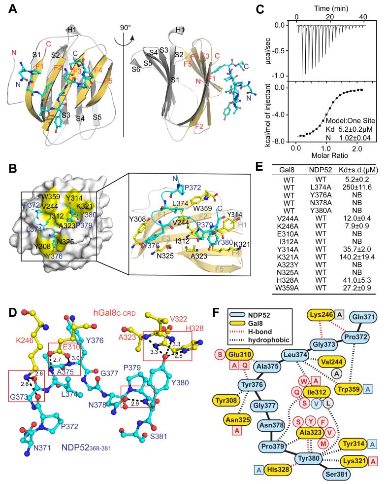
To characterize how galectins recruit effector proteins and to understand how specificity between NDP52 and galectin-8 is achieved, we solved the crystal structure of Gal8C-CRD (residues 228-359) in complex with NDP52368-381 at 2.0 ångströms (Å) resolution (Table 1). Similar to other galectins (13-15), Gal8C-CRD adopted a bent β-sandwich structure formed by two antiparallel β-sheets of six and five strands, which comprise the concave and convex surface, respectively (Fig. 1A, fig. S1). Galectins bind glycans through their concave surface (9), whereas NDP52371-381 bound to the opposite, convex site of Gal8C-CRD with a Kd of 5.2 μM, and this interaction involved an area of 496 Å2 (Fig. 1A-C, fig. S1, S2). A turn formed by Leu374-Ala-Tyr-Gly377 forces NDP52371-381 into a hook-like structure, the C-terminus of which is further stabilized by an intramolecular hydrogen bond formed between the main chain amino group of Tyr380 and the side chain of Asn378 (Fig. 1D). Mutation of Asn378 in NDP52368-381 (N378A) to prevent formation of this hydrogen bound resulted in a mutant lacking all detectable affinity for Gal8C-CRD (Fig. 1E, table S1). The hook-like structure of NDP52371-381 strategically positioned its side chains in contact with several hydrophobic areas of Gal8C-CRD: (i) NDP52 Pro372 contacted the indole ring of Gal8 Trp359, (ii) NDP52 Tyr376 engaged the side chains of Gal8 Tyr308 and Gal8 Asn325, (iii) NDP52 Leu374 inserted into a ridge formed by the Val244, Ile312, and Trp359 residues in Gal8, and (iv) the Pro379 and Tyr380 residues of NDP52 occupied a depression with hydrophobic walls formed by the Ile312, Tyr314, Lys321, and Ala323 of Gal8 (Fig. 1B, F). The importance of these hydrophobic interactions for the binding of NDP52368-381 to Gal8C-CRD was confirmed by mutational analysis (Fig. 1E, table S1). NDP52368-381 with any of the mutations L374A, Y376A, or Y380A had severely reduced affinities for Gal8C-CRD, as did Gal8C-CRD with mutations I312A, K321A, A323Y, or N325A for NDP52368-381. Gal8C-CRD Y314A and W359A bound NDP52368-381 with 7- and 5-fold reduced affinity compared to that of the wild-type Gal8C-CRD, respectively; whereas Gal8C-CRD V244A and K246A had no discernable change in binding affinity. Besides promoting hydrophobic contacts, the depression in Gal8 occupied by NDP52 Tyr380 also enabled the formation of hydrogen bonds between NDP52 Tyr380 and the side chain of Gal8 His328, as well as main chain atoms of Val322 and Ala323 in Gal8 (Fig. 1D, F). Confirming the importance of these hydrogen bonds, the affinity of NDP52368-381 Y380F and Gal8C-CRD H328A for wild-type Gal8C-CRD or NDP52368-381, respectively, was reduced 16- and 8-fold (Fig. 1E, Tab. S1). Furthermore, hydrogen bonds were formed between the main chain atoms of the Pro372-Gly373 and Ala375-Tyr376 residues in NDP52 and the side chains of the Lys246 and Glu310 residues in Gal8C-CRD, respectively (Fig. 1D, F). Mutational analysis of Gal8C-CRD revealed that Glu310, but not Lys246, was essential for binding to NDP52368-381 (Fig. 1E, Tab. S1). Altogether, NDP52371-381 adopted a hook-like conformation when bound to the convex surface of Gal8C-CRD, an interaction driven by hydrophobic contacts and supported by hydrogen bonds (Fig. 1F).
Table 1. Summary of diffraction data and structure refinement statistics.
| Galectin-8C-CRD-NDP52368-381 | |
|---|---|
| Data collection | |
| Space group | C222 |
| Cell dimensions | |
| a, b, c (Å) | 76.087 124.826 43.737 |
| α, β, γ (°) | 90.00 90.00 90.00 |
| Resolution(Å) | 50-2.02( 2.05-2.02)a |
| Rmerge(%) | 8.5(33.3) |
| I/σI | 20.34(6.79) |
| Completeness (%) | 99.9(100) |
| Redundancy | 5.3(4.8) |
| Refinement | |
| No. reflections (overall) | 13992 |
| No. reflections (test set) | 701 |
| Rwork/Rfree(%) | 17.67/21.61 |
| No. non-H atoms | |
| Gal8C-CRD | 1162 |
| NDP52 | 81 |
| Glycerol | 12 |
| Water | 119 |
| B-factors | |
| Gal8C-CRD | 21.59 |
| NDP52 | 27.15 |
| Glycerol | 20.45 |
| Water | 32.18 |
| R. m. s. deviations | |
| Bond lengths (Å) | 0.008 |
| Bond angles (°) | 1.257 |
Figure 1. The molecular basis of the interaction between Gal8C-CRD and NDP52368-381.
(A) Cartoon representation of Gal8C-CRD (yellow and grey) in complex with NDP52368-381 (cyan). F1-F5, S1-S6 numbering of β-strands. (B) Hydrophobic contacts between Gal8C-CRD and NDP52368-381. Residues of Gal8C-CRD involved in hydrophobic interaction are colored yellow and labeled in black. NDP52 in cyan. (C) ITC data for the interaction of NDP52368-381and Gal8C-CRD at the indicated peptide versus protein molar ratios. Top, raw data; bottom, integrated heat data. Kd, dissociation constant; N, reaction stoichiometry. (D) Hydrogen bonds between Gal8C-CRD (yellow) and NDP52368-381 (cyan) with distances measured in Ångströms (Å). (E) Dissociation constants for the indicated interactions obtained through ITC. . NB: binding not detectable. (F) Scheme of interactions between NDP52 (blue) and galectin-8 (yellow). Single letter abbreviations indicate mutations tested in this study with colors indicating affinity (red: Kd >120 μM, blue: 27< Kd <41 μM, black: Kd <12 μM). Circled residues occur in human galectin CRDs, squared residues are experimental mutations.
Recruitment of NDP52 to galectin-8 upon Salmonella infection
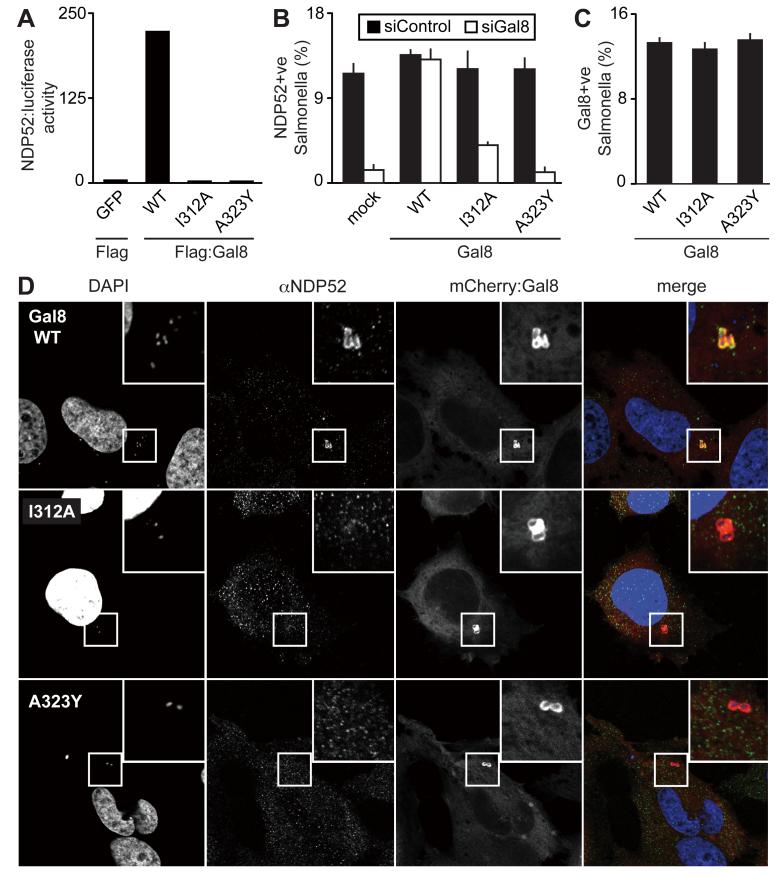
To test whether the binding site identified for NDP52368-381 in Gal8C-CRD also governs the interaction of the full-length proteins, we examined the importance of key mutations in galectin-8 by LUMIER assay. We found that galectin-8 harboring the amino acid mutations I312A or A323Y, in contrast to wild-type galectin-8, did not bind full-length NDP52 (Fig. 2A, fig. S3). To investigate the importance of the NDP52 binding site in galectin-8 in vivo, we analyzed the ability of cells to recruit NDP52 through galectin-8 to damaged vesicles containing S. Typhimurium as an essential step in inducing autophagy in response to bacterial infection. Consistent with our previous report (8), depletion of galectin-8 in HeLa cells abrogated NDP52 recruitment to damaged vesicles containing S. Typhimurium (Fig. 2B). Expression of siRNA-resistant wild-type galectin-8 restored this activity. In contrast, NDP52 recruitment was not restored in cells transfected with siRNA-resistant mutant galectin-8, Gal8 I312A or A323Y (Fig. 2B), even though both mutant proteins were fully functional in detecting S. Typhimurium--induced membrane damage (Fig. 2C, D). We conclude that the binding site in galectin-8 identified in our structure is essential for the recruitment of the autophagy receptor NDP52 to cytosol-exposed S. Typhimurium.
Figure 2. Functional importance of the NDP52 binding site in galectin-8 for anti-bacterial autophagy.
(A) LUMIER binding assay. Normalized ratio of luciferase activity bound to anti-Flag beads to luciferase activity in lysates from 293ET cells co-expressing NDP52 fused to luciferase and the indicated Flag-tagged wild-type (WT) or mutated (I312A, A323Y) galectin-8 proteins. (B-C) Percentage of S. Typhimurium exhibiting NDP52 (B) or galectin-8 (C) recruitment at 1 hour post-infection in Hela cells expressing the indicated siRNA-resistant WT or mutated galectin-8 alleles fused to mCherry as determined by fluorescence microscopy. Cells were treated with control siRNA (siControl) or siRNA against galectin-8 (siGal8) (B) or only siGal8 (C).. Mean and s.d. were quantified from quadruplicate cultures (n>200 bacteria per coverslip), and representative confocal images from siGal8-treated cells are shown (D).
Sterical hindrance between NDP52 and most nonbinding galectin CRDs
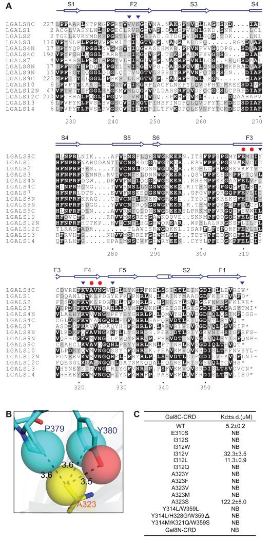
Having characterized the binding site for NDP52 in Gal8C-CRD, we investigated the features that prevent other human galectins from binding NDP52. A multiple sequence alignment of CRDs from human galectins revealed that amongst the amino acids essential for NDP52 binding, Ala323 is conserved in only five CRDs, including Gal8C-CRD (Fig. 3A). Inspection of the Gal8C-CRD–NDP52371-381 structure suggested that residues larger than Ala would interfere with binding (Fig. 3B). Indeed, replacing residue 323 with amino acids occurring in other galectins, such as Tyr, Phe, Met, or Val, abrogated binding of NDP52368-381 (Fig. 3C, table S1). Ala323 is therefore a key specificity determinant and most galectin CRDs do not bind NDP52 because of sterical hindrance in this position or, as is the case in the Gal12N-CRD mimetic Gal8C-CRD A323S, because hydrophobic contacts to NDP52P379 are disfavoured. In the four human galectin CRDs that harbor Ala323 but do not bind NDP52, the neighboring residue Ile312 is not conserved (Fig. 3A). Because Ile312 is a major contributor to the same hydrophobic pocket, we tested the impact of naturally occurring Ile312 replacements on NDP52 binding. Gal8C-CRD I312Q or I312W, which mimic the residues found in Gal3 or in the C-CRD of Gal9, respectively, did not bind NDP52368-381 (Fig. 3C, Tab. S1). In contrast, Gal8C-CRD variants carrying certain aliphatic residues in this position, such as I312L or I312V, which mimic the C-CRD of Gal12 or the N-CRD of Gal8, respectively, retained nearly wild-type affinity for NDP52368-381. Altogether, the lack of conservation of just two residues in galectin-8, the adjacently located Ile314 and Ala323, sufficed to explain the lack of NDP52 binding in all human galectin CRDs except the C-CRD of Gal12 and the N-CRD of Gal8. For these CRDs, the contribution of two aromatic residues, Tyr314 and Trp359, was investigated further because their individual replacement moderately decreased affinity for NDP52 (Fig. 1E, table S1). Indeed, Gal8C-CRD Y314L/H328G/W359Δ and Gal8C-CRD Y314M/K321Q/W359S, mimicking the C-CRD of Gal12 and the N-CRD of Gal8, respectively, showed no detectable binding to NDP52368-381 (Fig. 3C, table S1). Therefore, the data indicated that selectivity of NDP52 for Gal8C-CRD is primarily caused by sterical hindrance in the CRDs of other galectins that do not bind; whereas the lack of conserved contact residues in nonbinding CRDs contributed relatively less to this selectivity.
Figure 3. The molecular basis for the specificity of NDP52 for galectin-8.
(A) For galectins containing two CRDs, N- and C-terminal domains are indicated. Red dots and black triangles indicate residues in Gal8C-CRD that, if mutated, prevented or impaired binding to NDP52368-381, respectively.
(B) Close-up view of Gal8 Ala323 (yellow) in contact with the γ-carbon of the NDP52 Pro379 and the ε-carbon and hydroxyl oxygen of the NDP52 Tyr380 side chain (blue and red). Van der Waals volumes of key atoms rendered as transparent spheres.
(C) Dissociation constants for the interaction of NDP52368-381 with the indicated Gal8C-CRD variants obtained ITC. NB: binding not detectable.
Discussion
This study showed how information about defects in the integrity of vesicular membranes, revealed by the exposure of glycans otherwise hidden inside the vesicular lumen, is transduced with high selectivity from galectin-8 to NDP52. A crystal structure of galectin-8 bound to NDP52 revealed how the two proteins engage selectively. Binding of NDP52 to galectin-8 occurs through a hook-like structure in NDP52 that positions multiple side chains for mainly hydrophobic contacts with galectin-8. In contrast, binding of NDP52 to most other galectins is prevented by simple sterical hindrance. The key residue for the selective interaction with NDP52 is Gal8C-CRD Ala323, located at the bottom of a hydrophobic binding pocket, due to its low degree of conservation amongst galectin CRDs and an absolute requirement for a very small hydrophobic side chain in this position.
Inducing selective autophagy in response to vesicle damage removes potentially harmful membrane remnants from the cytosol (8, 11, 16). In addition, the ability of galectin-8 and NDP52 to detect and engulf pathogen-damaged vesicles bestows cells with a mechanism to deliver vesicle-damaging pathogens, such as S. Typhimurium, to autophagosomes for subsequent destruction (8). This pathway may impose a significant hurdle to professional cytosol-dwelling bacteria as their life style demands vacuolar damage during their translocation into the host cytosol. Galectin-mediated detection of membrane damage following bacterial infection also triggers inflammatory signaling, although the proteins involved and details of their interaction remain to be established (17). It will be interesting to determine which galectin(s) link vesicle damage to inflammatory signalling and whether advantage has been taken by pathogens, or could be taken therapeutically, to differentially manipulate individual pathways. The structure of galectin-8 bound to NDP52 provides the basis for experimental attempts to selectively manipulate galectin-8 signaling to autophagy.
Material and Methods
Antibodies
The antiserum against NDP52 used for immunofluorescence was a kind gift from John Kendrick-Jones (MRC LMB, Cambridge). Antibody detecting the Flag tag was purchased from Sigma.
Plasmids
M5P or closely related plasmids were used to produce recombinant MLV for the expression of galectin-8 in mammalian cells (18)..
Cell culture and infection with bacteria
HeLa cells, obtained from the European Collection of Cell Cultures, were grown in Iscove’s Modified Dulbecco’s Media (IMDM) supplemented with 10% FCS at 37°C in 5% CO2. S. Typhimurium (strain 12023), kindly provided by David Holden (Imperial College, London), was grown overnight in LB and sub-cultured (at a ratio of 1:33) in fresh LB for 3.5 hours prior to infection. HeLa cells in 24-well plates were infected with 20 μl of such cultures for 15 min. Following two washes with warm phosphate buffered saline (PBS, pH 7.4) cells were cultured for 45 min in IMDM containing 100 μg/ml gentamycin.
RNAi
5×10⁴ cells per well were seeded in 24-well plates. The following day cells were transfected with 40 pmol of siRNA against galectin-8 (siGal8#38; Invitrogen) using Lipofectamine 2000 (Invitrogen) in Optimem medium (Invitrogen). Optimem was replaced with complete IMDM medium after 4 hours and experiments were performed after 72 hours. siGal8#38 and galectin-8 resistant to siGal8#38 have been described previously (8).
LUMIER assay
The LUMIER assay was performed as previously described (19,20).. Binding assays with pairs of putative interactors, one fused to luciferase and the other fused to Flag, were performed in LUMIER lysis buffer [150 mM NaCl, 0.1% (v/v) Triton-X100, 20 mM Tris–HCl (pH 7.4), 5% (v/v) glycerol, 5 mM EDTA and Complete™ Protease Inhibitor Cocktail (Roche)]. After washing in lysis buffer, proteins were eluted with Flag-peptide in Renilla lysis buffer (Promega). Relative luciferase activity represents the ratio of activity eluted from beads to that from lysates.
Microscopy
HeLa cells were grown on glass cover slips prior to infection. Following infection, cells were washed twice with warm PBS and fixed in 4% paraformaldehyde in PBS for 30 min. Cells were washed twice in PBS and quenched with PBS (pH 7.4) containing 1 M glycine before being blocked and permeabilized for 1 hour in PBS containing 0.1% (w/v) saponin and 2% (w/v) BSA. Cover slips were incubated with antibody against NDP52 (1:500 in PBS containing 0.1% (w/v) saponin and 2% (w/v) BSA), followed with Alexa488-conjugated secondary antibody for 1 hour and mounted in medium containing DAPI (Vector Laboratories). Confocal images were captured with a −63X, 1.4 numerical aperture objective on a Zeiss 780 microscope.
Protein expression and purification
The open reading frame of the C-terminal CRD domain of human galectin-8 (residues 228-359) was amplified from a human brain cDNA library (Clontech) and inserted into a modified pET-28a(+) expression vector (Novagen). The modified pET-28a (+) plasmid contained an N-terminal 6×His tag, and a tobacco etch virus (TEV) protease cleavage site. Mutants were generated by site-directed mutagenesis and confirmed by sequencing. Proteins were expressed in Escherichia coli strain BL21(DE3) upon induction with 1 mM isopropyl-β-D-1-thiogalactopyranoside (IPTG) overnight at 16°C. Bacterial pellets were resuspended in buffer A (20 mM Tris-HCl, 500 mM NaCl, pH 8.0) and lysed by sonication on ice. Soluble proteins were purified using a Ni2+-chelating column (GE Healthcare), followed by gel-filtration (Hiload 16/60 Superdex 75 column, GE Healthcare). For crystallization trials, the purified protein was concentrated to approximately 10 mg ml−1 in buffer B (20 mM Tris-HCl, 200 mM NaCl, 1 mM EDTA , pH 7.0).
Peptide synthesis
Peptides were synthesized by GL Biochem (Shanghai), and purified by reverse-phase HPLC. Stock solutions (5-15 mM) were prepared in buffer B.
Isothermal titration calorimetry (ITC)
Proteins and peptides were dialyzed against buffer B, and adjusted to 0.13 mM and 1.3 mM, respectively. ITC experiments were carried out in a MicroCal iTC200 instrument (GE healthcare) at 25°C and analyzed using ITC data analysis module of Origin 7.5 (OriginLab Corporation) provided with the iTC200 calorimeter.
Crystallization, data collection, and structure determination
Gal8C-CRD and NDP52368-381, mixed at a 1:2 molar ratio, were crystallized in 2% PEG 400, 0.1 M HEPES sodium pH 7.5, 2.0 M ammonium sulfate, at 10°C. Crystals were soaked in cryoprotectant made of mother liquor supplemented with 25% glycerol, before being flash frozen in liquid nitrogen. Data sets were collected on beam line 17U at Shanghai Synchrotron Radiation Facility (SSRF). The structure of the NDP52368-381-hGal8C-CRD complex was solved by molecular replacement using the program MOLREP (21), employing the free hGal8C-CRD (PDB coordinate 4FQZ) as the search model. The NDP52368-381 peptide was then modeled in COOT (22)and the structure of NDP52368-381-hGal8C-CRD domain was refined using PHENIX.refine (23). Crystal diffraction data and refinement statistics are displayed in Table S1. Structure analysis was performed by using COOT and PyMOL (http://www.pymol.org/).
Supplementary Material
Figure S1
Stereo view of the hGal8C-CRD-NDP52371-381 complex.
Figure S2
Electron density map for NDP52371-381.
Figure S3
Expression of wild-type or mutant Flag-tagged Galectin 8 in transfected HeLa cells.
Table S1
Summary of thermodynamic parameters of ITC titrations.
Acknowledgements
We thank John Kendrick-Jones for the NDP52 antiserum, David Holden for S. Typhimurium 12023, Dr. Hongda Huang, Dr. Weiwei Wang and Juncheng Wang for helpful discussions, Dr. Jianye Zang, Minhao Wu, Zhenhua Shao, Yang Zou and Debiao Zhao for assistance with X-ray data collection and data processing, Dr. Congzhao Zhou and Wang Cheng for assistance in ITC experiments, and the staff at BL17U of Shanghai Synchrotron Radiation Facilities (SSRF) for assistance with X-ray data collection. Funding: Work in the group of YS is supported by the National Basic Research Program of China (973 Program, grants 2011CB966302, 2012CB917201 and 2011CB911104) and the Chinese National Natural Science Foundation (grant 30830031, 31170693). Work in the group of F.R. is supported by the Medical Research Council (U105170648) and The National Association for Colitis and Crohn’s Disease (M/11/3).
Footnotes
One-sentence summary A crystal structure of an autophagy receptor with a ligand reveals how Salmonella is specifically targeted for degradation.
Accession code. The structure coordinates and structure factors of Gal8C-CRD in complex with NDP52368-381 have been deposited in the Protein Data Bank (PDB) under accession numbers 4GXL.
Competing interests: The authors declare that they have no competing interests.
References
- 1.Mizushima N, Yoshimori T, Ohsumi Y. The role of atg proteins in autophagosome formation. Annu. Rev. Cell Dev. Biol. 2011;27:107–132. doi: 10.1146/annurev-cellbio-092910-154005. [DOI] [PubMed] [Google Scholar]
- 2.Levine B, Mizushima N, Virgin HW. Autophagy in immunity and inflammation. Nature. 2011;469:323–335. doi: 10.1038/nature09782. [DOI] [PMC free article] [PubMed] [Google Scholar]
- 3.Randow F, Münz C. Autophagy in the regulation of pathogen replication and adaptive immunity. Trends in Immunology. 2012;33:475–487. doi: 10.1016/j.it.2012.06.003. [DOI] [PMC free article] [PubMed] [Google Scholar]
- 4.Johansen T, Lamark T. Selective autophagy mediated by autophagic adapter proteins. Autophagy. 2011;7:279–296. doi: 10.4161/auto.7.3.14487. [DOI] [PMC free article] [PubMed] [Google Scholar]
- 5.Thurston TLM, Ryzhakov G, Bloor S, von Muhlinen N, Randow F. The TBK1 adaptor and autophagy receptor NDP52 restricts the proliferation of ubiquitin-coated bacteria. Nat Immunol. 2009;10:1215–1221. doi: 10.1038/ni.1800. [DOI] [PubMed] [Google Scholar]
- 6.Zheng YT, et al. The adaptor protein p62/SQSTM1 targets invading bacteria to the autophagy pathway. J Immunol. 2009;183:5909–5916. doi: 10.4049/jimmunol.0900441. [DOI] [PubMed] [Google Scholar]
- 7.Wild P, et al. Phosphorylation of the Autophagy Receptor Optineurin Restricts Salmonella Growth. Science. 2011;333:228–233. doi: 10.1126/science.1205405. [DOI] [PMC free article] [PubMed] [Google Scholar]
- 8.Thurston TLM, Wandel MP, von Muhlinen N, Foeglein A, Randow F. Galectin 8 targets damaged vesicles for autophagy to defend cells against bacterial invasion. Nature. 2012;482:414–418. doi: 10.1038/nature10744. [DOI] [PMC free article] [PubMed] [Google Scholar]
- 9.Houzelstein D, et al. Phylogenetic analysis of the vertebrate galectin family. Mol Biol Evol. 2004;21:1177–1187. doi: 10.1093/molbev/msh082. [DOI] [PubMed] [Google Scholar]
- 10.Rabinovich GA, Toscano MA. Turning “sweet” on immunity: galectin–glycan interactions in immune tolerance and inflammation. Nat. Rev. Immunol. 2009;9:338–352. doi: 10.1038/nri2536. [DOI] [PubMed] [Google Scholar]
- 11.Paz I, et al. Galectin-3, a marker for vacuole lysis by invasive pathogens. Cell Microbiol. 2009;12:530–544. doi: 10.1111/j.1462-5822.2009.01415.x. [DOI] [PubMed] [Google Scholar]
- 12.von Muhlinen N. LC3C, bound selectively by a non-canonical LIR motif in NDP52, is required for anti-bacterial autophagy. Mol Cell. 2012:1–56. doi: 10.1016/j.molcel.2012.08.024. in press. [DOI] [PMC free article] [PubMed] [Google Scholar]
- 13.Nagae M, et al. Crystal structure of the galectin-9 N-terminal carbohydrate recognition domain from Mus musculus reveals the basic mechanism of carbohydrate recognition. J Biol Chem. 2006;281:35884–35893. doi: 10.1074/jbc.M606648200. [DOI] [PubMed] [Google Scholar]
- 14.Seetharaman J. X-ray Crystal Structure of the Human Galectin-3 Carbohydrate Recognition Domain at 2.1-A Resolution. Journal of Biological Chemistry. 1998;273:13047–13052. doi: 10.1074/jbc.273.21.13047. [DOI] [PubMed] [Google Scholar]
- 15.Bourne Y, et al. Crosslinking of mammalian lectin (galectin-1) by complex biantennary saccharides. Nat Struct Biol. 1994;1:863–870. doi: 10.1038/nsb1294-863. [DOI] [PubMed] [Google Scholar]
- 16.Dupont N, Temime Smaali N, Lafont F. How ubiquitination and autophagy participate in the regulation of the cell response to bacterial infection. Biol. Cell. 2010;102:621–634. doi: 10.1042/BC20100101. [DOI] [PMC free article] [PubMed] [Google Scholar]
- 17.Dupont N, et al. Shigella Phagocytic Vacuolar Membrane Remnants Participate in the Cellular Response to Pathogen Invasion and Are Regulated by Autophagy. Cell Host and Microbe. 2009;6:137–149. doi: 10.1016/j.chom.2009.07.005. [DOI] [PubMed] [Google Scholar]
- 18.Randow F, Sale JE. Retroviral transduction of DT40. Subcell Biochem. 2006;40:383–6. doi: 10.1007/978-1-4020-4896-8_30. [DOI] [PubMed] [Google Scholar]
- 19.Barrios-Rodiles M, et al. High-throughput mapping of a dynamic signaling network in mammalian cells. Science. 2005;307:1621–5. doi: 10.1126/science.1105776. [DOI] [PubMed] [Google Scholar]
- 20.Ryzhakov G, Randow F. SINTBAD, a novel component of innate antiviral immunity, shares a TBK1-binding domain with NAP1 and TANK. EMBO J. 2007;26:3180–90. doi: 10.1038/sj.emboj.7601743. [DOI] [PMC free article] [PubMed] [Google Scholar]
- 21.Vagin A, Teplyakov A. Molecular replacement with MOLREP. Acta Crystallogr D Biol Crystallogr. 2010;66:22–5. doi: 10.1107/S0907444909042589. [DOI] [PubMed] [Google Scholar]
- 22.Emsley P, Lohkamp B, Scott WG, Cowtan K. Features and development of Coot. Acta Crystallogr D Biol Crystallogr. 2010;66:486–501. doi: 10.1107/S0907444910007493. [DOI] [PMC free article] [PubMed] [Google Scholar]
- 23.Adams PD, et al. PHENIX: a comprehensive Python-based system for macromolecular structure solution. Acta Crystallogr D Biol Crystallogr. 2010;66:213–21. doi: 10.1107/S0907444909052925. [DOI] [PMC free article] [PubMed] [Google Scholar]
Associated Data
This section collects any data citations, data availability statements, or supplementary materials included in this article.
Supplementary Materials
Figure S1
Stereo view of the hGal8C-CRD-NDP52371-381 complex.
Figure S2
Electron density map for NDP52371-381.
Figure S3
Expression of wild-type or mutant Flag-tagged Galectin 8 in transfected HeLa cells.
Table S1
Summary of thermodynamic parameters of ITC titrations.


