An electrical tuning mechanism in turtle cochlear hair cells (original) (raw)
Abstract
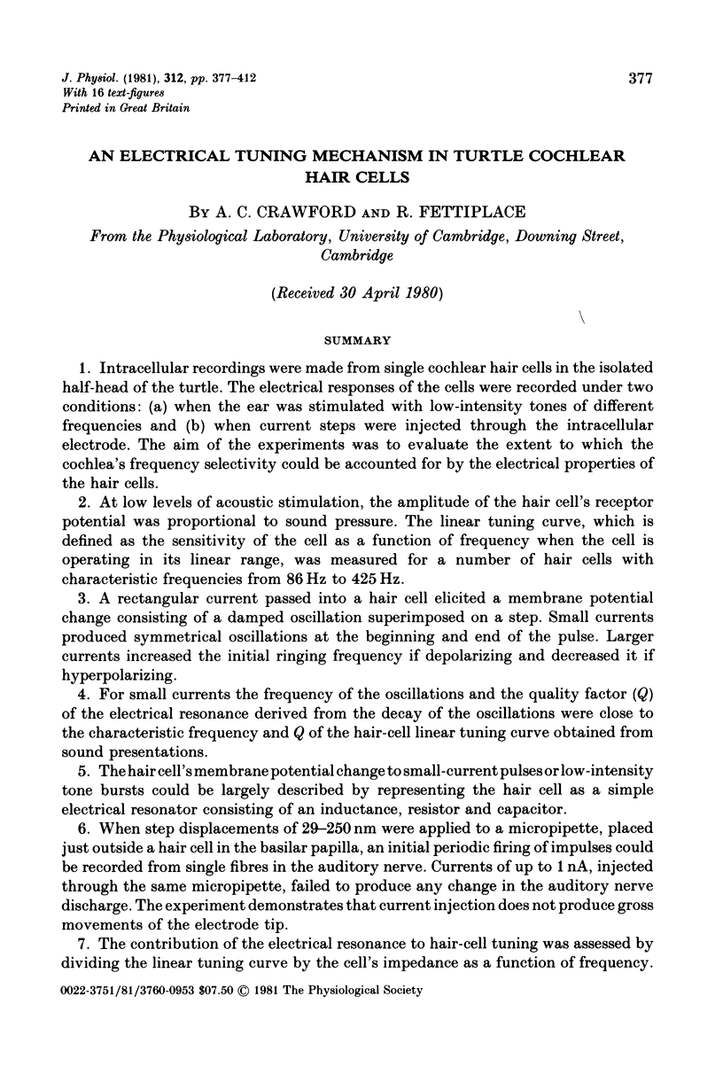
1. Intracellular recordings were made from single cochlear hair cells in the isolated half-head of the turtle. The electrical responses of the cells were recorded under two conditions: (a) when the ear was stimulated with low-intensity tones of different frequencies and (b) when current steps were injected through the intracellular electrode. The aim of the experiments was to evaluate the extent to which the cochlea's frequency selectivity could be accounted for by the electrical properties of the hair cells.
2. At low levels of acoustic stimulation, the amplitude of the hair cell's receptor potential was proportional to sound pressure. The linear tuning curve, which is defined as the sensitivity of the cell as a function of frequency when the cell is operating in its linear range, was measured for a number of hair cells with characteristic frequencies from 86 Hz to 425 Hz.
3. A rectangular current passed into a hair cell elicited a membrane potential change consisting of a damped oscillation superimposed on a step. Small currents produced symmetrical oscillations at the beginning and end of the pulse. Larger currents increased the initial ringing frequency if depolarizing and decreased it if hyperpolarizing.
4. For small currents the frequency of the oscillations and the quality factor (Q) of the electrical resonance derived from the decay of the oscillations were close to the characteristic frequency and Q of the hair-cell linear tuning curve obtained from sound presentations.
5. The hair cell's membrane potential change to small-current pulses or low-intensity tone bursts could be largely described by representing the hair cell as a simple electrical resonator consisting of an inductance, resistor and capacitor.
6. When step displacements of 29-250 nm were applied to a micropipette, placed just outside a hair cell in the basilar papilla, an initial periodic firing of impulses could be recorded from single fibres in the auditory nerve. Currents of up to 1 nA, injected through the same micropipette, failed to produce any change in the auditory nerve discharge. The experiment demonstrates that current injection does not produce gross movements of the electrode tip.
7. The contribution of the electrical resonance to hair-cell tuning was assessed by dividing the linear tuning curve by the cell's impedance as a function of frequency. The procedure assumes that the electrical resonance is independent of other filtering stages, and on this assumption the resonance can account for the tip of the acoustical tuning curve.
8. The residual filter produced by the division was broad; it exhibited a high-frequency roll-off with a corner frequency at 500-600 Hz, similar in all cells, and a low-frequency roll-off, with a corner frequency from 30 to 350 Hz which varied from cell to cell but was uncorrelated with the characteristic frequency of the cell.
9. The phase of the receptor potential relative to the sound pressure at the tympanum was measured in ten cells. For low intensities the phase characteristic was independent of the sound pressure. At low frequencies the receptor potential led the sound by 270-360°, and in the region of the characteristic frequency there was an abrupt phase lag of 90-180°; the abruptness of the phase change depended upon the Q of the cell.
10. The calculated phase shift of the electrical resonator as a function of frequency was subtracted from the phase characteristic of the receptor potential. The subtraction removed the sharp phase transition around the characteristic frequency, and in this frequency region the residual phase after subtraction was approximately constant at +180°. This is consistent with the idea that the hair cells depolarize in response to displacements of the basilar membrane towards the scala vestibuli. The high-frequency region of the residual phase characteristic was similar in all cells.
11. It is concluded that each hair cell contains its own electrical resonance mechanism which accounts for most of the frequency selectivity of the receptor potential. All cells also show evidence of a broad band-pass filter, the high frequency portion of which may be produced by the action of the middle ear.

Selected References
These references are in PubMed. This may not be the complete list of references from this article.
- Colburn T. R., Schwartz E. A. Linear voltage control of current passed through a micropipette with variable resistance. Med Biol Eng. 1972 Jul;10(4):504–509. doi: 10.1007/BF02474198. [DOI] [PubMed] [Google Scholar]
- Conti F. Nerve membrane electrical characteristics near the resting state. Biophysik. 1970;6(3):257–270. doi: 10.1007/BF01189086. [DOI] [PubMed] [Google Scholar]
- Crawford A. C., Fettiplace R. Ringing responses in cochlear hair cells of the turtle [proceedings]. J Physiol. 1978 Nov;284:120P–122P. [PubMed] [Google Scholar]
- Crawford A. C., Fettiplace R. The frequency selectivity of auditory nerve fibres and hair cells in the cochlea of the turtle. J Physiol. 1980 Sep;306:79–125. doi: 10.1113/jphysiol.1980.sp013387. [DOI] [PMC free article] [PubMed] [Google Scholar]
- Detwiler P. B., Hodgkin A. L., McNaughton P. A. Temporal and spatial characteristics of the voltage response of rods in the retina of the snapping turtle. J Physiol. 1980 Mar;300:213–250. doi: 10.1113/jphysiol.1980.sp013159. [DOI] [PMC free article] [PubMed] [Google Scholar]
- Evans E. F. The frequency response and other properties of single fibres in the guinea-pig cochlear nerve. J Physiol. 1972 Oct;226(1):263–287. doi: 10.1113/jphysiol.1972.sp009984. [DOI] [PMC free article] [PubMed] [Google Scholar]
- Fein H. Passing current through recording glass micro-pipette electrodes. IEEE Trans Biomed Eng. 1966 Oct;13(4):211–212. [PubMed] [Google Scholar]
- Fettiplace R., Crawford A. C. The coding of sound pressure and frequency in cochlear hair cells of the terrapin. Proc R Soc Lond B Biol Sci. 1978 Dec 4;203(1151):209–218. doi: 10.1098/rspb.1978.0101. [DOI] [PubMed] [Google Scholar]
- Fettiplace R., Crawford A. C. The origin of tuning in turtle cochlear hair cells. Hear Res. 1980 Jun;2(3-4):447–454. doi: 10.1016/0378-5955(80)90081-7. [DOI] [PubMed] [Google Scholar]
- Geisler C. D., Rhode W. S., Kennedy D. T. Responses to tonal stimuli of single auditory nerve fibers and their relationship to basilar membrane motion in the squirrel monkey. J Neurophysiol. 1974 Nov;37(6):1156–1172. doi: 10.1152/jn.1974.37.6.1156. [DOI] [PubMed] [Google Scholar]
- HODGKIN A. L., HUXLEY A. F. A quantitative description of membrane current and its application to conduction and excitation in nerve. J Physiol. 1952 Aug;117(4):500–544. doi: 10.1113/jphysiol.1952.sp004764. [DOI] [PMC free article] [PubMed] [Google Scholar]
- HUXLEY A. F. Ion movements during nerve activity. Ann N Y Acad Sci. 1959 Aug 28;81:221–246. doi: 10.1111/j.1749-6632.1959.tb49311.x. [DOI] [PubMed] [Google Scholar]
- Manley G. A. Some aspects of the evolution of hearing in vertebrates. Nature. 1971 Apr 23;230(5295):506–509. doi: 10.1038/230506a0. [DOI] [PubMed] [Google Scholar]
- Mauro A., Conti F., Dodge F., Schor R. Subthreshold behavior and phenomenological impedance of the squid giant axon. J Gen Physiol. 1970 Apr;55(4):497–523. doi: 10.1085/jgp.55.4.497. [DOI] [PMC free article] [PubMed] [Google Scholar]
- Miller M. R. Further scanning electron microscope studies of lizard auditory papillae. J Morphol. 1978 Jun;156(3):381–417. doi: 10.1002/jmor.1051560305. [DOI] [PubMed] [Google Scholar]
- Miller M. R. Scanning electron microscope studies of the papilla basilaris of some turtles and snakes. Am J Anat. 1978 Mar;151(3):409–435. doi: 10.1002/aja.1001510306. [DOI] [PubMed] [Google Scholar]
- Pfeiffer R. R. A model for two-tone inhibition of single cochlear-nerve fibers. J Acoust Soc Am. 1970 Dec;48(6 Suppl):1373+–1373+. doi: 10.1121/1.1912294. [DOI] [PubMed] [Google Scholar]
- Weiss T. F., Mulroy M. J., Turner R. G., Pike C. L. Tuning of single fibers in the cochlear nerve of the alligator lizard: relation to receptor morphology. Brain Res. 1976 Oct 8;115(1):71–90. doi: 10.1016/0006-8993(76)90823-4. [DOI] [PubMed] [Google Scholar]