In Vitro Studies on Rickettsia-Host Cell Interactions: Intracellular Growth Cycle of Virulent and Attenuated Rickettsia prowazeki in Chicken Embryo Cells in Slide Chamber Cultures (original) (raw)
Abstract
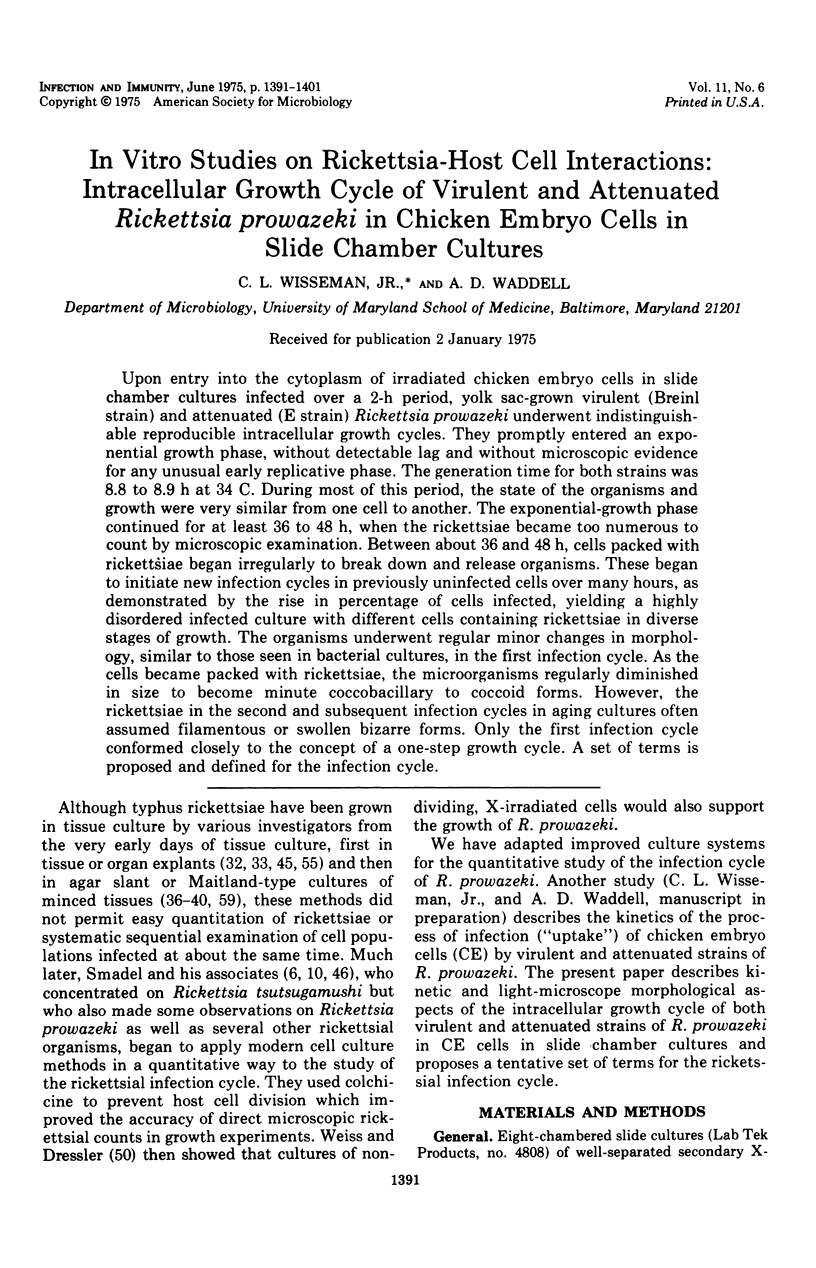
Upon entry into the cytoplasm of irradiated chicken embryo cells in slide chamber cultures infected over a 2-h period, yolk sac-grown virulent (Breinl strain) and attenuated (E strain) Rickettsia prowazeki underwent indistinguishable reproducible intracellular growth cycles. They promptly entered an exponential growth phase, without detectable lag and without microscopic evidence for any unusual early replicative phase. The generation time for both strains was 8.8 to 8.9 h at 34 C. During most of this period, the state of the organisms and growth were very similar from one cell to another. The exponential-growth phase continued for at least 36 to 48 h, when the rickettsiae became too numerous to count by microscopic examination. Between about 36 and 48 h, cells packed with rickettsiae began irregularly to break down and release organisms. These began to initiate new infection cycles in previously uninfected cells over many hours, as demonstrated by the rise in percentage of cells infected, yielding a highly disordered infected culture with different cells containing rickettsiae in diverse stages of growth. The organisms underwent regular minor changes in morphology, similar to those seen in bacterial cultures, in the first infection cycle. As the cells became packed with rickettsiae, the microorganisms regularly diminished in size to become minute coccobacillary to coccoid forms. However, the rickettsiae in the second and subsequent infection cycles in aging cultures often assumed filamentous or swollen bizarre forms. Only the first infection cycle conformed closely to the concept of a one-step growth cycle. A set of terms is proposed and defined for the infection cycle.

Images in this article
Selected References
These references are in PubMed. This may not be the complete list of references from this article.
- Anderson D. R., Hopps H. E., Barile M. F., Bernheim B. C. Comparison of the ultrastructure of several rickettsiae, ornithosis virus, and Mycoplasma in tissue culture. J Bacteriol. 1965 Nov;90(5):1387–1404. doi: 10.1128/jb.90.5.1387-1404.1965. [DOI] [PMC free article] [PubMed] [Google Scholar]
- BOZEMAN F. M., HOPPS H. E., DANAUSKAS J. X., JACKSON E. B., SMADEL J. E. Study on the growth of Rickettsiae. I. A tissue culture system for quantitative estimations of Rickettsia tsutsugamushi. J Immunol. 1956 Jun;76(6):475–488. [PubMed] [Google Scholar]
- Boese J. L., Wisseman C. L., Jr, Walsh W. T., Fiset P. Antibody and antibiotic action on Rickettsia prowazeki in body lice across the host-vector interface, with observations on strain virulence and retrieval mechanisms. Am J Epidemiol. 1973 Oct;98(4):262–282. doi: 10.1093/oxfordjournals.aje.a121556. [DOI] [PubMed] [Google Scholar]
- Burgdorfer W., Ormsbee R. A. Development of Rickettsia prowazeki in certain species of ixodid ticks. Acta Virol. 1968 Jan;12(1):36–40. [PubMed] [Google Scholar]
- COHN Z. A., BOZEMAN F. M., CAMPBELL J. M., HUMPHRIES J. W., SAWYER T. K. Study on growth of Rickettsia. V. Penetration of Rickettsia tsutsugamushi into mammalian cells in vitro. J Exp Med. 1959 Mar 1;109(3):271–292. doi: 10.1084/jem.109.3.271. [DOI] [PMC free article] [PubMed] [Google Scholar]
- GIMENEZ D. F. STAINING RICKETTSIAE IN YOLK-SAC CULTURES. Stain Technol. 1964 May;39:135–140. doi: 10.3109/10520296409061219. [DOI] [PubMed] [Google Scholar]
- GIROUD P., WEI W. P. Contribution à l'étude morphologique des corps homogènes constatés dans le poumon de lapin infecté de typhus epidémiqué. C R Seances Soc Biol Fil. 1950 Jun;144(11-12):794–795. [PubMed] [Google Scholar]
- Gambrill M. R., Wisseman C. L., Jr Mechanisms of immunity in typhus infections. I. Multiplication of typhus rickettsiae in human macrophage cell cultures in the nonimmune system: influence of virulence of rickettsial strains and of chloramphenicol. Infect Immun. 1973 Oct;8(4):519–527. doi: 10.1128/iai.8.4.519-527.1973. [DOI] [PMC free article] [PubMed] [Google Scholar]
- Golinevich E. M. Eksperimental'noe obosnovanie nalichiia infraform u rikketsii provacheka. Vestn Akad Med Nauk SSSR. 1969;24(10):56–63. [PubMed] [Google Scholar]
- Jadin J., Creemers J., Jadin J. M., Giroud P. Ultrastructure of Rickettsia prowazeki. Acta Virol. 1968 Jan;12(1):7–10. [PubMed] [Google Scholar]
- KORDOVA N., REHACEK J. MICROSCOPIC EXAMINATION OF THE ORGANS OF TICKS INFECTED WITH RICKETTSIA PROWAZEKI. Acta Virol. 1964 Sep;8:465–469. [PubMed] [Google Scholar]
- Kokorin I. N. Biological peculiarities of the development of rickettsiae. Acta Virol. 1968 Jan;12(1):31–35. [PubMed] [Google Scholar]
- Kordová N. Die Vermehrung der Rickettsia prowazeki in L-Zellen. I. Lichtmikroskopische Untersuchungen. Arch Gesamte Virusforsch. 1965;15(5):697–706. [PubMed] [Google Scholar]
- Kordová N., Kovácová E. Histochemical and fluorescent antibody studies on the early stages of infection of L cells with Coxiella burneti. Acta Virol. 1968 Jan;12(1):23–30. [PubMed] [Google Scholar]
- Kordová N., Kovácová E. Replication of Rickettsia prowazeki in L-cells as revealed by immunofluorescence. Acta Virol. 1967 May;11(3):252–255. [PubMed] [Google Scholar]
- Kordová N., Rosenberg M., Mrena E. Die Vermehrung der Rickettsia prowazeki in L-Zellen. II. Elektronenmikroskopische Untersuchungen an infizierten Gewebezellen in Dünnschnitten. Arch Gesamte Virusforsch. 1965;15(5):707–720. [PubMed] [Google Scholar]
- Pospísil V. F., Brezina R. Concerning the question of infraforms in Rickettsia prowazeki. Acta Virol. 1972 Jun;16(1):87–87. [PubMed] [Google Scholar]
- Rehácek J., Brezina R., Majerská M. Multiplication of rickettsiae in tick cells in vitro. Acta Virol. 1968 Jan;12(1):41–43. [PubMed] [Google Scholar]
- SCHAECHTER M., BOZEMAN F. M., SMADEL J. E. Study on the growth of Rickettsiae. II. Morphologic observations of living Rickettsiae in tissue culture cells. Virology. 1957 Feb;3(1):160–172. doi: 10.1016/0042-6822(57)90030-2. [DOI] [PubMed] [Google Scholar]
- WEISS E., DRESSLER H. R. Growth of Rickettsia prowazeki in irradiated monolayer cultures of chick embryo entodermal cells. J Bacteriol. 1958 May;75(5):544–552. doi: 10.1128/jb.75.5.544-552.1958. [DOI] [PMC free article] [PubMed] [Google Scholar]
- WEYER F. Explantationsversuche bei Läusen in Verbindung mit der Kultur von Rickettsien. Zentralbl Bakteriol Parasitenkd Infektionskr Hyg. 1952 Dec 20;159(1-2):13–22. [PubMed] [Google Scholar]
- Weiss E., Newman L. W., Grays R., Green A. E. Metabolism of Rickettsia typhi and Rickettsia akari in irradiated L cells. Infect Immun. 1972 Jul;6(1):50–57. doi: 10.1128/iai.6.1.50-57.1972. [DOI] [PMC free article] [PubMed] [Google Scholar]
- Wisseman C. L., Jr, Waddell A. D., Walsh W. T. In vitro studies of the action of antibiotics on Rickettsia prowazeki by two basic methods of cell culture. J Infect Dis. 1974 Dec;130(6):564–574. doi: 10.1093/infdis/130.6.564. [DOI] [PubMed] [Google Scholar]
- Wisseman C. L., Jr, Waddell A. D., Walsh W. T. Mechanisms of immunity in typhus infections. IV. Failure of chicken embryo cells in culture to restrict growth of antibody-sensitized Rickettsia prowazeki. Infect Immun. 1974 Mar;9(3):571–575. doi: 10.1128/iai.9.3.571-575.1974. [DOI] [PMC free article] [PubMed] [Google Scholar]