Distinct human immunodeficiency virus strains in the bone marrow are associated with the development of thrombocytopenia - PubMed (original) (raw)
Distinct human immunodeficiency virus strains in the bone marrow are associated with the development of thrombocytopenia
F Voulgaropoulou et al. J Virol. 1999 Apr.
Abstract
We analyzed bone marrow and blood from human immunodeficiency virus type 1 (HIV-1)-infected individuals and described the HIV-1 quasispecies in these cellular compartments. HIV-1 isolates from the bone marrow of thrombocytopenic patients contained distinct amino acids in the V3 loop and infected T-cell lines, implicating this virus in the development of thrombocytopenia.
Figures
FIG. 1
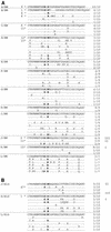
Alignment of V3 loop amino acid sequences of HIV-1 from bone marrow (A) and blood (B). The most prominent sequence for each patient is shown, using the one-letter amino acid code. The ratio to the right indicates the frequency of the sequence in each patient’s bone marrow or blood sample. Identical amino acids and deletions are represented by dots and dashes, respectively. Amino acid insertions are shown where they occur, but dashes are introduced into the rest of the sequences to optimize alignment. Boldfaced amino acids designate positions 11 and 13 of the V3 loop. Asterisks indicate the V3 loop sequences used to construct HIV-1 molecular clones, and the numbers next to them indicate the corresponding molecular clones. Roman numerals I, II, and III designate amino acid sequences that are identical in the bone marrow and blood of two of the patients.
FIG. 2
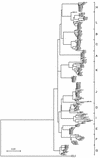
Rooted phylogenetic tree showing the V3 loop sequences of HIV-1 from the bone marrow of 12 patients and the blood of 3 of the patients. The scale bar indicates 5% divergence, at the nucleotide level, between any two sequences. BM, bone marrow; Bld, blood.
FIG. 3
The V3 loop consensus sequences from bone marrow HIV-1 sequences of patients with and without TP. Subscripts designate the frequency of each amino acid for every position in the V3 loop. The TP consensus was compiled from the 30 amino acid sequences of patients A to C. The non-TP consensus was compiled from the 83 amino acid sequences of patients D to L.
FIG. 4
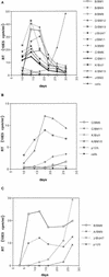
Replication kinetics of HIV-1 isolates in activated PBMC (A), primary macrophages (B), and MT-2 cells (C). p120 and p125 are T-cell-tropic and macrophage-tropic HIV-1 molecular clones, respectively. Activated PBMC and primary macrophages correspond to donor 2 of Table 2. Supernatants were assayed for reverse transcriptase (RT) activity (expressed as counts per minute per milliliter of supernatant) every 4 days. Each point represents the average of three independent measurements.
Similar articles
- Productive infection of CD34+-cell-derived megakaryocytes by X4 and R5 HIV-1 isolates.
Voulgaropoulou F, Pontow SE, Ratner L. Voulgaropoulou F, et al. Virology. 2000 Mar 30;269(1):78-85. doi: 10.1006/viro.2000.0193. Virology. 2000. PMID: 10725200 - Relationship between the V3 loop and the phenotypes of human immunodeficiency virus type 1 (HIV-1) isolates from children perinatally infected with HIV-1.
Mammano F, Salvatori F, Ometto L, Panozzo M, Chieco-Bianchi L, De Rossi A. Mammano F, et al. J Virol. 1995 Jan;69(1):82-92. doi: 10.1128/JVI.69.1.82-92.1995. J Virol. 1995. PMID: 7527089 Free PMC article. - Evidence of subtype B-like sequences in the V3 loop region of human immunodeficiency virus type 1 in Kilimanjaro, Tanzania.
Kiwelu IE, Nakkestad HL, Shao J, Sommerfelt MA. Kiwelu IE, et al. AIDS Res Hum Retroviruses. 2000 Aug 10;16(12):1191-5. doi: 10.1089/088922200415054. AIDS Res Hum Retroviruses. 2000. PMID: 10954896 - Inhibition of human immunodeficiency virus type 1 infection and syncytium formation in human cells by V3 loop synthetic peptides from gp120.
Nehete PN, Arlinghaus RB, Sastry KJ. Nehete PN, et al. J Virol. 1993 Nov;67(11):6841-6. doi: 10.1128/JVI.67.11.6841-6846.1993. J Virol. 1993. PMID: 7692087 Free PMC article. - T-tropic human immunodeficiency virus type 1 (HIV-1)-derived V3 loop peptides directly bind to CXCR-4 and inhibit T-tropic HIV-1 infection.
Sakaida H, Hori T, Yonezawa A, Sato A, Isaka Y, Yoshie O, Hattori T, Uchiyama T. Sakaida H, et al. J Virol. 1998 Dec;72(12):9763-70. doi: 10.1128/JVI.72.12.9763-9770.1998. J Virol. 1998. PMID: 9811711 Free PMC article.
Cited by
- Compartmentalization of the gut viral reservoir in HIV-1 infected patients.
van Marle G, Gill MJ, Kolodka D, McManus L, Grant T, Church DL. van Marle G, et al. Retrovirology. 2007 Dec 4;4:87. doi: 10.1186/1742-4690-4-87. Retrovirology. 2007. PMID: 18053211 Free PMC article. - Compartmentalization of human immunodeficiency virus type 1 between blood monocytes and CD4+ T cells during infection.
Fulcher JA, Hwangbo Y, Zioni R, Nickle D, Lin X, Heath L, Mullins JI, Corey L, Zhu T. Fulcher JA, et al. J Virol. 2004 Aug;78(15):7883-93. doi: 10.1128/JVI.78.15.7883-7893.2004. J Virol. 2004. PMID: 15254161 Free PMC article. - Human immunodeficiency virus type 1 genetic diversity in the nervous system: evolutionary epiphenomenon or disease determinant?
van Marle G, Power C. van Marle G, et al. J Neurovirol. 2005 Apr;11(2):107-28. doi: 10.1080/13550280590922838. J Neurovirol. 2005. PMID: 16036790 Review. - HIV-1 determinants of thrombocytopenia at the stage of CD34+ progenitor cell differentiation in vivo lie in the viral envelope gp120 V3 loop region.
Zhang M, Evans S, Yuan J, Ratner L, Koka PS. Zhang M, et al. Virology. 2010 Jun 5;401(2):131-6. doi: 10.1016/j.virol.2010.03.005. Epub 2010 Mar 24. Virology. 2010. PMID: 20338611 Free PMC article. - Higher levels of Zidovudine resistant HIV in the colon compared to blood and other gastrointestinal compartments in HIV infection.
van Marle G, Church DL, Nunweiler KD, Cannon K, Wainberg MA, Gill MJ. van Marle G, et al. Retrovirology. 2010 Sep 13;7:74. doi: 10.1186/1742-4690-7-74. Retrovirology. 2010. PMID: 20836880 Free PMC article.
References
- Bahner I, Kearns K, Coutinho S, Leonard E H, Kohn D B. Infection of human marrow stroma by human immunodeficiency virus-1 (HIV-1) is both required and sufficient for HIV-1-induced hematopoietic suppression in vitro: demonstration by gene modification of primary human stroma. Blood. 1997;90:1787–1798. - PubMed
- Ballem P J, Belzberg A, Devine D V, Lyster D, Spruston B, Chambers H, Doubroff P, Mikulash K. Kinetic studies of the mechanism of thrombocytopenia in patients with human immunodeficiency virus infection. N Engl J Med. 1992;327:1779–1784. . (Comments, 327:1812–1813, 1992, and 328:1785–1786, 1993.) - PubMed
- Boyar A, Beall G. HIV-seropositive thrombocytopenia: the action of zidovudine. AIDS. 1991;5:1351–1356. - PubMed
- Carrillo A, Trowbridge D B, Westervelt P, Ratner L. Identification of HIV-1 determinants for T lymphoid cell line infection. Virology. 1993;197:817–824. - PubMed
Publication types
MeSH terms
Substances
LinkOut - more resources
Full Text Sources
Medical
Molecular Biology Databases


