Wnt3a-/--like phenotype and limb deficiency in Lef1(-/-)Tcf1(-/-) mice - PubMed (original) (raw)
Wnt3a-/--like phenotype and limb deficiency in Lef1(-/-)Tcf1(-/-) mice
J Galceran et al. Genes Dev. 1999.
Abstract
Members of the LEF-1/TCF family of transcription factors have been implicated in the transduction of Wnt signals. However, targeted gene inactivations of Lef1, Tcf1, or Tcf4 in the mouse do not produce phenotypes that mimic any known Wnt mutation. Here we show that null mutations in both Lef1 and Tcf1, which are expressed in an overlapping pattern in the early mouse embryo, cause a severe defect in the differentiation of paraxial mesoderm and lead to the formation of additional neural tubes, phenotypes identical to those reported for Wnt3a-deficient mice. In addition, Lef1(-/-)Tcf1(-/-) embryos have defects in the formation of the placenta and in the development of limb buds, which fail both to express Fgf8 and to form an apical ectodermal ridge. Together, these data provide evidence for a redundant role of LEF-1 and TCF-1 in Wnt signaling during mouse development.
Figures
Figure 1
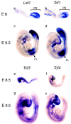
Expression of members of the LEF-1/TCF family of transcription factors in early mouse development. Expression was analyzed at embryonic day 8.5 (E8.5) and E9.5 by whole mount in situ hybridization with cDNA probes for Lef1 (a,c),Tcf1 (b,d), Tcf3 (e,g), and Tcf4(f,h). In E8.5 embryos, expression of _Lef1_and Tcf1 is detected predominantly in the primitive streak (PS) and in E9.5 embryos expression is detected in the primitive streak and unsegmented presomitic mesoderm, the forelimb bud (FL) and the branchial arches. Tcf3 expression is detected in newly formed somites and in the primordia of the fore-, mid-, and hindbrain. Expression of Tcf4 at E9.0 (f) is detected in the midbrain, around the optic placode, in the P2 region of the diencephalon, and the forming hindgut.
Figure 2
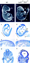
Caudal defects in embryos carrying targeted null mutations in both Lef1 and Tcf1 genes. (a,b) Scanning electron microscopy (SEM) of E9.5 wild-type (a) and_Lef1−/−Tcf1−/−_(b) littermates. In the mutant embryo, somites can be detected up to the forelimb (FL), but not in the caudal region. The caudal extremity of the tail (T) shows an abnormal morphology, is deformed and comparably smaller than the wild type. The mutant embryo also has a smaller telencephalic vesicle (TE) and a less pronounced isthmus (I) between midbrain and the hindbrain. The mass of cells labeled A corresponds to the remnants of the allantois, which is not fused to the placenta (data not shown). Histology of sagital sections of E9.5 wild-type (wt; c,e) and_Lef1−/−Tcf1−/−_(d,f) embryos. Somites and a well-developed heart (H) can be seen in the wild-type embryo. In the mutant embryo, no identifiable somites can be detected in the disformed caudal half of the embryo, which contains multiple neural tube-like structures (NT). The mutant embryo has, however, a heart (d). In the region anterior to the forelimb level (bracket in c,d) somites can be detected in both wild-type and mutant embryos (e,f). However, somites in the mutant embryo have incomplete structure and lack clear segmental boundaries. (g,h) Transverse sections of E9.5 wild-type and mutant embryos at a caudal level at which normally the first somites form. In the mutant embryo (h) three neural tubes are detected. Normal lateral mesoderm (LM) and visceral mesoderm (VM) are formed in the mutant embryo. (Arrow) Position of the notochord. (a–f) Rostral is to the left and caudal to the_right_.
Figure 3
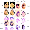
Expression of mesoderm and neural markers in wild-type and_Lef1−/−Tcf1−/−embryos. Analysis of molecular markers by whole-mount in situ hybridization on E9.5 embryos. The sclerotome marker, Pax1, shows a regular pattern of somitic expression throughout the wild-type embryo, whereas weak expression is detected only in nine somites anterior to the forelimb level (arrowhead) in the mutant embryo (a,b). Pax3, normally expressed in the dorsal neural tube and the dermamyotome is expressed in the mutant embryo in a nonsegmental pattern caudal to the forelimb level (arrowhead; c,d). Transverse sections of the caudal region of these embryos (thin line) are shown in e and f. Pax3 expression is detected in the dermamyotome and in the dorsal neural tube of a wild-type embryo and in three neural tubes of the mutant embryo. Expression of Notch1 in the unsegmented presomitic mesoderm (PM; bracket) is observed in wild-type but not in mutant embryos (g,h). Notch1 expression is also detected in the forelimb bud of the wild-type embryo (arrowhead in_g), but it is absent in the forelimb bud of the mutant embryo (arrowhead; h). Expression of Notch1 in the neural tube is detected in transverse sections of both wild-type and mutant embryos (i,j). In the mutant embryo, one of the neural tubes is not closed. Expression of Wnt5a in the presomitic mesoderm is detected in the wild-type embryo (k) but not in the mutant embryo(l). Expression of_Wnt5a_ is also detected in the forelimb bud (arrowhead) of the wild-type, but not mutant embryo. (m–p) Pattern of expression of the dorsal CNS marker, Wnt1, in whole-mount hybridization and in transverse sections of the caudal region at the level indicated by a thin line. Wnt1 is expressed in the brain of wild-type and mutant embryos at a similar level, but expression is increased in the CNS of the mutant embryo. In the region posterior to the forelimb level of the mutant embryo, additional signals can be detected in extra bands and patches of cells (arrowheads; n) that represent an open neural tube and three additional neural tubes (p). The presomitic mesoderm marker, Tbx6, is expressed in the tailbud and presomitic mesoderm of the wild-type, but not mutant embryo (q,r).
Figure 4
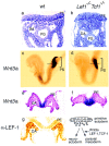
Ectopic formation of neural tissue in_Lef1−/−Tcf1−/−_embryos at the expense of paraxial mesoderm. (a,b) Histological analysis of the primitive streak region in wild-type and mutant E8.5 embryos. The cells underlying the primitive ectoderm (PE) have mesenchymal morphology in the wild-type embryos (a) and compact epithelial morphology in the mutant embryo (b). The positions of the dorsal aorta (DA) and primitive gut (PG) are shown. (c–f). Expression of Wnt3a in the primitive streak (PS) region of E8.0 wild-type (c) and mutant embryos (d). In transverse sections at a level indicated by a horizontal line, Wnt3a expression can be detected in the primitive ectoderm (PE) and in the epithelial cell mass underlying the primitive ectoderm of the mutant embryo (d). (g) Expression of Lef1 in the presomitic mesoderm. Detection of LEF-1 protein by immunohistochemistry with a polyclonal anti-LEF-1 serum in a transverse section of an E8.5 wildtype embryo in the primitive streak region. Schematic diagram of the fate of cells delaminating from the primitive ectoderm in the absence or presence of Wnt3a, and LEF-1 and TCF-1.
Figure 5
Limb bud defects in_Lef1−/− Tcf1−/−embryos. (a,b) Morphology of the forelimb field in wild-type and mutant embryos. SEM pictures of E9.5 embryos showing the cervico-thoracic region at the forelimb level. A well formed limb bud (LB) is seen in the wild-type embryo, whereas the_Lef1−/−Tcf1−/−_embryo only shows indications of a protrusion in the lateral plate mesoderm (LPM) in the forelimb bud region. The mutant embryo shows somites rostral, but not caudal to the prospective forelimb bud. The somites (S) of the mutant embryo are covered with ectoderm that resembles that covering the dorsal part of the neural tube. (c,d) Transverse sections of wild-type and mutant embryos at the level of the forelimb bud (FL) that were hybridized with_Pax3. Both wild-type and mutant embryos show migration of_Pax3_ expressing myogenic precursors into the lateral plate mesoderm. (NT) The neural tube. (e–l) Analysis of molecular markers in E9.5 wild-type and mutant embryos by whole mount in situ hybridization. Expression of Fgf8, an early marker of the AER in the developing forelimb bud is detected in a wildtype (e) but not mutant embryo (f). (arrowhead) Position of the forelimb bud in these and the other panels. (g,h) Expression of En1 is detected in the ventral region of the wild-type but not mutant forelimb bud. However,En1 expression is detected at the mid-hindbrain boundary. (i,j) Expression of Lmx1b, a marker of the dorsal mesenchyme of the emerging limb buds (Cygan et al. 1997) is detected in both the wild-type and mutant embryos. However, the region of Lmx1b expression is broadened in the mutant limb bud and the level of Lmx1b expression is lower relative to the wild type limb bud. The expression of Lmx1b at the mid-hindbrain boundary is not affected in the mutant embryo. (k,l) Transverse sections at the level of the limb bud of the embryos shown in (i and j) stained with fast neutral red.Lmx1b expression is restricted to the dorsal mesenchyme of the wild-type limb bud (i) but is extended along the entire limb margin in the mutant embryo (j).
Similar articles
- Rescue of a Wnt mutation by an activated form of LEF-1: regulation of maintenance but not initiation of Brachyury expression.
Galceran J, Hsu SC, Grosschedl R. Galceran J, et al. Proc Natl Acad Sci U S A. 2001 Jul 17;98(15):8668-73. doi: 10.1073/pnas.151258098. Epub 2001 Jul 10. Proc Natl Acad Sci U S A. 2001. PMID: 11447280 Free PMC article. - Beta-catenin-dependent Wnt signaling in apical ectodermal ridge induction and FGF8 expression in normal and limbless mutant chick limbs.
McQueeney K, Soufer R, Dealy CN. McQueeney K, et al. Dev Growth Differ. 2002 Aug;44(4):315-25. doi: 10.1046/j.1440-169x.2002.00647.x. Dev Growth Differ. 2002. PMID: 12175366 - Hindgut defects and transformation of the gastro-intestinal tract in Tcf4(-/-)/Tcf1(-/-) embryos.
Gregorieff A, Grosschedl R, Clevers H. Gregorieff A, et al. EMBO J. 2004 Apr 21;23(8):1825-33. doi: 10.1038/sj.emboj.7600191. Epub 2004 Apr 1. EMBO J. 2004. PMID: 15057272 Free PMC article. - Hippocampus formation: an intriguing collaboration.
Roelink H. Roelink H. Curr Biol. 2000 Apr 6;10(7):R279-81. doi: 10.1016/s0960-9822(00)00407-3. Curr Biol. 2000. PMID: 10753739 Review. - TCF/LEF Transcription Factors: An Update from the Internet Resources.
Hrckulak D, Kolar M, Strnad H, Korinek V. Hrckulak D, et al. Cancers (Basel). 2016 Jul 20;8(7):70. doi: 10.3390/cancers8070070. Cancers (Basel). 2016. PMID: 27447672 Free PMC article. Review.
Cited by
- Intrinsic properties of Tcf1 and Tcf4 splice variants determine cell-type-specific Wnt/β-catenin target gene expression.
Wallmen B, Schrempp M, Hecht A. Wallmen B, et al. Nucleic Acids Res. 2012 Oct;40(19):9455-69. doi: 10.1093/nar/gks690. Epub 2012 Aug 2. Nucleic Acids Res. 2012. PMID: 22859735 Free PMC article. - Stage-specific regulation of reprogramming to induced pluripotent stem cells by Wnt signaling and T cell factor proteins.
Ho R, Papp B, Hoffman JA, Merrill BJ, Plath K. Ho R, et al. Cell Rep. 2013 Jun 27;3(6):2113-26. doi: 10.1016/j.celrep.2013.05.015. Epub 2013 Jun 20. Cell Rep. 2013. PMID: 23791530 Free PMC article. - Generation of human muscle fibers and satellite-like cells from human pluripotent stem cells in vitro.
Chal J, Al Tanoury Z, Hestin M, Gobert B, Aivio S, Hick A, Cherrier T, Nesmith AP, Parker KK, Pourquié O. Chal J, et al. Nat Protoc. 2016 Oct;11(10):1833-50. doi: 10.1038/nprot.2016.110. Epub 2016 Sep 1. Nat Protoc. 2016. PMID: 27583644 - Functional interactions between Dlx2 and lymphoid enhancer factor regulate Msx2.
Diamond E, Amen M, Hu Q, Espinoza HM, Amendt BA. Diamond E, et al. Nucleic Acids Res. 2006;34(20):5951-65. doi: 10.1093/nar/gkl689. Epub 2006 Oct 26. Nucleic Acids Res. 2006. PMID: 17068080 Free PMC article. - Foxg1 confines Cajal-Retzius neuronogenesis and hippocampal morphogenesis to the dorsomedial pallium.
Muzio L, Mallamaci A. Muzio L, et al. J Neurosci. 2005 Apr 27;25(17):4435-41. doi: 10.1523/JNEUROSCI.4804-04.2005. J Neurosci. 2005. PMID: 15858069 Free PMC article.
References
- Behrens J, von Kries JP, Kühl M, Bruhn L, Weklich D, Grosschedl R, Birchmeier W. Functional interaction of β-catenin with the transcription factor LEF-1. Nature. 1996;382:638–642. - PubMed
- Bruhn L, Munnerlyn A, Grosschedl R. ALY, a context-dependent coactivator of LEF-1 and AML-1, is required for TCRα enhancer function. Genes & Dev. 1997;11:640–653. - PubMed
- Brunner E, Peter O, Schweizer L, Basler K. Pangolin encodes a Lef-1 homologue that acts downstream of Armadillo to transduce the wingless signal in Drosophila. Nature. 1997;385:829–833. - PubMed
- Cadigan KM, Nusse R. Wnt signaling: A common theme in animal development. Genes & Dev. 1997;11:3286–3305. - PubMed
Publication types
MeSH terms
Substances
LinkOut - more resources
Full Text Sources
Other Literature Sources
Molecular Biology Databases


