Cyclopentenone prostaglandins suppress activation of microglia: down-regulation of inducible nitric-oxide synthase by 15-deoxy-Delta12,14-prostaglandin J2 - PubMed (original) (raw)
Cyclopentenone prostaglandins suppress activation of microglia: down-regulation of inducible nitric-oxide synthase by 15-deoxy-Delta12,14-prostaglandin J2
T V Petrova et al. Proc Natl Acad Sci U S A. 1999.
Abstract
Mechanisms leading to down-regulation of activated microglia and astrocytes are poorly understood, in spite of the potentially detrimental role of activated glia in neurodegeneration. Prostaglandins, produced both by neurons and glia, may serve as mediators of glial and neuronal functions. We examined the influence of cyclopentenone prostaglandins and their precursors on activated glia. As models of glial activation, production of inducible nitric-oxide synthase (iNOS) was studied in lipopolysaccharide-stimulated rat microglia, a murine microglial cell line BV-2, and IL-1beta-stimulated rat astrocytes. Cyclopentenone prostaglandins were potent inhibitors of iNOS induction and were more effective than their precursors, prostaglandins E2 and D2. 15-Deoxy-Delta12,14-prostaglandin J2 (15d-PGJ2) was the most potent prostaglandin among those tested. In activated microglia, 15d-PGJ2 suppressed iNOS promoter activity, iNOS mRNA, and protein levels. The action of 15d-PGJ2 does not appear to involve its nuclear receptor peroxisome proliferator-activated receptor gamma (PPARgamma) because troglitazone, a specific ligand of PPARgamma, was unable to inhibit iNOS induction, and neither troglitazone nor 15d-PGJ2 could stimulate the activity of a PPAR-dependent promoter in the absence of cotransfected PPARgamma. 15d-PGJ2 did not block nuclear translocation or DNA-binding activity of the transcription factor NFkappaB, but it did inhibit the activity of an NFkappaB reporter construct, suggesting that the mechanism of suppression of microglial iNOS by 15d-PGJ2 may involve interference with NFkappaB transcriptional activity in the nucleus. Thus, our data suggest the existence of a novel pathway mediated by cyclopentenone prostaglandins, which may represent part of a feedback mechanism leading to the cessation of inflammatory glial responses in the brain.
Figures
Figure 1
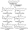
Structures and relationships among the prostaglandins. PGE2 and PGD2, major prostaglandin products of activated microglia, are produced enzymatically from arachidonic acid by the action of cyclooxygenase-2 and corresponding synthases, whereas cyclopentenone prostaglandins PGA2, 15d-PGA2, PGJ2, and 15d-PGJ2 are the products of nonenzymatic conversion of the precursor prostaglandins.
Figure 2
Influence of prostaglandins on nitrite production in the murine microglial cell line BV-2 (A and B), rat microglial cells (C), and rat astrocytes (D). Cells pretreated with the indicated concentrations of prostaglandins were stimulated with 0.4 ng/ml of LPS (microglia), 80 ng/ml of LPS (BV-2 and astrocytes), or 100 ng/ml of IL-1β (astrocytes). Control cultures were treated with diluent alone (C) or prostaglandins alone. Conditioned medium was collected at 12 h (microglia), 24 h (BV-2), or 48 h (astrocytes), and nitrite level was determined. Results are the mean ± SEM of n = 3 (BV-2) and n = 4 (microglia) independent experiments. For astrocytes, results of one of three representative experiments are shown. For BV-2 experiments, inhibition is expressed as percentage of control values, where the control is the nitrite level in cells stimulated with LPS alone.
Figure 3
15d-PGJ2 is not toxic to microglia. (A) MTT reduction assay. BV-2 cells were treated with 1 μM 15d-PGJ2, 80 ng/ml of LPS, 15d-PGJ2 + LPS, or the control buffer for 24 h, then the MTT assay was performed. Results are mean ± SEM of n = 3 independent experiments, each of which was done in triplicate. Asterisk indicates significantly different from other samples (P < 0.05). Statistics here and throughout have been calculated by using Student’s t test with significance determined as P < 0.05. (B) Phase-contrast images of BV-2 cells after treatment with LPS or LPS + 0.5 μM 15d-PGJ2 for 24 h. (Bar = 100 μm.)
Figure 4
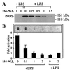
15d-PGJ2 inhibits production of iNOS protein and mRNA in LPS-stimulated BV-2 cells. (A) Cells were incubated with 80 ng/ml of LPS alone, LPS + 0.25, 0.5, 1, or 1.5 μM 15d-PGJ2, 15d-PGJ2 alone, or control buffer. Cell lysates were prepared at 9 h after treatment, and iNOS protein levels were determined by Western blotting. (B) Cells were incubated with 80 ng/ml of LPS alone, LPS + 2, 1, or 0.1 μM 15d-PGJ2, the control buffer, or 2 μM 15d-PGJ2 and total RNA isolated at 12h. One representative slot blot is shown. Levels of iNOS mRNA were normalized to glyceraldehyde-3-phosphate dehydrogenase levels and expressed as relative increase compared with the control. Values correspond to the mean ± SEM of four independent experiments.
Figure 5
15d-PGJ2 inhibits iNOS and 3xRel reporter activity. (A) Cells were transfected with murine wild-type (WT) iNOS-luc plasmid or iNOS/mutNFκB-luc plasmid, pretreated with 1 μM 15d-PGJ2 (15d) or vehicle, and stimulated with 80 ng/ml of LPS for 9 h. Results are the mean ± SEM of n = 7 (WT) and n = 5 (iNOS/mutNFκB) independent experiments, each done in triplicate. (B) Cells were transfected with 3xRel-luc plasmid, pretreated with the indicated concentrations of 15d-PGJ2 or vehicle, and stimulated with 80 ng/ml of LPS for 7 h. Results are mean ± SEM of n = 6 independent experiments. Control values in A and B are normalized to 1.0. Asterisk indicates significantly different from LPS-treated samples (P < 0.05).
Figure 6
15d-PGJ2 does not inhibit nuclear translocation or DNA-binding ability of NFκB. (A) Nuclear extracts were prepared from untreated BV-2 cells (NT) or BV-2 cells treated with the control buffer (1), 80 ng/ml of LPS (2), LPS + 1 μM 15d-PGJ2 (3), or 15d-PGJ2 alone (4) for 1 and 3 h. p65 protein levels were determined by Western blotting. (B) The same nuclear extracts as in A were analyzed for the presence of NFκB-binding activity in a gel-shift assay. Arrow indicates the position of the shifted band. Experiments were repeated three (A) and two (B) times with similar results, and data from one representative of each experiment are shown.
Figure 7
Action of 15d-PGJ2 in BV-2 cells is PPARγ-independent. (A) Cotransfection with PPARγ expression vector is necessary to observe PPRE-luc activity in troglitazone- or 15d-PGJ2-stimulated BV-2. Cells were transfected with PPRE-luc plasmid + control vector or PPARγ expression vector and treated with 5 μM troglitazone (tro), 2 μM 15d-PGJ2 (15d), or control buffer (c) for 24 h. Results are mean ± SEM of n = 7 independent experiments. Control values are normalized to 1.0. Asterisk indicates significantly different from the control (P < 0.05). (B) Troglitazone is not an effective inhibitor of LPS-stimulated nitrite production in BV-2 cells compared with 15d-PGJ2. Cells were preincubated with troglitazone or 15d-PGJ2 and then treated with 80 ng/ml of LPS for 24 h, and nitrite level in conditioned medium was determined. Results are mean ± SEM of n = 3 (15d-PGJ2) and n = 2 (troglitazone) independent experiments, with each point done in triplicate. Data are expressed as percentage of control nitrite levels, where control is the nitrite level from cells stimulated with LPS alone.
Similar articles
- Physiological and Pathological Roles of 15-Deoxy-Δ12,14-Prostaglandin J2 in the Central Nervous System and Neurological Diseases.
Yagami T, Yamamoto Y, Koma H. Yagami T, et al. Mol Neurobiol. 2018 Mar;55(3):2227-2248. doi: 10.1007/s12035-017-0435-4. Epub 2017 Mar 16. Mol Neurobiol. 2018. PMID: 28299574 Review. - Prostaglandin J2 family and the cardiovascular system.
Sasaguri T, Miwa Y. Sasaguri T, et al. Curr Vasc Pharmacol. 2004 Apr;2(2):103-14. doi: 10.2174/1570161043476384. Curr Vasc Pharmacol. 2004. PMID: 15320511 Review.
Cited by
- MicroRNAs in shaping the resolution phase of inflammation.
Naqvi RA, Gupta M, George A, Naqvi AR. Naqvi RA, et al. Semin Cell Dev Biol. 2022 Apr;124:48-62. doi: 10.1016/j.semcdb.2021.03.019. Epub 2021 Apr 29. Semin Cell Dev Biol. 2022. PMID: 33934990 Free PMC article. Review. - 15-deoxy-Delta12,14-prostaglandin J2 (15d-PGJ2) and ciglitazone modulate Staphylococcus aureus-dependent astrocyte activation primarily through a PPAR-gamma-independent pathway.
Phulwani NK, Feinstein DL, Gavrilyuk V, Akar C, Kielian T. Phulwani NK, et al. J Neurochem. 2006 Dec;99(5):1389-1402. doi: 10.1111/j.1471-4159.2006.04183.x. J Neurochem. 2006. PMID: 17074064 Free PMC article. - The organization and consequences of eicosanoid signaling.
Soberman RJ, Christmas P. Soberman RJ, et al. J Clin Invest. 2003 Apr;111(8):1107-13. doi: 10.1172/JCI18338. J Clin Invest. 2003. PMID: 12697726 Free PMC article. Review. No abstract available. - Inhibition of IkappaB kinase and IkappaB phosphorylation by 15-deoxy-Delta(12,14)-prostaglandin J(2) in activated murine macrophages.
Castrillo A, Díaz-Guerra MJ, Hortelano S, Martín-Sanz P, Boscá L. Castrillo A, et al. Mol Cell Biol. 2000 Mar;20(5):1692-8. doi: 10.1128/MCB.20.5.1692-1698.2000. Mol Cell Biol. 2000. PMID: 10669746 Free PMC article.
References
- Itagaki S, McGeer P L, Akiyama H, Zhu S, Selkoe D. J Neuroimmunol. 1989;24:173–182. - PubMed
- Stewart W F, Kawas C, Corrada M, Metter E J. Neurology. 1997;48:626–632. - PubMed
Publication types
MeSH terms
Substances
Grants and funding
- GM08061/GM/NIGMS NIH HHS/United States
- AG13939/AG/NIA NIH HHS/United States
- T32 GM008061/GM/NIGMS NIH HHS/United States
- R37 AG013939/AG/NIA NIH HHS/United States
- R01 AG013939/AG/NIA NIH HHS/United States
LinkOut - more resources
Full Text Sources
Other Literature Sources
Research Materials


