Conservation of sequence and structure flanking the mouse and human beta-globin loci: the beta-globin genes are embedded within an array of odorant receptor genes - PubMed (original) (raw)
Conservation of sequence and structure flanking the mouse and human beta-globin loci: the beta-globin genes are embedded within an array of odorant receptor genes
M Bulger et al. Proc Natl Acad Sci U S A. 1999.
Erratum in
- Proc Natl Acad Sci U S A 1999 Jul 6;96(14):8307. von Doorninck JH [corrected to van Doorninck JH]
Abstract
In mouse and human, the beta-globin genes reside in a linear array that is associated with a positive regulatory element located 5' to the genes known as the locus control region (LCR). The sequences of the mouse and human beta-globin LCRs are homologous, indicating conservation of an essential function in beta-globin gene regulation. We have sequenced regions flanking the beta-globin locus in both mouse and human and found that homology associated with the LCR is more extensive than previously known, making up a conserved block of approximately 40 kb. In addition, we have identified DNaseI-hypersensitive sites within the newly sequenced regions in both mouse and human, and these structural features also are conserved. Finally, we have found that both mouse and human beta-globin loci are embedded within an array of odorant receptor genes that are expressed in olfactory epithelium, and we also identify an olfactory receptor gene located 3' of the beta-globin locus in chicken. The data demonstrate an evolutionarily conserved genomic organization for the beta-globin locus and suggest a possible role for the beta-globin LCR in control of expression of these odorant receptor genes and/or the presence of mechanisms to separate regulatory signals in different tissues.
Figures
Figure 1
Mouse (Upper) and human (Lower) β-globin loci. The cap sites of the human ɛ-globin gene and of the mouse Ey gene are set as +1. Sequences reported here are represented by a wider horizontal bar. Globin genes are represented by filled arrows, olfactory receptor ORFs by shaded arrows, and pseudogenes by filled or shaded boxes. HSs are shown as vertical arrows. The lengths of the arrows illustrate the relative intensities of the HS that they represent; the arrow indicating mouse 5′ HS7 is lighter to indicate that this site is still hypothetical. The lower, filled lines for both mouse and human loci designate repetitive elements. Horizontal arrows pointing outward from these sequences indicate the presence of additional olfactory receptor ORFs located beyond the regions shown here. Brackets beneath the sequences show the extent of deletions within the β-globin locus that are discussed in the text.
Figure 2
PIP analyses of mouse and human sequences. (A) PIP of the region 5′ of the β-globin LCR. The mouse sequence is represented at the top, and the percent identity of gap-free aligning segments from human is plotted beneath. The odorant receptor genes MOR5′β1 and MOR5′β2 are indicated as boxes, and repetitive elements shown as triangles. The positions of HSs and the 5′ breakpoint of the human Hispanic deletion are indicated. Distance from the hypothetical Ey cap site in kb is shown on the bottom. For reference, the homologous human sequences that give rise to the alignments shown here correspond to HOR5′β1 and HOR5′β2 and surrounding regions as shown in Fig. 1. (B) PIP of the region of homology 3′ of the β-globin genes. The mouse sequence is represented at the top, with comparison made to the human 3′ HS1 sequence (24). The A/T-rich region is indicated by the shaded box.
Figure 3
DNaseI-HS mapping within the newly sequenced β-globin-flanking regions. (A) Mapping of human 5′ HS6 and 5′ HS7. A Southern blot of an _Eco_RI-digested DNase series prepared from human fetal liver was hybridized with a probe corresponding to an _Eco_RI/_Eco_0109I restriction fragment located at −23.4 to −22.8 kb. Arrows indicate DNaseI subbands evident on the autoradiogram and their corresponding positions on the human sequence represented below it. Filled regions on the diagram indicate significant (>50% identity) homology to mouse sequence, as shown in Fig. 2. (B) DNaseI-HSs located 3′ of mouse β-globin genes. A Southern blot of an _Sph_I-digested DNase series prepared from phenylhydrazine-treated mouse spleen was hybridized with a probe corresponding to an _Acc_I/_Nsi_I restriction fragment located from +69.9 to +70.5 kb.
Figure 4
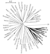
Phylogenetic tree of ORGs. An unrooted phylogenetic tree was generated by using 82 polypeptide sequences corresponding to ORGs from human, mouse, rat, dog, gibbon, chicken, catfish, zebrafish, and goldfish, along with 14 of the ORGs found proximal to the β-globin loci of human, mouse, and chicken. The genes known to be proximal to the β-globin loci are indicated by bold type. The tree was confirmed by 1,000 rounds of “bootstrapping.” Only intact, full-length sequences were used in the generation of the tree. Not all of the genes are labeled (see Materials and Methods). β2 represents the β-2-adrenergic receptor and is shown here to indicate the likely position of the root (outgroup). The bar corresponds to the graphical distance equivalent to 20% divergence.
Figure 5
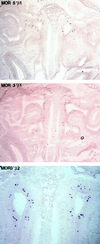
In situ hybridization of mouse olfactory epithelium by using β-globin-proximal odorant receptor genes as probes. Coronal sections through the mouse nasal cavity were hybridized with digoxigenin-labeled antisense RNA probes prepared from the odorant receptor genes indicated (also see Fig. 1). The sections shown are oriented with the dorsal region toward the top and ventral toward the bottom; the nasal septum is visible down the center of each panel.
Similar articles
- Homology of a 130-kb region enclosing the alpha-globin gene cluster, the alpha-locus controlling region, and two non-globin genes in human and mouse.
Kielman MF, Smits R, Devi TS, Fodde R, Bernini LF. Kielman MF, et al. Mamm Genome. 1993;4(6):314-23. doi: 10.1007/BF00357090. Mamm Genome. 1993. PMID: 8318735 - Comparative structural and functional analysis of the olfactory receptor genes flanking the human and mouse beta-globin gene clusters.
Bulger M, Bender MA, van Doorninck JH, Wertman B, Farrell CM, Felsenfeld G, Groudine M, Hardison R. Bulger M, et al. Proc Natl Acad Sci U S A. 2000 Dec 19;97(26):14560-5. doi: 10.1073/pnas.97.26.14560. Proc Natl Acad Sci U S A. 2000. PMID: 11121057 Free PMC article. - The complete sequences of the galago and rabbit beta-globin locus control regions: extended sequence and functional conservation outside the cores of DNase hypersensitive sites.
Slightom JL, Bock JH, Tagle DA, Gumucio DL, Goodman M, Stojanovic N, Jackson J, Miller W, Hardison R. Slightom JL, et al. Genomics. 1997 Jan 1;39(1):90-4. doi: 10.1006/geno.1996.4458. Genomics. 1997. PMID: 9027490 - Use of long sequence alignments to study the evolution and regulation of mammalian globin gene clusters.
Hardison R, Miller W. Hardison R, et al. Mol Biol Evol. 1993 Jan;10(1):73-102. doi: 10.1093/oxfordjournals.molbev.a039991. Mol Biol Evol. 1993. PMID: 8383794 Review. - Locus control regions of mammalian beta-globin gene clusters: combining phylogenetic analyses and experimental results to gain functional insights.
Hardison R, Slightom JL, Gumucio DL, Goodman M, Stojanovic N, Miller W. Hardison R, et al. Gene. 1997 Dec 31;205(1-2):73-94. doi: 10.1016/s0378-1119(97)00474-5. Gene. 1997. PMID: 9461381 Review.
Cited by
- Flanking HS-62.5 and 3' HS1, and regions upstream of the LCR, are not required for beta-globin transcription.
Bender MA, Byron R, Ragoczy T, Telling A, Bulger M, Groudine M. Bender MA, et al. Blood. 2006 Aug 15;108(4):1395-401. doi: 10.1182/blood-2006-04-014431. Epub 2006 Apr 27. Blood. 2006. PMID: 16645164 Free PMC article. - An RNA architectural locus control region involved in Dscam mutually exclusive splicing.
Wang X, Li G, Yang Y, Wang W, Zhang W, Pan H, Zhang P, Yue Y, Lin H, Liu B, Bi J, Shi F, Mao J, Meng Y, Zhan L, Jin Y. Wang X, et al. Nat Commun. 2012;3:1255. doi: 10.1038/ncomms2269. Nat Commun. 2012. PMID: 23212384 Free PMC article. - Appearance of the pituitary factor Pit-1 increases chromatin remodeling at hypersensitive site III in the human GH locus.
Yang X, Jin Y, Cattini PA. Yang X, et al. J Mol Endocrinol. 2010 Jul;45(1):19-32. doi: 10.1677/JME-10-0017. Epub 2010 Apr 15. J Mol Endocrinol. 2010. PMID: 20395397 Free PMC article. - Genome architecture of the human beta-globin locus affects developmental regulation of gene expression.
Harju S, Navas PA, Stamatoyannopoulos G, Peterson KR. Harju S, et al. Mol Cell Biol. 2005 Oct;25(20):8765-78. doi: 10.1128/MCB.25.20.8765-8778.2005. Mol Cell Biol. 2005. PMID: 16199858 Free PMC article. - Molecular basis of polycomb group protein-mediated fetal hemoglobin repression.
Qin K, Lan X, Huang P, Saari MS, Khandros E, Keller CA, Giardine B, Abdulmalik O, Shi J, Hardison RC, Blobel GA. Qin K, et al. Blood. 2023 Jun 1;141(22):2756-2770. doi: 10.1182/blood.2022019578. Blood. 2023. PMID: 36893455 Free PMC article.
References
- Hardison R, Slightom J L, Gumucio D L, Goodman M, Stojanovic N, Miller W. Gene. 1997;205:73–94. - PubMed
- Grosveld F, Dillon N, Higgs D. Baillieres Clin Haematol. 1993;6:31–55. - PubMed
- Martin D I K, Fiering S, Groudine M. Curr Opin Genet Dev. 1996;6:488–495. - PubMed
- Forrester W C, Epner E, Driscoll M C, Enver T, Brice M, Papayannopoulou T, Groudine M. Genes Dev. 1990;4:1637–1649. - PubMed
- Epner E, Reik A, Cimbora D, Telling A, Bender M A, Fiering S, Enver T, Martin D I K, Kennedy M, Keller G, Groudine M. Mol Cell. 1998;2:447–455. - PubMed
Publication types
MeSH terms
Substances
LinkOut - more resources
Full Text Sources
Other Literature Sources
Molecular Biology Databases


