Properties and plasticity of excitatory synapses on dopaminergic and GABAergic cells in the ventral tegmental area - PubMed (original) (raw)
Properties and plasticity of excitatory synapses on dopaminergic and GABAergic cells in the ventral tegmental area
A Bonci et al. J Neurosci. 1999.
Abstract
Excitatory inputs to the ventral tegmental area (VTA) influence the activity of both dopaminergic (DA) and GABAergic (GABA) cells, yet little is known about the basic properties of excitatory synapses on these two cell types. Using a midbrain slice preparation and whole-cell recording techniques, we found that excitatory synapses on DA and GABA cells display several differences. Synapses on DA cells exhibit a depression in response to repetitive activation, are minimally affected by the GABAB receptor agonist baclofen, and express NMDA receptor-dependent long-term potentiation (LTP). In contrast, synapses on GABA cells exhibit a facilitation in response to repetitive activation, are depressed significantly by baclofen, and do not express LTP. The relative contribution of NMDA and non-NMDA receptors to the synaptic currents recorded from the two cell types is the same as is the depression of synaptic transmission elicited by the application of adenosine, serotonin, or methionine enkephalin (met-enkephalin). The significant differences in the manner in which excitatory synaptic inputs to DA and GABA cells in the VTA can be modulated have potentially important implications for understanding the behavior of VTA neurons during normal behavior and during pathological states such as addiction.
Figures
Fig. 1.
Dopaminergic (DA) neurons display paired-pulse depression, whereas GABAergic (GABA) neurons show paired-pulse facilitation. A, Sample EPSCs in response to paired stimuli (50 msec interstimulus interval) for a DA (A 1) and GABA (A 2) neuron (traces are the average of 20 consecutive sweeps). B, Average of the paired-pulse ratio for DA (n = 9) and GABA (n = 12) neurons. C, Graph of the paired-pulse ratio as a function of the first EPSC amplitude. The amount of paired-pulse facilitation or depression is independent of the amplitude of the EPSC. Each symbol represents a distinct cell. D, The paired-pulse ratio for DA and GABA neurons as a function of interstimulus interval.
Fig. 2.
Repetitive stimulation produces a depression in DA neurons and a facilitation in GABA neurons. A, Sample EPSCs in response to 10 stimuli at 25 Hz for a DA (A 1) and GABA (A 2) neuron (traces are the average of 10 consecutive episodes). B, Graphs show the changes in EPSC amplitude during the 10 pulse train for DA (B 1, n = 5) and GABA neurons (B 2, n = 5).
Fig. 3.
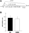
EPSCs recorded from DA and GABA neurons express a similar AMPA/NMDA ratio. A, Sample EPSCs recorded from a DA neuron under control conditions, in the presence of
d
-APV (50 μ
m
), and in the presence of
d
-APV (50 μ
m
) plus CNQX (10 μ
m
). (Each trace is an average of 20 consecutive sweeps.)B, Summary graph of the AMPA/NMDA ratio for DA (n = 7) and GABA (n = 8) neurons.
Fig. 4.
The magnitude of depression of EPSCs caused by adenosine, serotonin, and met-enkephalin is similar in DA and GABA neurons. A, Graph shows examples of the effects of adenosine (100 μ
m
) in a DA (●) and GABA (○) neuron.B, Examples of the effects of serotonin (20 μ
m
) in a DA (●) and GABA (○) neuron.C, Examples of the effects of met-enkephalin (30 μ
m
) in a DA (●) and GABA (○) neuron.D, Summary of the effects on EPSCs of adenosine, serotonin, and met-enkephalin in DA and GABA neurons (n = 3 for each column).
Fig. 5.
Baclofen depresses synaptic transmission in GABA neurons but has minimal effect on DA neurons. A,B, Examples of the effects of baclofen (1 μ
m
) on EPSCs recorded from a DA (A) and GABA (B) neuron. Baclofen caused a marked depression of EPSC amplitude in a GABA neuron while having minimal effect on the DA neuron. C, D, Dose–response curves displaying the EPSC inhibition as a function of baclofen concentration for DA (C) and GABA (D) neurons. Each point is an average of at least four cells.
Fig. 6.
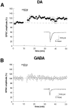
LTP can be elicited at excitatory synapses on DA neurons, but not on GABA neurons. A, B, Time course of the effects of a pairing protocol (+10 mV, 200 stimuli at 1 Hz) on EPSC amplitude for DA (A,n = 9) and GABA (B,n = 6) neurons. All recordings were made with the perforated patch-clamp recording technique.
Fig. 7.
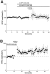
The triggering of LTP in DA cells requires activation of NMDA receptors, but not metabotropic glutamate receptors (mGluRs). A, In the presence of the NMDA receptor antagonist
d
-APV (50 μ
m
) and the mGluR antagonist MCPG (500 μ
m
), a pairing protocol fails to elicit LTP (n = 6). B, Application of MCPG (500 μ
m
) alone does not prevent LTP in DA neurons (n = 6).
Similar articles
- The endocannabinoid 2-arachidonoylglycerol inhibits long-term potentiation of glutamatergic synapses onto ventral tegmental area dopamine neurons in mice.
Kortleven C, Fasano C, Thibault D, Lacaille JC, Trudeau LE. Kortleven C, et al. Eur J Neurosci. 2011 May;33(10):1751-60. doi: 10.1111/j.1460-9568.2011.07648.x. Epub 2011 Mar 17. Eur J Neurosci. 2011. PMID: 21410793 - High-frequency afferent stimulation induces long-term potentiation of field potentials in the ventral tegmental area.
Nugent FS, Hwong AR, Udaka Y, Kauer JA. Nugent FS, et al. Neuropsychopharmacology. 2008 Jun;33(7):1704-12. doi: 10.1038/sj.npp.1301561. Epub 2007 Sep 12. Neuropsychopharmacology. 2008. PMID: 17851541 - Spike timing-dependent plasticity at GABAergic synapses in the ventral tegmental area.
Kodangattil JN, Dacher M, Authement ME, Nugent FS. Kodangattil JN, et al. J Physiol. 2013 Oct 1;591(19):4699-710. doi: 10.1113/jphysiol.2013.257873. Epub 2013 Jul 29. J Physiol. 2013. PMID: 23897235 Free PMC article. - LTP of GABAergic synapses in the ventral tegmental area and beyond.
Nugent FS, Kauer JA. Nugent FS, et al. J Physiol. 2008 Mar 15;586(6):1487-93. doi: 10.1113/jphysiol.2007.148098. Epub 2007 Dec 13. J Physiol. 2008. PMID: 18079157 Free PMC article. Review. - VTA dopamine neuron plasticity - the unusual suspects.
Xin W, Edwards N, Bonci A. Xin W, et al. Eur J Neurosci. 2016 Dec;44(12):2975-2983. doi: 10.1111/ejn.13425. Epub 2016 Nov 1. Eur J Neurosci. 2016. PMID: 27711998 Free PMC article. Review.
Cited by
- Ethanol blocks a novel form of iLTD, but not iLTP of inhibitory inputs to VTA GABA neurons.
Nufer TM, Wu BJ, Boyce Z, Steffensen SC, Edwards JG. Nufer TM, et al. Neuropsychopharmacology. 2023 Aug;48(9):1396-1408. doi: 10.1038/s41386-023-01554-y. Epub 2023 Mar 10. Neuropsychopharmacology. 2023. PMID: 36899030 Free PMC article. - Amphetamine blocks long-term synaptic depression in the ventral tegmental area.
Jones S, Kornblum JL, Kauer JA. Jones S, et al. J Neurosci. 2000 Aug 1;20(15):5575-80. doi: 10.1523/JNEUROSCI.20-15-05575.2000. J Neurosci. 2000. PMID: 10908593 Free PMC article. - CB1-Dependent Long-Term Depression in Ventral Tegmental Area GABA Neurons: A Novel Target for Marijuana.
Friend L, Weed J, Sandoval P, Nufer T, Ostlund I, Edwards JG. Friend L, et al. J Neurosci. 2017 Nov 8;37(45):10943-10954. doi: 10.1523/JNEUROSCI.0190-17.2017. Epub 2017 Oct 16. J Neurosci. 2017. PMID: 29038246 Free PMC article. - Nicotinic cholinergic synaptic mechanisms in the ventral tegmental area contribute to nicotine addiction.
Pidoplichko VI, Noguchi J, Areola OO, Liang Y, Peterson J, Zhang T, Dani JA. Pidoplichko VI, et al. Learn Mem. 2004 Jan-Feb;11(1):60-9. doi: 10.1101/lm.70004. Learn Mem. 2004. PMID: 14747518 Free PMC article. - Regulation of neuronal PLCgamma by chronic morphine.
Wolf DH, Nestler EJ, Russell DS. Wolf DH, et al. Brain Res. 2007 Jul 2;1156:9-20. doi: 10.1016/j.brainres.2007.04.059. Epub 2007 May 4. Brain Res. 2007. PMID: 17524370 Free PMC article.
References
- Bashir ZI, Bortolotto ZA, Davies CH, Berretta N, Irving AJ, Seal AJ, Henley JM, Jane DE, Watkins JC, Collingridge GL. Induction of LTP in the hippocampus needs synaptic activation of glutamate metabotropic receptors. Nature. 1993;363:347–350. - PubMed
- Bliss TVP, Collingridge GL. A synaptic model of memory: long-term potentiation in the hippocampus. Nature. 1993;361:31–39. - PubMed
- Buonomano DV, Merzenich MM. Temporal information transformed into a spatial code by a neural network with realistic properties. Science. 1995;267:1028–1030. - PubMed
Publication types
MeSH terms
Substances
LinkOut - more resources
Full Text Sources
Miscellaneous


