MRI anatomy of schizophrenia - PubMed (original) (raw)
Review
MRI anatomy of schizophrenia
R W McCarley et al. Biol Psychiatry. 1999.
Abstract
Structural magnetic resonance imaging (MRI) data have provided much evidence in support of our current view that schizophrenia is a brain disorder with altered brain structure, and consequently involving more than a simple disturbance in neurotransmission. This review surveys 118 peer-reviewed studies with control group from 1987 to May 1998. Most studies (81%) do not find abnormalities of whole brain/intracranial contents, while lateral ventricle enlargement is reported in 77%, and third ventricle enlargement in 67%. The temporal lobe was the brain parenchymal region with the most consistently documented abnormalities. Volume decreases were found in 62% of 37 studies of whole temporal lobe, and in 81% of 16 studies of the superior temporal gyrus (and in 100% with gray matter separately evaluated). Fully 77% of the 30 studies of the medial temporal lobe reported volume reduction in one or more of its constituent structures (hippocampus, amygdala, parahippocampal gyrus). Despite evidence for frontal lobe functional abnormalities, structural MRI investigations less consistently found abnormalities, with 55% describing volume reduction. It may be that frontal lobe volume changes are small, and near the threshold for MRI detection. The parietal and occipital lobes were much less studied; about half of the studies showed positive findings. Most studies of cortical gray matter (86%) found volume reductions were not diffuse, but more pronounced in certain areas. About two thirds of the studies of subcortical structures of thalamus, corpus callosum and basal ganglia (which tend to increase volume with typical neuroleptics), show positive findings, as do almost all (91%) studies of cavum septi pellucidi (CSP). Most data were consistent with a developmental model, but growing evidence was compatible also with progressive, neurodegenerative features, suggesting a "two-hit" model of schizophrenia, for which a cellular hypothesis is discussed. The relationship of clinical symptoms to MRI findings is reviewed, as is the growing evidence suggesting structural abnormalities differ in affective (bipolar) psychosis and schizophrenia.
Figures
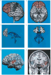
Figure 1
Illustrations, in healthy subjects, of brain Regions of Interest (ROI) frequently examined in schizophrenia. A: Left lateral view of a three–dimensional reconstruction of the prefrontal gray matter (shown in peach) in relationship to the precentral and postcentral gyri (shown in purple). The blue line within the prefrontal gray corresponds to the posterior landmark used for delineation of the white matter. B: Ventral view of the same brain as in A, and illustrates the orbitofrontal cortex. C: Left lateral view of a three–dimensional reconstruction of the prefrontal white matter (defined as in A) in relationship to the ventricles (same brain as in A). Note that the temporal horns of the lateral ventricles are mainly “virtual spaces” in this healthy subject. The open space in the area of the third ventricle is occupied by the massa intermedia, the midline thalamus. D: Posterior view of the same white matter and ventricles as in Part C. A, B, C, and D are adapted from Wible et al (1995). E: Left lateral view of a three dimensional reconstruction of the cortex, with pink indicating the anterior portion and red the posterior portion of the superior temporal gyrus (STG). F: SPGR coronal image (1.5 mm thick, 0.937×0.937 mm in plane voxels), showing the manually drawn outlines of regions of interest. This anterior–posterior location of this coronal slice is just posterior to the onset of the posterior portion of the STG in E, and is from the same subject. The regions of interest outlined are: the gray matter of the superior temporal gyrus (STG; subject left, red; subject right, green); more medially, the amygdala–hippocampal complex is shown (left, orange; right, blue) with the parahippocampal gyrus underneath (left, pink; right, purple). Adapted from Hirayasu et al (1998a).
Similar articles
- A review of MRI findings in schizophrenia.
Shenton ME, Dickey CC, Frumin M, McCarley RW. Shenton ME, et al. Schizophr Res. 2001 Apr 15;49(1-2):1-52. doi: 10.1016/s0920-9964(01)00163-3. Schizophr Res. 2001. PMID: 11343862 Free PMC article. Review. - Morphometric brain abnormalities in schizophrenia in a population-based sample: relationship to duration of illness.
Tanskanen P, Ridler K, Murray GK, Haapea M, Veijola JM, Jääskeläinen E, Miettunen J, Jones PB, Bullmore ET, Isohanni MK. Tanskanen P, et al. Schizophr Bull. 2010 Jul;36(4):766-77. doi: 10.1093/schbul/sbn141. Epub 2008 Nov 17. Schizophr Bull. 2010. PMID: 19015212 Free PMC article. - Abnormalities of the left temporal lobe and thought disorder in schizophrenia. A quantitative magnetic resonance imaging study.
Shenton ME, Kikinis R, Jolesz FA, Pollak SD, LeMay M, Wible CG, Hokama H, Martin J, Metcalf D, Coleman M, et al. Shenton ME, et al. N Engl J Med. 1992 Aug 27;327(9):604-12. doi: 10.1056/NEJM199208273270905. N Engl J Med. 1992. PMID: 1640954 - Temporal lobe gray matter in schizophrenia spectrum: a volumetric MRI study of the fusiform gyrus, parahippocampal gyrus, and middle and inferior temporal gyri.
Takahashi T, Suzuki M, Zhou SY, Tanino R, Hagino H, Niu L, Kawasaki Y, Seto H, Kurachi M. Takahashi T, et al. Schizophr Res. 2006 Oct;87(1-3):116-26. doi: 10.1016/j.schres.2006.04.023. Epub 2006 Jun 5. Schizophr Res. 2006. PMID: 16750349 - [Neurodevelopmental hypothesis in schizophrenia].
Gourion D, Gourevitch R, Leprovost JB, Olié H lôo JP, Krebs MO. Gourion D, et al. Encephale. 2004 Mar-Apr;30(2):109-18. doi: 10.1016/s0013-7006(04)95421-8. Encephale. 2004. PMID: 15107713 Review. French.
Cited by
- Mapping pathological changes in brain structure by combining T1- and T2-weighted MR imaging data.
Ganzetti M, Wenderoth N, Mantini D. Ganzetti M, et al. Neuroradiology. 2015 Sep;57(9):917-28. doi: 10.1007/s00234-015-1550-4. Epub 2015 Jun 24. Neuroradiology. 2015. PMID: 26104102 Free PMC article. - Auditory steady state response in the schizophrenia, first-degree relatives, and schizotypal personality disorder.
Rass O, Forsyth JK, Krishnan GP, Hetrick WP, Klaunig MJ, Breier A, O'Donnell BF, Brenner CA. Rass O, et al. Schizophr Res. 2012 Apr;136(1-3):143-9. doi: 10.1016/j.schres.2012.01.003. Epub 2012 Jan 28. Schizophr Res. 2012. PMID: 22285558 Free PMC article. - Comorbid mood, psychosis, and marijuana abuse disorders: a theoretical review.
Wilson N, Cadet JL. Wilson N, et al. J Addict Dis. 2009 Oct;28(4):309-19. doi: 10.1080/10550880903182960. J Addict Dis. 2009. PMID: 20155601 Free PMC article. Review. - Large CSF volume not attributable to ventricular volume in schizotypal personality disorder.
Dickey CC, Shenton ME, Hirayasu Y, Fischer I, Voglmaier MM, Niznikiewicz MA, Seidman LJ, Fraone S, McCarley RW. Dickey CC, et al. Am J Psychiatry. 2000 Jan;157(1):48-54. doi: 10.1176/ajp.157.1.48. Am J Psychiatry. 2000. PMID: 10618012 Free PMC article. - Are Bipolar Disorder and Schizophrenia Neuroanatomically Distinct? An Anatomical Likelihood Meta-analysis.
Yu K, Cheung C, Leung M, Li Q, Chua S, McAlonan G. Yu K, et al. Front Hum Neurosci. 2010 Oct 26;4:189. doi: 10.3389/fnhum.2010.00189. eCollection 2010. Front Hum Neurosci. 2010. PMID: 21103008 Free PMC article.
References
- American College of Neuropsychopharmacology Satellite Meeting. Longitudinal perspectives on the pathophysiology of schizophrenia. Examining the neurodevelopmental versus neurodegenerative hypotheses. Schizophr Res. 1991;5(3):183–210. - PubMed
- Anderson JE, O’Donnell BF, McCarley RW, Shenton ME. Progressive changes in Schizophrenia: Do they exist and what do they mean? Restor Neurol Neurosci. 1998;12:1–10. - PubMed
- Andreasen NC, Nasrallah HA, Dunn V, Olson SC, Grove WM, Ehrhardt JC, et al. Structural abnormalities in the frontal system in Schizophrenia. Arch Gen Psychiatry. 1986;43:136. - PubMed
- Andreasen NC, Ehrhardt JC, Swayze VW, II, Alliger RJ, Yuh WTC, Cohen G, et al. Magnetic resonance imaging of the brain in Schizophrenia: The pathophysiologic significance of structural abnormalities. Arch Gen Psychiatry. 1990;47:35–44. - PubMed
- Andreasen NC, Arndt S, Swayze VW, II, Cizadlo T, Flaum M, O’Leary D, et al. Thalamic abnormalities in schizophrenia visualized through magnetic resonance image averaging. Science. 1994a;266:294–298. - PubMed
Publication types
MeSH terms
Grants and funding
- R01 MH040799/MH/NIMH NIH HHS/United States
- R01 MH050740/MH/NIMH NIH HHS/United States
- R01 MH050740-06/MH/NIMH NIH HHS/United States
- NIMH 50747/MH/NIMH NIH HHS/United States
- NIMH 01110/MH/NIMH NIH HHS/United States
- K02 MH001110-06/MH/NIMH NIH HHS/United States
- R01 MH040799-13/MH/NIMH NIH HHS/United States
- NIMH 40977/MH/NIMH NIH HHS/United States
LinkOut - more resources
Full Text Sources
Medical
