Autoregulation of pituitary corticotroph SOCS-3 expression: characterization of the murine SOCS-3 promoter - PubMed (original) (raw)
Autoregulation of pituitary corticotroph SOCS-3 expression: characterization of the murine SOCS-3 promoter
C J Auernhammer et al. Proc Natl Acad Sci U S A. 1999.
Abstract
Pituitary corticotroph SOCS-3 is a novel intracellular regulator of leukemia inhibitory factor (LIF)-mediated proopiomelanocortin gene expression and adrenocorticotropic hormone (ACTH) secretion, inhibiting LIF-activated Janus kinase-signal transducers and activators of transcription (STAT) signaling in a negative autoregulatory loop. We now demonstrate in corticotroph AtT-20 cells that LIF-stimulated endogenous SOCS-3 mRNA expression is blocked in stable transfectants of SOCS-3 wild type or in dominant negative STAT-3 mutants, respectively. We characterized approximately 3.8-kb genomic 5' sequence of murine SOCS-3, including approximately 2.9-kb sequence upstream of the transcription start site (+1), which was determined by 5' rapid amplification of cDNA ends and RNase protection assay. Different 5' constructs were cloned into the pGL3Basic vector, and luciferase activity was assayed in transiently transfected ACTH-secreting corticotroph AtT-20 cells. A STAT-1/STAT-3 binding element, located at nucleotides -72 to -64, was essential for LIF stimulation of SOCS-3 promoter activity. LIF induced 10-fold increased luciferase activity in a wild-type construct spanning -2757 to +929 bases. However, deletion or point mutation of the STAT-1/STAT-3 binding element abrogated LIF action (2- to 3-fold). Electrophoretic mobility-shift assay analysis confirmed specific binding of STAT-1 and STAT-3 to this region. These results characterize the genomic 5' region of murine SOCS-3 and identify an important STAT-1/STAT-3 binding element therein. Thus, LIF-stimulated SOCS-3 gene expression is at least in part mediated by STAT-3 and STAT-1. The cytokine inhibitor SOCS-3 acts in a negative loop to autoregulate its own gene expression, thus limiting its accumulation in the corticotroph cell. These results demonstrate a mechanism for corticotroph plasticity with rapid "on" and "off" ACTH induction in response to neuro-immuno-endocrine stimuli, such as LIF.
Figures
Figure 1
Nucleotide sequence of the full-length ≈ 3.8-kb genomic 5′ region of murine SOCS-3. Exons are single underlined. The transcription start site is defined as +1. The translation initiation codon ATG and a putative TATA-box are indicated in bold letters. Two putative STAT binding elements are indicated in bold and underlined. The complete available sequence of the genomic 5′ region of murine SOCS-3 has been deposited in GenBank (accession no. AF117732).
Figure 2
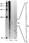
Determination of the transcription start site of murine SOCS-3 mRNA by RNase protection assay. As described in Materials and Methods, a 32P-labeled antisense probe spanning nucleotides +160 to −273 of the murine SOCS-3 gene was hybridized at 42°C with 10 μg yeast RNA (lanes 2 and 3) or 10 μg total RNA derived from LIF-stimulated AtT-20 cells (lane 4). Lane 2 shows the undigested full-length probe. RNase A/RNase T1 mix was added for digestion of unprotected fragments to samples of lane 3 (negative control) and lane 4. Lanes 5 and 6 show 33P-sequencing of A and C with antisense primer (+160 to +143). Arrows indicate a protected band in lane 4 and the corresponding nucleotide sequence.
Figure 3
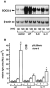
Stimulation of murine SOCS-3 mRNA and SOCS-3 reporter gene activity by different cytokines in corticotroph AtT-20 cells. (A) AtT-20 cells were treated with 0.5 × 10⁻⁹ M LIF, IL-6, or IL-11 for 60 and 120 min. Northern blot analysis was performed with 25 μg total RNA per lane. (Upper) SOCS-3 mRNA. (Lower) β-actin mRNA. (B) Luciferase activity of pGL3Basic alone and a −2757/+929 murine SOCS-3 promoter-pGL3Basic construct (clone 6) was measured in AtT-20 cells. Cells were treated with 0.5 × 10⁻⁹ M LIF, IL-6, or IL-11. Relative light units were calculated from four independently performed experiments. Each experiment was performed with n = 3 wells per group. * indicate in-group significance of untreated (−) vs. treated (+); ∗, P < 0.05; ∗∗, P < 0.01.
Figure 4
Effect of overexpressed dominant negative STAT-3 mutants or wt SOCS-3 on LIF-induced SOCS-3 promoter activity and gene expression. Corticotroph AtT-20 cells overexpressing wt STAT-3 (AtT-20W) or dominant negative STAT-3 mutants (AtT-20F and AtT-20D), as well as wt SOCS-3 (AtT-20S) and mock-transfected (AtT-20M), were isolated after stable transfection, as described (15, 28). (A and D) Cells were treated with 0.5 × 10⁻⁹ M LIF for 45 min. Northern blot analysis was performed with 15 μg total RNA per lane; shown is a representative experiment. (Upper) SOCS-3 mRNA. (Lower) β-actin mRNA. (B and E) Northern blot signals for SOCS-3 mRNA were analyzed by quantitative densitometry and normalized for β-actin. The relative increase of LIF-induced SOCS-3 mRNA was calculated from three independently performed experiments. Each experiment was performed with three different clones per group. (C and F) Luciferase activity of a −2757/+929 murine SOCS-3 promoter-pGL3Basic construct (clone 6) was measured in different cell clones and treated with 0.5 × 10⁻⁹ M LIF. LIF-induced luciferase activity was normalized to the untreated control for each clone. Relative induction of luciferase activity after stimulation was calculated from three independently performed experiments. Each experiment was performed with three independent clones per group.
Figure 5
Luciferase activity of different constructs of the genomic 5′ region of murine SOCS-3. Different constructs were obtained as described in Materials and Methods. After transient transfection, luciferase activity of each construct was measured in untreated (filled bars) and LIF-stimulated (empty bars) AtT-20 cells. Relative luciferase activity was normalized to the activity of pGL3Basic alone in untreated AtT-20 cells, which was defined as 1.0. Crossed lines indicate deletions of STAT binding elements in clones 6D1 and 6D2, in between the named nucleotides. Dotted line indicates a mutation of the wt STAT binding sequence (5′-TTCCAGGAA-3′) with mutant (5′-ATCGACGAT-3′) in clone 6M1.
Figure 6
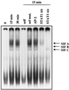
Gel shift analysis with nuclear cell extracts (20 μg) from AtT-20 cells. A 32P-labeled ds oligonucleotide (STAT oligo), containing the STAT binding consensus sequence (sense 5′-−75CAGTTCCAGGAATCGGGGGGC−55-3′) was used as a probe. Cells were either untreated or treated with 1 nM LIF for 15 min or 30 min. By using nuclear cell extract from 15-min LIF-treated AtT-20 cells, competition of the probe with a 100-fold excess of the unlabeled STAT oligo or the unlabeled STAT oligo mutated at positions −72, −69, −67, and −64, or an unrelated AP-2 oligo could be demonstrated. A presumably unspecific band, unaltered by competition with unlabeled STAT oligo, is shown at the bottom of each lane. Incubation with a STAT-1 antibody or with a STAT-3 antibody abolished DNA binding of specific complexes as evidenced by the absence of specific bands.
Figure 7
Model of interaction and negative autoregulatory feedback of SOCS-3 protein on POMC and SOCS-3 gene expression. The LIF-induced signaling cascade in the corticotroph cell envolves tyrosine phosphorylation of gp130, STAT-3, and STAT-1 (15, 23, 26, 27). LIF induces gene expression of POMC (28) and SOCS-3 in the corticotroph cell in a STAT-3 dependent manner. Although the region from nucleotides −173 to −160 in the rat POMC promoter is important for synergy of LIF and corticotropin-releasing hormone, it does not involve STAT protein binding (25); the putative STAT binding element in the POMC promoter has not yet been characterized. The murine SOCS-3 promoter has a functionally critical STAT-1/STAT-3 binding region at −72 to −64. SOCS-3 inhibits Jak2 activity by binding to its JH1 domain (9) and thus inhibits LIF-induced tyrosine phosphorylation of gp130 and STAT-3 in the corticotroph cell (15). By inhibiting Jak-STAT signaling, SOCS-3 negatively regulates LIF-induced POMC gene expression and ACTH secretion and also exerts a negative autoregulatory feedback on its own gene expression. This negative autoregulatory feedback of SOCS-3 on its own gene expression limits the accumulation of SOCS-3 protein in the corticotroph cell.
Similar articles
- Pituitary corticotroph SOCS-3: novel intracellular regulation of leukemia-inhibitory factor-mediated proopiomelanocortin gene expression and adrenocorticotropin secretion.
Auernhammer CJ, Chesnokova V, Bousquet C, Melmed S. Auernhammer CJ, et al. Mol Endocrinol. 1998 Jul;12(7):954-61. doi: 10.1210/mend.12.7.0140. Mol Endocrinol. 1998. PMID: 9658400 - SOCS proteins: modulators of neuroimmunoendocrine functions. Impact on corticotroph LIF signaling.
Auernhammer CJ, Bousquet C, Chesnokova V, Melmed S. Auernhammer CJ, et al. Ann N Y Acad Sci. 2000;917:658-64. doi: 10.1111/j.1749-6632.2000.tb05431.x. Ann N Y Acad Sci. 2000. PMID: 11268394 Review. - SOCS proteins: negative regulators of cytokine signaling.
Krebs DL, Hilton DJ. Krebs DL, et al. Stem Cells. 2001;19(5):378-87. doi: 10.1634/stemcells.19-5-378. Stem Cells. 2001. PMID: 11553846 Review.
Cited by
- C/EBPβ mediates growth hormone-regulated expression of multiple target genes.
Cui TX, Lin G, LaPensee CR, Calinescu AA, Rathore M, Streeter C, Piwien-Pilipuk G, Lanning N, Jin H, Carter-Su C, Qin ZS, Schwartz J. Cui TX, et al. Mol Endocrinol. 2011 Apr;25(4):681-93. doi: 10.1210/me.2010-0232. Epub 2011 Feb 3. Mol Endocrinol. 2011. PMID: 21292824 Free PMC article. - Cellular transcriptional profiling in influenza A virus-infected lung epithelial cells: the role of the nonstructural NS1 protein in the evasion of the host innate defense and its potential contribution to pandemic influenza.
Geiss GK, Salvatore M, Tumpey TM, Carter VS, Wang X, Basler CF, Taubenberger JK, Bumgarner RE, Palese P, Katze MG, García-Sastre A. Geiss GK, et al. Proc Natl Acad Sci U S A. 2002 Aug 6;99(16):10736-41. doi: 10.1073/pnas.112338099. Epub 2002 Jul 29. Proc Natl Acad Sci U S A. 2002. PMID: 12149435 Free PMC article. - Acute myeloid leukemia-associated Mkl1 (Mrtf-a) is a key regulator of mammary gland function.
Sun Y, Boyd K, Xu W, Ma J, Jackson CW, Fu A, Shillingford JM, Robinson GW, Hennighausen L, Hitzler JK, Ma Z, Morris SW. Sun Y, et al. Mol Cell Biol. 2006 Aug;26(15):5809-26. doi: 10.1128/MCB.00024-06. Mol Cell Biol. 2006. PMID: 16847333 Free PMC article. - The central role of SOCS-3 in integrating the neuro-immunoendocrine interface.
Auernhammer CJ, Melmed S. Auernhammer CJ, et al. J Clin Invest. 2001 Dec;108(12):1735-40. doi: 10.1172/JCI14662. J Clin Invest. 2001. PMID: 11748254 Free PMC article. Review. No abstract available. - Leptin and leptin receptor in anterior pituitary function.
Lloyd RV, Jin L, Tsumanuma I, Vidal S, Kovacs K, Horvath E, Scheithauer BW, Couce ME, Burguera B. Lloyd RV, et al. Pituitary. 2001 Jan-Apr;4(1-2):33-47. doi: 10.1023/a:1012982626401. Pituitary. 2001. PMID: 11824506 Review.
References
- Hirano T, Nakajima K, Hibi M. Cytokine Growth Factor Rev. 1997;8:241–252. - PubMed
- Carter-Su C, Smit L S. Recent Prog Horm Res. 1998;53:61–82. - PubMed
- Haque S J, Wiliams B R G. Semin Oncol. 1998;25, Suppl. 1:14–22. - PubMed
- Starr R, Willson T A, Viney E M, Murray L J, Rayner J R, Jenkins B J, Gonda T J, Alexander W S, Metcalf D, Nicola N A, Hilton D J. Nature (London) 1997;387:917–921. - PubMed
Publication types
MeSH terms
Substances
LinkOut - more resources
Full Text Sources
Other Literature Sources
Molecular Biology Databases
Research Materials
Miscellaneous


