Clinical experience over 48 years with pheochromocytoma - PubMed (original) (raw)
. 1999 Jun;229(6):755-64; discussion 764-6.
doi: 10.1097/00000658-199906000-00001.
J A O'Neill Jr, G W Holcomb 3rd, W M Morgan 3rd, W W Neblett 3rd, J A Oates, N Brown, J Nadeau, B Smith, D L Page, N N Abumrad, H W Scott Jr
Affiliations
- PMID: 10363888
- PMCID: PMC1420821
- DOI: 10.1097/00000658-199906000-00001
Clinical experience over 48 years with pheochromocytoma
R E Goldstein et al. Ann Surg. 1999 Jun.
Abstract
Objective: To analyze the presentation, localization, surgical management, pathology, and long-term outcome of a large series of patients with pheochromocytomas.
Summary background data: There are several areas of controversy pertaining to pheochromocytomas. Although many studies report a higher rate of malignancy for extraadrenal pheochromocytomas than for adrenal pheochromocytomas, the number of patients with the former tumor are small and statistical analysis is lacking. There has also been recent debate as to whether microscopic features of the tumor may be predictive of future behavior.
Methods: From 1950 to 1998, the authors observed 108 pheochromocytomas in 104 patients. The outcome of these patients has been followed prospectively. The medical records of these patients were reviewed for data on the presentation, localization, surgical management, pathology, and outcome. Patient survival was analyzed using Kaplan-Meier survival distributions.
Results: This study included 66 female patients and 38 male patients. The average age at surgery was 42.3 years. Sporadic cases accounted for 84% of the patients; the other 16% had multiple endocrine neoplasia type 2, von Recklinghausen's disease, von Hippel-Lindau disease, or Carney's syndrome. Of 64 adrenal tumors, 55 were initially considered benign, 6 had microscopic malignant features, and 3 had malignant disease. Mean patient follow-up was 12.6 years. To date, in five additional patients (none with microscopic disease) malignant disease developed (13% overall rate of malignancy). Recurrence occurred as late as 15 years after resection. Of 26 extraadrenal pheochromocytomas, 14 were initially considered benign, 8 had microscopic malignant features, and 4 had malignant disease. Thus, 46% of patients had either malignant disease or tumors with malignant features. Mean patient follow-up was 11.5 years. In one patient with benign disease and in one patient with malignant features, malignant disease developed (23% overall rate of malignancy). The difference in the rate of malignancy was not statistically significant between adrenal and extraadrenal pheochromocytomas. Patients with adrenal and extraadrenal pheochromocytomas also had similar rates of survival (p = NS).
Conclusions: The data suggest that patients with extraadrenal pheochromocytomas have the same risk of malignancy and the same overall survival as patients with adrenal pheochromocytomas. Lifelong follow-up of these patients is mandatory.
Figures
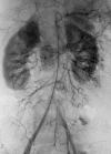
Figure 1. Angiogram demonstrating the typical radiographic blush of an extraadrenal pheochromocytoma to the patient’s right of the aorta. This is also the angiogram that did not detect a second extraadrenal pheochromocytoma located on the patient’s left side; it was successfully resected at a second operation.
Figure 2. A representative noncontrast CT scan of the abdomen demonstrating a 5-cm left adrenal pheochromocytoma.
Figure 3. Overall actual survival. This figure depicts the Kaplan–Meier survival curve for all patients with pheochromocytoma dying from all causes. The patients were divided into categories based on whether they underwent bilateral adrenalectomies, unilateral adrenalectomies, or resection of extraadrenal pheochromocytomas. The survival times of the individual groups were compared using the log-rank test; p = 0.82, indicating that there was no statistical difference in the survival time between groups.
Figure 4. Cause-specific survival. This figure depicts the Kaplan–Meier survival curve for all patients with pheochromocytoma dying specifically from pheochromocytoma. The patients were divided into categories based on whether they underwent bilateral adrenalectomies, unilateral adrenalectomies, or resection of extraadrenal pheochromocytomas. The survival times of the individual groups were compared using the log-rank test; p = 0.31, indicating that there was no statistical difference in the survival time between groups.
Similar articles
- Twenty-five-year surgical experience with pheochromocytoma in children.
Reddy VS, O'Neill JA Jr, Holcomb GW 3rd, Neblett WW 3rd, Pietsch JB, Morgan WM 3rd, Goldstein RE. Reddy VS, et al. Am Surg. 2000 Dec;66(12):1085-91; discussion 1092. Am Surg. 2000. PMID: 11149577 - Pheochromocytomas, multiple endocrine neoplasia type 2, and von Hippel-Lindau disease.
Neumann HP, Berger DP, Sigmund G, Blum U, Schmidt D, Parmer RJ, Volk B, Kirste G. Neumann HP, et al. N Engl J Med. 1993 Nov 18;329(21):1531-8. doi: 10.1056/NEJM199311183292103. N Engl J Med. 1993. PMID: 8105382 - Extraadrenal and multiple pheochromocytomas. Are there really any differences in pathophysiology and outcome?
Lumachi F, Polistina F, Favia G, D'Amico DF. Lumachi F, et al. J Exp Clin Cancer Res. 1998 Sep;17(3):303-5. J Exp Clin Cancer Res. 1998. PMID: 9894766 - Pheochromocytomas and secreting paragangliomas.
Plouin PF, Gimenez-Roqueplo AP. Plouin PF, et al. Orphanet J Rare Dis. 2006 Dec 8;1:49. doi: 10.1186/1750-1172-1-49. Orphanet J Rare Dis. 2006. PMID: 17156452 Free PMC article. Review. - Incidental adrenal pheochromocytoma. Report on 5 operated patients and update of the literature.
Porcaro AB, Novella G, Ficarra V, D'Amico A, Antoniolli SZ, Curti P. Porcaro AB, et al. Arch Ital Urol Androl. 2003 Dec;75(4):217-25. Arch Ital Urol Androl. 2003. PMID: 15005498 Review.
Cited by
- [Preoperative α-adrenoceptor block in asymptomatic pheochromocytoma? Pro].
Bracker L, Rath S, Dralle H, Bucher M. Bracker L, et al. Chirurg. 2012 Jun;83(6):546-50. doi: 10.1007/s00104-011-2195-4. Chirurg. 2012. PMID: 22466760 Review. German. - Metastatic disease and major adverse cardiovascular events preceding diagnosis are the main determinants of disease-specific survival of pheochromocytoma/paraganglioma: long-term follow-up of 303 patients.
Raber W, Schendl R, Arikan M, Scheuba A, Mazal P, Stadlmann V, Lehner R, Zeitlhofer P, Baumgartner-Parzer S, Gabler C, Esterbauer H. Raber W, et al. Front Endocrinol (Lausanne). 2024 Aug 21;15:1419028. doi: 10.3389/fendo.2024.1419028. eCollection 2024. Front Endocrinol (Lausanne). 2024. PMID: 39234504 Free PMC article. - Pheochromocytoma: an uncommon presentation of an asymptomatic and biochemically silent adrenal incidentaloma.
Kota SK, Kota SK, Panda S, Modi KD. Kota SK, et al. Malays J Med Sci. 2012 Apr;19(2):86-91. Malays J Med Sci. 2012. PMID: 22973143 Free PMC article. - Pheochromocytoma-induced atrial tachycardia leading to cardiogenic shock and cardiac arrest: resolution with atrioventricular node ablation and pacemaker placement.
Shawa H, Bajaj M, Cunningham GR. Shawa H, et al. Tex Heart Inst J. 2014 Dec 1;41(6):660-3. doi: 10.14503/THIJ-13-3692. eCollection 2014 Dec. Tex Heart Inst J. 2014. PMID: 25593537 Free PMC article. Review. - Recurrence of Phaeochromocytoma and Abdominal Paraganglioma After Initial Surgical Intervention.
Johnston PC, Mullan KR, Atkinson AB, Eatock FC, Wallace H, Gray M, Hunter SJ. Johnston PC, et al. Ulster Med J. 2015 May;84(2):102-6. Ulster Med J. 2015. PMID: 26170485 Free PMC article.
References
- Wilbourn RB. Early surgical history of phaeochromocytoma. Br J Surg 1987; 74: 594–596. - PubMed
- O’Riordain DS, Young WF Jr, Grant CS, et al. Clinical spectrum and outcome of functional extraadrenal paraganglioma. World J Surg 1996; 20: 916–922. - PubMed
- Pommier RF, Vetto JT, Billingsly K, et al. Comparison of adrenal and extraadrenal pheochromocytomas. Surgery 1993; 114: 1160–1166. - PubMed
- Proye CAG, Vix H, Jansson S, et al. “The” pheochromocytoma: a benign, intra-adrenal, hypertensive, sporadic unilateral tumor. Does it exist? World J Surg 1994; 18: 467–472. - PubMed
- Scott HW Jr, Reynolds V, Green N, et al. Clinical experience with malignant pheochromocytomas. Surg Gynecol Obstet 1982; 154: 801–818. - PubMed
MeSH terms
LinkOut - more resources
Full Text Sources
Medical
Miscellaneous


