Critical roles for IL-4, IL-5, and eosinophils in chronic skin allograft rejection - PubMed (original) (raw)
Critical roles for IL-4, IL-5, and eosinophils in chronic skin allograft rejection
A Le Moine et al. J Clin Invest. 1999 Jun.
Abstract
C57BL/6 mice injected with the 145-2C11 anti-CD3 mAb and grafted with MHC class II disparate bm12 skin develop a chronic rejection characterized by interstitial dermal fibrosis, a marked eosinophil infiltrate, and an obliterative intimal vasculopathy. Because these changes occur in the absence of alloreactive antibodies, we examined the contribution of cytokines in their pathogenesis. Chronically rejected grafts showed a marked accumulation of both IL-4 and IL-5 mRNA. Mixed lymphocyte reaction experiments established that mice undergoing chronic rejection were primed for IL-4, IL-5, and IL-10 secretion. In vivo administration of anti-IL-4 mAb completely prevented allograft vasculopathy as well as graft eosinophil infiltration and dermal fibrosis. Injection of anti-IL-5 mAb or the use of IL-5-deficient mice as recipients also resulted in the lack of eosinophil infiltration or dermal fibrosis, but these mice did develop allograft vasculopathy. Administration of anti-IL-10 mAb did not influence any histologic parameter of chronic rejection. Thus, in this model, IL-4- and IL-5-mediated tissue allograft eosinophil infiltration is associated with interstitial fibrosis. IL-4, but not eosinophils, is also required for the development of obliterative graft arteriolopathy.
Figures
Figure 1
Cytokine gene expression within chronically rejected allografts. Cytokine gene expression was assayed within pooled syngeneic C57BL/6 grafts (n = 4) or bm12 allografts undergoing chronic rejection at either day 20 (n = 4) or day 60 (n = 4) after transplantation. Lymph node cells from C57BL/6 mice stimulated in vivo with the 2C11 anti-CD3 mAb served as positive controls. Similar results were observed in 2 other separate experiments.
Figure 2
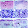
Allograft eosinophil infiltrate: effect of anti–IL-4 and anti–IL-5 mAb administration. (a) Syngeneic skin graft 40 days after transplantation. The dermis, below the epidermal layer (E), is free of inflammatory cells. Sebaceous glands (S) and hair follicles (H) show a normal appearance (hematoxylin and eosin, ×200). (b) bm12 skin allografts at day 40 from a mouse that received anti-CD3 and the control mAb. There is a massive infiltration of the dermis by leukocytes. Up to 35% of the infiltrating leukocytes were identified as eosinophils (refer to c). The number of sebaceous glands and hair follicles is reduced, and those remaining are being engulfed by inflammatory cells (arrow) (hematoxylin and eosin, ×100). (c) bm12 skin allografts at day 40 from a mouse that received anti-CD3 and the control mAb. The dermis is heavily infiltrated by eosinophils, recognizable by the red spots in the cytoplasm (arrows) (hematoxylin and eosin, ×1,000). (d) bm12 skin allografts at day 40 from a mouse that received anti-CD3 and the control mAb. A shrinking hair follicle is being infiltrated by numerous eosinophils (arrows) (hematoxylin and eosin, ×400). (e) bm12 skin allografts at day 40 from a mouse that received anti-CD3 and anti–IL-4 mAb. The aspect is similar to that of a syngeneic graft. The dermis is free of eosinophils. (f) bm12 skin allografts at day 40 from a mouse that received anti-CD3 and anti–IL-5 mAb. There is only a small number of leukocytes present in the dermis. The structure of hair follicles (H) and sebaceous glands (S) are well preserved (compare with b) (hematoxylin and eosin, ×200).
Figure 3
Allograft interstitial fibrosis: effect of IL-4 and IL-5 blockade. (a) A syngeneic skin graft 40 days after transplantation. The collagen-dense deposits normally present within syngeneic tail skin grafts are stained blue. (b) bm12 skin allograft at day 40 from a mouse that received anti-CD3 and the control mAb. The thickness of the dermis is increased, mainly because of dense collagen deposits. (c and d) bm12 skin allografts at day 40 in mice that received the anti-CD3 mAb and either anti–IL-4 mAb (c) or anti–IL-5 mAb (d). In both settings (c and d), the collagen-dense deposits and the thickness of the dermis are comparable to that observed in syngeneic skin grafts.
Figure 4
Allograft vasculopathy: effect of anti–IL-4 and anti–IL-5 mAb administration. (a) Normal appearance of a small artery in a syngeneic skin graft 60 days after transplantation. The lamina elastica interna, stained brown (arrow), separates the media from the intima (orcein stain, ×1,000). (b) bm12 skin allograft at day 40 from a mouse that received anti-CD3 and the control mAb. An increased cellularity of the intima, leading to a reduction of the vessel lumen, is observed (orcein staining, ×400). (c) bm12 skin allograft at day 40 from a mouse that received anti-CD3 and the anti–IL-4 mAb. The vessel is normal, with no intimal proliferation (orcein staining, ×400). (d and e) bm12 skin allograft at day 40 from a wild-type mouse that received anti-CD3 and the anti–IL-5 mAb (d), or an IL-5–deficient mouse that received anti-CD3 mAb (e). An obliterative vasculopathy is observed in both settings.
Figure 5
Effect of IL-5 neutralization on the production of TGF-β 1 mRNA in chronically rejected skin allografts. Quantitative competitive RT-PCR assay for TGF-β1 mRNA was performed 40 days after transplantation in syngeneic skin grafts (cross-hatched bar), in mice that received anti-CD3 and control mAb (filled bar), and in mice that received anti-CD3 and anti–IL-5 mAb (hatched bar). Results are expressed as femtograms of TGF-β mRNA per microgram of cellular RNA. Each experimental group represents a pool of 3 skin grafts. Similar results were obtained in another separate experiment.
Figure 6
Immunostaining for TGF-β1 expression in chronically rejected skin allografts. (a) TGF-β1 staining (brown) is present within eosinophils (arrows) and other inflammatory cells (×1,000). (b) Immunostaining of the same sample after preincubation of the anti–TGF-β antibody with the TGF-β–specific blocking peptide as a control procedure. Only faint background staining can still be seen (×1,000). Similar staining patterns were observed in 5 other skin allografts.
References
- Orosz CG, Pelletier RP. Chronic remodeling pathology in grafts. Curr Opin Immunol. 1997;9:676–680. - PubMed
- Russell PS, Chase CM, Winn HJ, Colvin RB. Coronary atherosclerosis in transplanted mouse hearts. III. Effects of recipient treatment with a monoclonal antibody to interferon-gamma. Transplantation. 1994;57:1367–1371. - PubMed
Publication types
MeSH terms
Substances
LinkOut - more resources
Full Text Sources
Other Literature Sources
Research Materials


