Interaction between astrocytes and adult subventricular zone precursors stimulates neurogenesis - PubMed (original) (raw)
Interaction between astrocytes and adult subventricular zone precursors stimulates neurogenesis
D A Lim et al. Proc Natl Acad Sci U S A. 1999.
Abstract
Neurogenesis continues in the mammalian subventricular zone (SVZ) throughout life. However, the signaling and cell-cell interactions required for adult SVZ neurogenesis are not known. In vivo, migratory neuroblasts (type A cells) and putative precursors (type C cells) are in intimate contact with astrocytes (type B cells). Type B cells also contact each other. We reconstituted SVZ cell-cell interactions in a culture system free of serum or exogenous growth factors. Culturing dissociated postnatal or adult SVZ cells on astrocyte monolayers-but not other substrates-supported extensive neurogenesis similar to that observed in vivo. SVZ precursors proliferated rapidly on astrocytes to form colonies containing up to 100 type A neuroblasts. By fractionating the SVZ cell dissociates with differential adhesion to immobilized polylysine, we show that neuronal colony-forming precursors were concentrated in a fraction enriched for type B and C cells. Pure type A cells could migrate in chains but did not give rise to neuronal colonies. Because astrocyte-conditioned medium alone was not sufficient to support SVZ neurogenesis, direct cell-cell contact between astrocytes and SVZ neuronal precursors may be necessary for the production of type A cells.
Figures
Figure 1
Astrocyte monolayers support the proliferation and differentiation of SVZ cells. (A and C) Differential interference contrast (DIC) images of typical colonies from postnatal SVZ precursors. (B and D) Epifluorescent images of the postnatal SVZ precursors depicted in A and C, respectively. (B) Colonies are immunopositive for the neuronal marker, neuron-specific tubulin (Tuj1; red), and have incorporated BrdUrd (green) between 4 and 5 days in vitro (DIV). (D) Colonies are also immunopositive for neuronal marker MAP-2. (E) Time course of SVZ neurogenesis. Tuj1+ cell density in coculture vs. culture on different substrates is reported at 2-day intervals. Error bars = SEM of triplicate cultures. Astrocyte monolayers alone did not produce Tuj1+ cells at any time point. (F) DIC image of a neuronal colony from the adult SVZ. (G) Respective epifluorescence showing BrdUrd incorporation (yellow-green) and Tuj1 immunoreactivity (red). All above cultures were photographed at 5 DIV. (Bars = 10 μm.) (Inset) Schematic of SVZ cellular architecture as seen in a coronal section. The ventricular cavity would be to the right of the ependymal cell layer (gray). Type A migratory neuroblasts (red) migrate through glial tubes formed by type B cells (blue). The direction of migration would be perpendicular to the plane of the page. Type C cells (green) also are in intimate contact with type B astrocytes. Type A, B2, and C are mitotic.
Figure 2
Fractionation of SVZ cells by differential adhesion yields populations of cells with distinct characteristics. (A–C) Fraction 1, type A cells. See Results and Fig. 3 for details. (A) DIC image of isolated type A cells. (B) Epifluorescent image showing Tuj1 staining (red). (C) Purified type A cells are PSA-NCAM+ (red) and can migrate in chains. (D) Aggregate of type A cells immediately after they were embedded in Matrigel. (E) Chain migration from aggregates after 6 h in culture. The culture in E was double-stained for Tuj1 (F) and GFAP (G). (H) A positive control for GFAP staining (green) in Matrigel culture. (I–K) Fraction 4, B/C cells. Most of the adherent cells had a flattened, spread, phase-dark appearance, and ≈30% of these cells were GFAP+ (see Fig. 3 Inset). Nuclei are counterstained with Hoechst 33258 (blue). (Bar = 10 μm.)
Figure 3
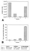
The four fractions of SVZ cells have different yields and abilities to generate Tuj1+ colonies. (A) Fractionation yield. Fraction 4 was collected after treatment with trypsin. (B) Equal numbers of cells from the different fractions in A were plated onto astrocyte monolayers. Cultures were fixed at 5 DIV. Clusters of more than four Tuj1+ cells were counted as colonies. Error bars = SEM of triplicate cultures. (Inset) Immunocytochemistry of fraction 1 (type A cells) and fraction 4 (type B/C cells) purity. For every SVZ fractionation, an aliquot of each fraction was plated for immunostaining for type A markers (Tuj1 and PSA-NCAM) and a type B marker (GFAP). Between 500 and 1,000 cells were counted in each fraction. Standard deviation is indicated in parentheses. Note that in fraction 4, almost half of the cells were immunonegative for all three markers; these are putative type C cells. There is no marker specific for type C cells at present.
Figure 4
Fraction 1 (type A cells) and fraction 4 (type B/C cells) in astrocyte coculture. (A) Time course of a cell–astrocyte coculture vs. culture on PDK. Error bars = SEM of triplicate cultures. (B) Tuj1+ cells stained with diaminobenzidine in a cell–astrocyte coculture at 5 DIV. (C) Time course of B/C cell–astrocyte coculture vs. culture on PDK. (D) Typical colony of Tuj1+ cells stained with diaminobenzidine in astrocyte coculture at 5 DIV. (E and F) Cocultures were exposed to BrdUrd from 2 to 4 DIV. (E) DIC image of a neuronal colony. (F) Same colony double-stained for Tuj1 (red) and BrdUrd (green). DIC (G) and epifluorescent (H) images of a neuronal colony stained for PSA-NCAM. (Bars = 10 μm.)
Similar articles
- EphA4 Regulates Neuroblast and Astrocyte Organization in a Neurogenic Niche.
Todd KL, Baker KL, Eastman MB, Kolling FW, Trausch AG, Nelson CE, Conover JC. Todd KL, et al. J Neurosci. 2017 Mar 22;37(12):3331-3341. doi: 10.1523/JNEUROSCI.3738-16.2017. Epub 2017 Mar 3. J Neurosci. 2017. PMID: 28258169 Free PMC article. - Chain formation and glial tube assembly in the shift from neonatal to adult subventricular zone of the rodent forebrain.
Peretto P, Giachino C, Aimar P, Fasolo A, Bonfanti L. Peretto P, et al. J Comp Neurol. 2005 Jul 11;487(4):407-27. doi: 10.1002/cne.20576. J Comp Neurol. 2005. PMID: 15906315 - Multiple cell populations in the early postnatal subventricular zone take distinct migratory pathways: a dynamic study of glial and neuronal progenitor migration.
Suzuki SO, Goldman JE. Suzuki SO, et al. J Neurosci. 2003 May 15;23(10):4240-50. doi: 10.1523/JNEUROSCI.23-10-04240.2003. J Neurosci. 2003. PMID: 12764112 Free PMC article. - The subventricular zone: source of neuronal precursors for brain repair.
Alvarez-Buylla A, Herrera DG, Wichterle H. Alvarez-Buylla A, et al. Prog Brain Res. 2000;127:1-11. doi: 10.1016/s0079-6123(00)27002-7. Prog Brain Res. 2000. PMID: 11142024 Review. - In vivo targeting of subventricular zone astrocytes.
Mamber C, Verhaagen J, Hol EM. Mamber C, et al. Prog Neurobiol. 2010 Sep;92(1):19-32. doi: 10.1016/j.pneurobio.2010.04.007. Epub 2010 May 2. Prog Neurobiol. 2010. PMID: 20441785 Review.
Cited by
- Astrocyte Regulation of Neuronal Function and Survival in Stroke Pathophysiology.
Boyle BR, Berghella AP, Blanco-Suarez E. Boyle BR, et al. Adv Neurobiol. 2024;39:233-267. doi: 10.1007/978-3-031-64839-7_10. Adv Neurobiol. 2024. PMID: 39190078 Review. - Glial cells in the mammalian olfactory bulb.
Zhao D, Hu M, Liu S. Zhao D, et al. Front Cell Neurosci. 2024 Jul 16;18:1426094. doi: 10.3389/fncel.2024.1426094. eCollection 2024. Front Cell Neurosci. 2024. PMID: 39081666 Free PMC article. Review. - Comprehensive characterization of the neurogenic and neuroprotective action of a novel TrkB agonist using mouse and human stem cell models of Alzheimer's disease.
Charou D, Rogdakis T, Latorrata A, Valcarcel M, Papadogiannis V, Athanasiou C, Tsengenes A, Papadopoulou MA, Lypitkas D, Lavigne MD, Katsila T, Wade RC, Cader MZ, Calogeropoulou T, Gravanis A, Charalampopoulos I. Charou D, et al. Stem Cell Res Ther. 2024 Jul 6;15(1):200. doi: 10.1186/s13287-024-03818-w. Stem Cell Res Ther. 2024. PMID: 38971770 Free PMC article. - Dissecting Intra-tumor Heterogeneity in the Glioblastoma Microenvironment Using Fluorescence-Guided Multiple Sampling.
García-Montaño LA, Licón-Muñoz Y, Martinez FJ, Keddari YR, Ziemke MK, Chohan MO, Piccirillo SGM. García-Montaño LA, et al. Mol Cancer Res. 2023 Aug 1;21(8):755-767. doi: 10.1158/1541-7786.MCR-23-0048. Mol Cancer Res. 2023. PMID: 37255362 Free PMC article. Review. - The Adult Neurogenesis Theory of Alzheimer's Disease.
Abbate C. Abbate C. J Alzheimers Dis. 2023;93(4):1237-1276. doi: 10.3233/JAD-221279. J Alzheimers Dis. 2023. PMID: 37182879 Free PMC article.
References
- Alvarez-Buylla A, Lois C. Stem Cells. 1995;13:263–272. - PubMed
- García-Verdugo J M, Doetsch F, Wichterle H, Lim D A, Alvarez-Buylla A. J Neurobiol. 1998;36:234–248. - PubMed
- McKay R. Science. 1997;276:66–71. - PubMed
- Weiss S, Reynolds B A, Vescovi A L, Morshead C, Craig C G, Van der Kooy D. Trends Neurosci. 1996;19:387–393. - PubMed
- Gage F H, Ray J, Fisher L J. Annu Rev Neurosci. 1995;18:159–192. - PubMed
Publication types
MeSH terms
Grants and funding
- R01 NS028478/NS/NINDS NIH HHS/United States
- GM07739/GM/NIGMS NIH HHS/United States
- NS28478/NS/NINDS NIH HHS/United States
- T32 GM007739/GM/NIGMS NIH HHS/United States
- R37 NS028478/NS/NINDS NIH HHS/United States
LinkOut - more resources
Full Text Sources
Other Literature Sources


