Leukemic HRX fusion proteins inhibit GADD34-induced apoptosis and associate with the GADD34 and hSNF5/INI1 proteins - PubMed (original) (raw)
Leukemic HRX fusion proteins inhibit GADD34-induced apoptosis and associate with the GADD34 and hSNF5/INI1 proteins
H T Adler et al. Mol Cell Biol. 1999 Oct.
Abstract
One of the most common chromosomal abnormalities in acute leukemia is a reciprocal translocation involving the HRX gene (also called MLL, ALL-1, or HTRX) at chromosomal locus 11q23, resulting in the formation of HRX fusion proteins. Using the yeast two-hybrid system and human cell culture coimmunoprecipitation experiments, we show here that HRX proteins interact directly with the GADD34 protein. We have found that transfected cells overexpressing GADD34 display a significant increase in apoptosis after treatment with ionizing radiation, indicating that GADD34 expression not only correlates with apoptosis but also can enhance apoptosis. The amino-terminal third of the GADD34 protein was necessary for this observed increase in apoptosis. Furthermore, coexpression of three different HRX fusion proteins (HRX-ENL, HRX-AF9, and HRX-ELL) had an anti-apoptotic effect, abrogating GADD34-induced apoptosis. In contrast, expression of wild-type HRX gave rise to an increase in apoptosis. The difference observed here between wild-type HRX and the leukemic HRX fusion proteins suggests that inhibition of GADD34-mediated apoptosis may be important to leukemogenesis. We also show here that GADD34 binds the human SNF5/INI1 protein, a member of the SNF/SWI complex that can remodel chromatin and activate transcription. These studies demonstrate, for the first time, a gain of function for leukemic HRX fusion proteins compared to wild-type protein. We propose that the role of HRX fusion proteins as negative regulators of post-DNA-damage-induced apoptosis is important to leukemia progression.
Figures
FIG. 1
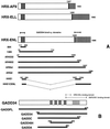
HRX fusion proteins and deletion mutants of HRX-ENL and GADD34. (A) Schematic representation of full-length human HRX-AF9, HRX-ELL, and HRX-ENL fusion proteins (top) and various HRX-ENL deletion constructs (bottom). Numbers represent amino acid residues. The three thick vertical lines designate the AT hooks. Shaded areas represent C-terminal residues donated by each of the respective fusion partners. Fusion sites are delineated by single thick vertical lines. (B) Schematic representation of the GADD34 protein (top) and GADD34 deletion constructs (bottom). The vertical stripes represent the four repeated 20- to 23-amino-acid motifs, FXXXWXYRPGXXTEEEEXXX, in GADD34. The area designated by diagonal lines represents the conserved 63-amino-acid region present in the carboxy terminus of GADD34, MyD116, and HSV ICP34.5.
FIG. 2
HRX proteins interact with GADD34 in vivo. (A) An anti-FLAG Western blot of anti-myc immunoprecipitates showing the amount of coimmunoprecipitated FLAG-tagged amino-truncated GADD34 protein, pSGADD34 (open triangle), bound to myc-tagged HRX fusion proteins and HRX-ENL deletion proteins expressed in the transfections described below. Solid triangles show cross-reacting murine immunoglobin bands in coimmunoprecipitates. (B) An anti-myc Western blot of cell lysates, using an anti-myc antibody, showing expression levels of the myc-tagged HRX fusion proteins and HRX-ENL deletion proteins expressed in the transfections described below. Longer exposure of lane 6 showed expression of the appropriately sized HRX-ENL protein (not shown). pSGADD34 (amino-truncated FLAG-tagged GADD34) was cotransfected with the following myc-tagged constructs: pCSmyc-TEL, pCS884, pCSAR4332, pCSAR4602, pCSARBst, pCSARQ2, pCSHRXAF9, and pCSHRXELL. (C and D) Anti-FLAG Western blot of anti-myc immunoprecipitates showing the amount of coimmunoprecipitated FLAG-tagged amino-truncated GADD34 protein, pSGADD34 (open triangle), bound to myc-tagged HRX-ENL, HRX N-terminal deletion proteins, and wild-type HRX expressed in the transfections described below. Solid triangles show cross-reacting murine immunoglobin bands in coimmunoprecipitates. The results shown here in are from two separate sets of transfections. pSGADD34 (amino-truncated FLAG-tagged GADD34) was cotransfected with the following myc-tagged constructs: pCSHMG-C, pCSARQ2, pCS1385, pCS884, and pCS2+MT vector only. (E to G) HRX fusion proteins bind endogenous GADD34 in vivo. (E) An anti-myc Western blot of anti-GADD34 immunoprecipitates showing the amount of coimmunoprecipitated myc-tagged proteins expressed in the transfections described below. Lane 1a is a longer exposure of lane 1. (F) An anti-myc Western blot of lysates from the transfections described below. (G) An anti-GADD34 Western blot of anti-GADD34 immunoprecipitates showing the amount of coimmunoprecipitated proteins from the transfections with the following constructs: pCSARQ2 (lane 1), pCSHHE1CENL (lane 2), pCSGADDFL (lane 3), pCSGADD34 (lane 4), myc-TEL (lane 5), pCSSET (lane 6), and pCSHHE1 (lane 7). The open arrow delineates endogenous GADD34 protein, shown here as a 90-kDa doublet.
FIG. 3
GADD34 deletion proteins interact with HRX-ENL in vivo. (A) Anti-FLAG Western blot of anti-myc immunoprecipitates, showing the amount of coimmunoprecipitated FLAG-tagged GADD34 deletion proteins bound to the myc-tagged HRX-ENL fusion protein (pCSARQ2) expressed in the transfections described below. A solid triangle shows a cross-reacting murine immunoglobin band in the coimmunoprecipitates. (B) Anti-FLAG Western blot of cell lysates with an anti-FLAG antibody, showing expression levels of the FLAG-tagged GADD34 deletion proteins expressed in the transfections described below. pCSARQ2 (myc-tagged HRX-ENL) was cotransfected with the following FLAG-tagged constructs: pSGADD34, pSGADDA, pSGADDC, and pSGADD484.
FIG. 4
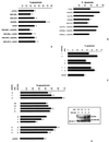
GADD34 induces apoptosis in SW480 cells. HRX fusion proteins abrogate GADD34-induced apoptosis and wild-type HRX induces apoptosis of SW480 cells after transfection and treatment with ionizing radiation. The mean percent apoptosis is shown for the transfected constructs below. Each column represents three separate experiments. (A) GADD34 induces apoptosis in SW480 cells, and the amino terminus is necessary for this effect. HRX fusion proteins, HRX-ENL and HRX-AF9, abrogate GADD34-induced apoptosis. The percent apoptosis of SW480 cells without transfection and without IR is approximately 8%, and after transfection of pSGADD34FL without IR it is approximately 17% (results of one trial only). GADD34FL encodes the full-length cDNA, and GADD34 encodes an amino-terminal deletion GADD34 protein (the clone retrieved from the yeast two-hybrid screen). (B) The HRX fusion protein HRX-ELL abrogates GADD34- induced apoptosis. (C) Wild-type HRX induces apoptosis in a dose-dependent fashion and does not inhibit GADD34-induced apoptosis. To the left is designated, in micrograms, the amount of wild-type HRX and GADD34 plasmid transfected. (D) GADD34 induces apoptosis in a dose-dependent fashion. HRX-ENL inhibits GADD34-induced apoptosis in a dose-dependent fashion. The inset shows an anti-myc Western blot from a SDS-PAGE gel (6% polyacrylamide) in a series of cotransfection experiments with increasing amounts of transfected pCSHRX plasmid (encoding myc-tagged wild-type HRX protein) with constant amounts of transfected pCSARQ2 plasmid (encoding myc-tagged HRX-ENL). The amounts of transfected plasmids are shown above in micrograms.
FIG. 5
HRX-ENL and GADD34 interact with hSNF5/INI1 in vivo. (A) Anti-FLAG Western blot of anti-myc immunoprecipitates showing the amount of coimmunoprecipitated FLAG-tagged hSNF5/INI1 (lanes 1 to 7) and FLAG-tagged hSNF5/INI1 and GADD34 (lane 8) proteins bound to the myc-tagged proteins expressed in the transfections described below. (B) Anti-FLAG Western blot of cell lysates with an anti-FLAG antibody, showing expression levels of full-length FLAG-tagged GADD34 and FLAG-tagged hSNF5/INI1 proteins expressed in the transfections described below. pSGSnf5 (full length FLAG-tagged hSNF5/INI1) was cotransfected with the following constructs: myc-TEL (negative control) (lane 1), pCSAR4332 (lane 2), pCSARQ2 (HRX-ENL) (lane 3), pCSHHE1 (lane 4), pCSHHE1CENL (lane 5), pCSGADDA (lane 6), pCSGADDFL (lane 7), and pCSARQ2 plus pSGADD34FL (lane 8).
FIG. 6
In vitro binding of GADD34 to GST-hSNF5/INI1. 35S-labeled GADD34 was transcribed and translated in vitro from pSGFL2GADD34FL (lane 1) and used directly to bind agarose (lane 2), GST-agarose (lanes 3, 5, and 7), and GST-hSNF5-agarose (lanes 4, 6, and 8) in binding buffers with the NaCl and NP-40 concentrations shown. After three washes with binding buffer, the bound 35S-labeled GADD34 was eluted, subjected to SDS-PAGE, and visualized by autoradiography.
Similar articles
- The human SNF5/INI1 protein facilitates the function of the growth arrest and DNA damage-inducible protein (GADD34) and modulates GADD34-bound protein phosphatase-1 activity.
Wu DY, Tkachuck DC, Roberson RS, Schubach WH. Wu DY, et al. J Biol Chem. 2002 Aug 2;277(31):27706-15. doi: 10.1074/jbc.M200955200. Epub 2002 May 16. J Biol Chem. 2002. PMID: 12016208 - HRX leukemic fusion proteins form a heterocomplex with the leukemia-associated protein SET and protein phosphatase 2A.
Adler HT, Nallaseth FS, Walter G, Tkachuk DC. Adler HT, et al. J Biol Chem. 1997 Nov 7;272(45):28407-14. doi: 10.1074/jbc.272.45.28407. J Biol Chem. 1997. PMID: 9353299 - The HRX proto-oncogene product is widely expressed in human tissues and localizes to nuclear structures.
Butler LH, Slany R, Cui X, Cleary ML, Mason DY. Butler LH, et al. Blood. 1997 May 1;89(9):3361-70. Blood. 1997. PMID: 9129043 - Molecular mechanisms of leukemogenesis mediated by MLL fusion proteins.
Ayton PM, Cleary ML. Ayton PM, et al. Oncogene. 2001 Sep 10;20(40):5695-707. doi: 10.1038/sj.onc.1204639. Oncogene. 2001. PMID: 11607819 Review.
Cited by
- Growth arrest and DNA damage-inducible protein (GADD34) enhanced liver inflammation and tumorigenesis in a diethylnitrosamine (DEN)-treated murine model.
Chen N, Nishio N, Ito S, Tanaka Y, Sun Y, Isobe K. Chen N, et al. Cancer Immunol Immunother. 2015 Jun;64(6):777-89. doi: 10.1007/s00262-015-1690-8. Epub 2015 Apr 2. Cancer Immunol Immunother. 2015. PMID: 25832002 Free PMC article. - The Drosophila SNR1 (SNF5/INI1) subunit directs essential developmental functions of the Brahma chromatin remodeling complex.
Marenda DR, Zraly CB, Feng Y, Egan S, Dingwall AK. Marenda DR, et al. Mol Cell Biol. 2003 Jan;23(1):289-305. doi: 10.1128/MCB.23.1.289-305.2003. Mol Cell Biol. 2003. PMID: 12482982 Free PMC article. - An upstream open reading frame regulates translation of GADD34 during cellular stresses that induce eIF2alpha phosphorylation.
Lee YY, Cevallos RC, Jan E. Lee YY, et al. J Biol Chem. 2009 Mar 13;284(11):6661-73. doi: 10.1074/jbc.M806735200. Epub 2009 Jan 8. J Biol Chem. 2009. PMID: 19131336 Free PMC article. - In vitro nuclear interactome of the HIV-1 Tat protein.
Gautier VW, Gu L, O'Donoghue N, Pennington S, Sheehy N, Hall WW. Gautier VW, et al. Retrovirology. 2009 May 19;6:47. doi: 10.1186/1742-4690-6-47. Retrovirology. 2009. PMID: 19454010 Free PMC article. - A systems biological analysis of the ATF4-GADD34-CHOP regulatory triangle upon endoplasmic reticulum stress.
Márton M, Bánhegyi G, Gyöngyösi N, Kálmán EÉ, Pettkó-Szandtner A, Káldi K, Kapuy O. Márton M, et al. FEBS Open Bio. 2022 Nov;12(11):2065-2082. doi: 10.1002/2211-5463.13484. Epub 2022 Sep 27. FEBS Open Bio. 2022. PMID: 36097827 Free PMC article.
References
- Adler H T, Nallaseth F S, Walter G, Tkachuk D C. HRX leukemic fusion proteins form a heterocomplex with the leukemia-associated protein SET and protein phosphatase 2A. J Biol Chem. 1997;272:28407–28414. - PubMed
- Akao Y, Mizoguchi H, Misiura K, Stec W J, Seto M, Ohishi N, Yagi K. Antisense oligodeoxyribonucleotide against the MLL-LTG19 chimeric transcript inhibits cell growth and induces apoptosis in cells of an infantile leukemia cell line carrying the t(11;19) chromosomal translocation. Cancer Res. 1998;58:3773–3776. - PubMed
- Bernard O A, Berger R. Molecular basis of 11q23 rearrangements in hematopoietic malignant proliferations. Genes Chromosomes Cancer. 1995;13:75–85. - PubMed
- Borkhardt A, Repp R, Haas O A, Leis T, Harbott J, Kreuder J, Hammermann J, Henn T, Lampert F. Cloning and characterization of AFX, the gene that fuses to MLL in acute leukemias with a t(X;11)(q13;q23) Oncogene. 1997;14:195–202. - PubMed
Publication types
MeSH terms
Substances
LinkOut - more resources
Full Text Sources
Other Literature Sources
Medical
Molecular Biology Databases


