Targeted disruption of mouse Yin Yang 1 transcription factor results in peri-implantation lethality - PubMed (original) (raw)
Targeted disruption of mouse Yin Yang 1 transcription factor results in peri-implantation lethality
M E Donohoe et al. Mol Cell Biol. 1999 Oct.
Abstract
Yin Yang 1 (YY1) is a zinc finger-containing transcription factor and a target of viral oncoproteins. To determine the biological role of YY1 in mammalian development, we generated mice deficient for YY1 by gene targeting. Homozygosity for the mutated YY1 allele results in embryonic lethality in the mouse. YY1 mutants undergo implantation and induce uterine decidualization but rapidly degenerate around the time of implantation. A subset of YY1 heterozygote embryos are developmentally retarded and exhibit neurulation defects, suggesting that YY1 may have additional roles during later stages of mouse embryogenesis. Our studies demonstrate an essential function for YY1 in the development of the mouse embryo.
Figures
FIG. 1
Generation of a targeted mutation at the mouse YY1 locus and identification of the YY1 mutant allele. (A) Genomic organization of the mouse YY1 locus and design of the targeting construct. An 18-kb YY1 fragment was obtained from a genomic library of the mouse strain 129/Sv. The YY1 fragment targeted for gene replacement contains exon I and the sequences 11 kb 5′ and 6 kb 3′ to exon I. A _Xho_I/_Hin_dIII fragment (approximately 2 kb) containing the entire exon I including both the transcription and the translational start sites was replaced with the neo cassette. The arrow marked “Txn start” denotes the transcriptional start site. The arrow labeled “ATG” denotes the translational start site. The probe used in Southern hybridization to detect homologous recombination is indicated by a thick bar and labeled as such. The size of a 1-kb sequence is also indicated. B, _Bam_HI; H, _Hin_dIII; K, _Kpn_I; RV, _Eco_RV; S, _Sac_I; X, _Xho_I; RI, _Eco_RI; PGK, phosphoglycerate kinase. (B) Identification of ES cells containing a mutant YY1 allele. The YY1 targeting vector was introduced into ES cells and selected with G418. Southern blot hybridization was performed on genomic DNA extracted from ES cells, digested with _Bam_HI, and hybridized to a radioactively labeled neo and external YY1 probe as shown in panel A. Wild-type (wt) ES cells contain a single 25-kb YY1 fragment, while YY1+/− ES cells contain the 25-kb and the mutant (mut) 19-kb YY1 fragment. The 6-kb fragment containing the neo gene is detected for the YY1+/− ES cells only. The genotypes of the ES cells are indicated above the lanes. The size of the hybridized bands is indicated on the right.
FIG. 2
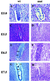
Histological examination of in utero embryos obtained from YY1 heterozygous matings. The uteri of female YY1+/− mice were dissected between 5.0 and 7.5 days after intercross mating as described in Materials and Methods. All uterine decidua (wild-type and mutant) were sectioned transversely at a 7-μm thickness and stained with hematoxylin and eosin. Wild-type or heterozygous embryos are shown in the column labeled wt, and presumptive YY1−/− mutants are shown in the column labeled mut. (A and B) E5.0, early egg cylinder stage; (C and D) E5.5, egg cylinder stage; (E and F) E6.5, late egg cylinder stage; (G and H) E7.5, late primitive streak stage. The wild-type embryos show a differentiating early egg cylinder stage embryo at E5.0 (A) and E5.5 (C), whereas the mutant embryos show decreased proliferation and a lack of organized embryos (B and D) at these embryonic stages. Note the appearance of a proamniotic cavity in the E6.5 elongated late-egg-cylinder wild-type embryo (E) including the formation of the embryonic ectoderm (ee) and extraembryonic ectoderm (eee) and the lack of these clearly differentiated tissues in the mutant embryos (F). Wild-type embryos at the late primitive streak stage (G) show clearly defined yolk sac (ys), ectoplacental cavity (ec), extraembryonic coelomic cavity (eec), amniotic cavities (ac), and allantois (al). In contrast, the presumptive YY1−/− mutant at E7.5 is nearly completely resorbed as shown by the appearance of an empty uterine crypt (H).
FIG. 3
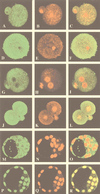
Expression of YY1 protein in wild-type murine preimplantation embryos. Immunofluorescent detection of YY1 protein was performed on wild-type preimplantation embryos by confocal microscopy. Embryos were stained with monoclonal and polyclonal (not shown) anti-YY1 antibodies and polyclonal anti-TBP and anti-Sp1 antibodies. The leftmost column represents embryos stained with these primary antibodies and visualized with FITC-conjugated secondary antibodies. The middle column represents the same embryos stained with propidium iodide that visualizes DNA. The rightmost column shows superimposed images of the antibody and DNA staining. (A to C) YY1 expression in a one-cell unfertilized oocyte; (D to F) TBP expression in a one-cell unfertilized oocyte; (G to I) YY1 expression in a one-cell fertilized embryo; (J to L) YY1 expression in a two-cell embryo. Note the pronounced YY1 expression in the nucleus as shown by the intense yellow signal as a result of the colocalization of the YY1 signal (green) and the DNA signal (red) (L). (M to O) YY1 expression in the inner cell mass and trophoblast of an E3.5 blastocyst; (P to R) Sp1 expression in the nucleus of a blastocyst. Note the intense yellow signal as a result of colocalization of the Sp1 signal (green) with DNA signal (red) (R).
FIG. 4
YY1 has a widespread expression pattern in the developing embryo. Whole-mount in situ hybridization of E7.5 (A), E8.5 (B), E9.5 (C), and E12.5 (D) wild-type embryos with a YY1 antisense riboprobe. Hybridization of an E12.5 embryo with a YY1 sense riboprobe was included as a negative control (E). The Brachyury (T) riboprobe was used as a positive control (5a).
FIG. 5
Exencephaly and growth retardation in a subset of YY1+/− mice. (A) E13.5 littermates obtained from mixed-strain heterozygous intercrosses (129/Sv × C57BL/6) genotyped as YY1+/+ on the left and YY1+/− on the right. The YY1+/− embryo that is exencephalic with an open brain is shown on the right. (B) A coronal histological section (4-μm thickness) of the exencephalic YY1+/− embryo observed in panel A stained with hematoxylin and eosin as described in Materials and Methods shows pseudoventricles and asymmetry. (C) An entire litter of E10.5 mice obtained from a YY1+/− male crossed to a YY1+/+ wild-type female on the inbred C57BL/6 genetic background. The two smaller embryos in the right-hand corner indicated by arrows were genotyped as YY1+/− whereas the other embryos were either YY1+/− or YY1+/+.
Similar articles
- Expression, activity, and subcellular localization of the Yin Yang 1 transcription factor in Xenopus oocytes and embryos.
Ficzycz A, Eskiw C, Meyer D, Marley KE, Hurt M, Ovsenek N. Ficzycz A, et al. J Biol Chem. 2001 Jun 22;276(25):22819-25. doi: 10.1074/jbc.M011188200. Epub 2001 Apr 6. J Biol Chem. 2001. PMID: 11294833 - Essential dosage-dependent functions of the transcription factor yin yang 1 in late embryonic development and cell cycle progression.
Affar el B, Gay F, Shi Y, Liu H, Huarte M, Wu S, Collins T, Li E, Shi Y. Affar el B, et al. Mol Cell Biol. 2006 May;26(9):3565-81. doi: 10.1128/MCB.26.9.3565-3581.2006. Mol Cell Biol. 2006. PMID: 16611997 Free PMC article. - Everything you have ever wanted to know about Yin Yang 1.....
Shi Y, Lee JS, Galvin KM. Shi Y, et al. Biochim Biophys Acta. 1997 Apr 18;1332(2):F49-66. doi: 10.1016/s0304-419x(96)00044-3. Biochim Biophys Acta. 1997. PMID: 9141463 Review. No abstract available. - The Yin and Yang of YY1 in tumor growth and suppression.
Khachigian LM. Khachigian LM. Int J Cancer. 2018 Aug 1;143(3):460-465. doi: 10.1002/ijc.31255. Epub 2018 Jan 31. Int J Cancer. 2018. PMID: 29322514 Review.
Cited by
- Essential and unexpected role of Yin Yang 1 to promote mesodermal cardiac differentiation.
Gregoire S, Karra R, Passer D, Deutsch MA, Krane M, Feistritzer R, Sturzu A, Domian I, Saga Y, Wu SM. Gregoire S, et al. Circ Res. 2013 Mar 15;112(6):900-10. doi: 10.1161/CIRCRESAHA.113.259259. Epub 2013 Jan 10. Circ Res. 2013. PMID: 23307821 Free PMC article. - Cistrome analysis of YY1 uncovers a regulatory axis of YY1:BRD2/4-PFKP during tumorigenesis of advanced prostate cancer.
Xu C, Tsai YH, Galbo PM, Gong W, Storey AJ, Xu Y, Byrum SD, Xu L, Whang YE, Parker JS, Mackintosh SG, Edmondson RD, Tackett AJ, Huang J, Zheng D, Earp HS, Wang GG, Cai L. Xu C, et al. Nucleic Acids Res. 2021 May 21;49(9):4971-4988. doi: 10.1093/nar/gkab252. Nucleic Acids Res. 2021. PMID: 33849067 Free PMC article. - YY1 safeguard multidimensional epigenetic landscape associated with extended pluripotency.
Dong X, Guo R, Ji T, Zhang J, Xu J, Li Y, Sheng Y, Wang Y, Fang K, Wen Y, Liu B, Hu G, Deng H, Yao H. Dong X, et al. Nucleic Acids Res. 2022 Nov 28;50(21):12019-12038. doi: 10.1093/nar/gkac230. Nucleic Acids Res. 2022. PMID: 35425987 Free PMC article. - Co-binding by YY1 identifies the transcriptionally active, highly conserved set of CTCF-bound regions in primate genomes.
Schwalie PC, Ward MC, Cain CE, Faure AJ, Gilad Y, Odom DT, Flicek P. Schwalie PC, et al. Genome Biol. 2013 Dec 31;14(12):R148. doi: 10.1186/gb-2013-14-12-r148. Genome Biol. 2013. PMID: 24380390 Free PMC article.
References
- Brown J L, Lesley D, Whiteley M, Dirksen M L, Kassis J A. The Drosophila polycomb group gene pleiohomeotic encodes a DNA binding protein with homology to the transcription factor YY1. Mol Cell. 1998;1:1057–1064. - PubMed
- Bushmeyer S, Park K-S, Atchison M L. Characterization of functional domains within the multifunctional transcription factor, YY1. J Biol Chem. 1996;270:30213–30220. - PubMed
- Campbell L R, Datton D H, Sohal G S. Neural tube defects: a review of human and animal studies on the etiology of neural tube defects. Teratology. 1986;34:171–187. - PubMed
- Copp A J. Deaths before birth: clues from gene knockouts and mutations. Trends Genet. 1995;11:87–93. - PubMed
- DeGregori J, Russ A, von Melchner H, Rayburn H, Priyaranjan P, Jenkins N A, Copeland N G, Ruley H E. A murine homolog of the yeast RNA1 gene is required for postimplantation development. Genes Dev. 1994;8:265–276. - PubMed
Publication types
MeSH terms
Substances
LinkOut - more resources
Full Text Sources
Other Literature Sources
Molecular Biology Databases


