Rescue of influenza A virus from recombinant DNA - PubMed (original) (raw)
Rescue of influenza A virus from recombinant DNA
E Fodor et al. J Virol. 1999 Nov.
Abstract
We have rescued influenza A virus by transfection of 12 plasmids into Vero cells. The eight individual negative-sense genomic viral RNAs were transcribed from plasmids containing human RNA polymerase I promoter and hepatitis delta virus ribozyme sequences. The three influenza virus polymerase proteins and the nucleoprotein were expressed from protein expression plasmids. This plasmid-based reverse genetics technique facilitates the generation of recombinant influenza viruses containing specific mutations in their genes.
Figures
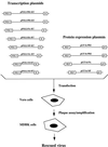
FIG. 1
Schematic representation of the plasmid-based rescue system for influenza A virus. The pGT-h set of protein expression plasmids was constructed by inserting the open reading frames of PB1, PB2, PA, and NP proteins into the _Bcl_I cloning site of the pGT-h plasmid (2). The PB1 and PA genes were derived from influenza A/WSN/33 virus. The PB2 and NP genes were derived from influenza A/PR/8/34 virus. The viral genomic sequences of influenza A/WSN/33 virus were cloned into pUC18- or pUC19-based plasmids between a truncated human RNA Pol I promoter (nt −250 to −1) (19) and sequences of the hepatitis delta virus ribozyme in an analogous way as described for pPOLI-CAT-RT (28). Genetic tags were inserted into the HA- and NA-encoding plasmids by using conventional mutagenic techniques. For viral rescue, 5 μg of each of the polymerase protein expression plasmids (pGT-h-PB1, pGT-h-PB2, and pGT-h-PA), 10 μg of the NP-expressing pGT-h-NP, and 3 μg of each of the eight vRNA-coding transcription plasmids (pPOLI-PB2-RT, pPOLI-PB1-RT, pPOLI-PA-RT, pPOLI-HA-RT, pPOLI-NP-RT, pPOLI-NA-RT, pPOLI-M-RT, and pPOLI-NS-RT) were diluted to a concentration of 0.1 μg/μl in 20 mM HEPES buffer (pH 7.5). The DNA solution was added to diluted DOTAP liposomal transfection reagent (Boehringer) containing 240 μl of DOTAP and 720 μl of 20 mM HEPES buffer (pH 7.5). The transfection mixture was incubated at room temperature for 15 min, mixed with 6.5 ml of MEM containing 0.5% fetal calf serum, 0.3% bovine serum albumin, penicillin, and streptomycin and added to near-confluent Vero cells washed with phosphate-buffered saline in 8.5-cm dishes (about 107 cells). At 24 h after transfection, the transfection mixture was removed and the cells were incubated with 8 ml of fresh medium, which was replaced daily for 4 days. The harvested medium from transfected dishes was screened for rescued influenza virus by plaquing and amplification on MDBK cells. POL I, truncated human RNA polymerase I promoter; R, genomic hepatitis virus ribozyme; MLP, adenovirus type 2 major late promoter; pA, polyadenylation sequence from SV40.
FIG. 2
Demonstration of the presence of genetic tags in the HA and NA vRNA segments of the rescued virus by RT-PCR and restriction enzyme analysis. vRNA of the rescued virus was isolated from medium of infected MDBK cells. One hundred microliters of the medium was treated with 5 U of RNase-free DNase to remove any residual plasmid DNA carried over. After 15 min at 37°C, vRNA was isolated by using the RNeasy Mini Kit (Qiagen), following the manufacturer’s instructions. vRNA from authentic wild-type A/WSN/33 virus was isolated from purified virus as described previously (10). The first 149 nt at the 3′ end of the HA vRNA were amplified by RT-PCR using oligonucleotide primers 5′-GCGCTCTAGAGCAAAAGCAGGGGAAAATAA-3′ (corresponding to nt 1 to 21) and 5′-CGCGAAGCTTCTCGAATATTGTGTCAAC-3′ (corresponding to nt 129 to 149), resulting in a 165-nt-long PCR product. To amplify the sequence containing the genetic tag from the NA segment, primers 5′-TGGACTAGTGGGAGCATCAT-3′ (corresponding to nt 1280 to 1309) and 5′-GAACAAACTACTTGTCAATGGT-3′ (corresponding to nt 1367 to 1388) were used in RT-PCR to produce a 108-nt PCR product. The HA- and NA-specific PCR products were incubated for 2 h at 37°C in the presence (+) or absence (−) of 10 U of _Spe_I and _Sac_I restriction enzymes, respectively. Samples were analyzed on 16% polyacrylamide gels and stained with ethidium bromide. Lanes: 1, DNA size markers (sizes in nucleotides are indicated); 2, 3, 7, and 8, PCR products from the rescued virus; 4 and 9, control reactions omitting reverse transcriptase; 5, 6, 10, and 11, PCR products from the authentic wild-type A/WSN virus.
Similar articles
- A DNA transfection system for generation of influenza A virus from eight plasmids.
Hoffmann E, Neumann G, Kawaoka Y, Hobom G, Webster RG. Hoffmann E, et al. Proc Natl Acad Sci U S A. 2000 May 23;97(11):6108-13. doi: 10.1073/pnas.100133697. Proc Natl Acad Sci U S A. 2000. PMID: 10801978 Free PMC article. - A reverse-genetics system for Influenza A virus using T7 RNA polymerase.
de Wit E, Spronken MIJ, Vervaet G, Rimmelzwaan GF, Osterhaus ADME, Fouchier RAM. de Wit E, et al. J Gen Virol. 2007 Apr;88(Pt 4):1281-1287. doi: 10.1099/vir.0.82452-0. J Gen Virol. 2007. PMID: 17374773 - A plasmid-based reverse genetics system for influenza A virus.
Pleschka S, Jaskunas R, Engelhardt OG, Zürcher T, Palese P, García-Sastre A. Pleschka S, et al. J Virol. 1996 Jun;70(6):4188-92. doi: 10.1128/JVI.70.6.4188-4192.1996. J Virol. 1996. PMID: 8648766 Free PMC article. - Reverse genetics and influenza virus research.
Kiraly J, Kostolansky F. Kiraly J, et al. Acta Virol. 2009;53(4):217-24. doi: 10.4149/av_2009_04_217. Acta Virol. 2009. PMID: 19941384 Review. No abstract available. - Plasmid-only rescue of influenza A virus vaccine candidates.
Schickli JH, Flandorfer A, Nakaya T, Martinez-Sobrido L, García-Sastre A, Palese P. Schickli JH, et al. Philos Trans R Soc Lond B Biol Sci. 2001 Dec 29;356(1416):1965-73. doi: 10.1098/rstb.2001.0979. Philos Trans R Soc Lond B Biol Sci. 2001. PMID: 11779399 Free PMC article. Review.
Cited by
- Rescue of Infectious Salmon Anemia Virus (ISAV) from Cloned cDNA.
Toro-Ascuy D, Cárdenas M, Vásquez-Martínez Y, Cortez-San Martín M. Toro-Ascuy D, et al. Methods Mol Biol. 2024;2733:87-99. doi: 10.1007/978-1-0716-3533-9_6. Methods Mol Biol. 2024. PMID: 38064028 - The short stalk length of highly pathogenic avian influenza H5N1 virus neuraminidase limits transmission of pandemic H1N1 virus in ferrets.
Blumenkrantz D, Roberts KL, Shelton H, Lycett S, Barclay WS. Blumenkrantz D, et al. J Virol. 2013 Oct;87(19):10539-51. doi: 10.1128/JVI.00967-13. Epub 2013 Jul 17. J Virol. 2013. PMID: 23864615 Free PMC article. - Recombinant influenza A viruses with enhanced levels of PB1 and PA viral protein expression.
Belicha-Villanueva A, Rodriguez-Madoz JR, Maamary J, Baum A, Bernal-Rubio D, Minguito de la Escalera M, Fernandez-Sesma A, García-Sastre A. Belicha-Villanueva A, et al. J Virol. 2012 May;86(10):5926-30. doi: 10.1128/JVI.06384-11. Epub 2012 Mar 7. J Virol. 2012. PMID: 22398284 Free PMC article. - Structure-based discovery of the novel antiviral properties of naproxen against the nucleoprotein of influenza A virus.
Lejal N, Tarus B, Bouguyon E, Chenavas S, Bertho N, Delmas B, Ruigrok RW, Di Primo C, Slama-Schwok A. Lejal N, et al. Antimicrob Agents Chemother. 2013 May;57(5):2231-42. doi: 10.1128/AAC.02335-12. Epub 2013 Mar 4. Antimicrob Agents Chemother. 2013. PMID: 23459490 Free PMC article. - Cell culture-based influenza vaccines: A necessary and indispensable investment for the future.
Hegde NR. Hegde NR. Hum Vaccin Immunother. 2015;11(5):1223-34. doi: 10.1080/21645515.2015.1016666. Hum Vaccin Immunother. 2015. PMID: 25875691 Free PMC article. Review.
References
- Berg D T, McClure D B, Grinnel B W. High-level expression of secreted proteins from cells adapted to serum-free suspension culture. BioTechniques. 1993;14:972–978. - PubMed
- Collins P L, Hill M G, Camargo E, Grosfeld H, Chanock R M, Murphy B R. Production of infectious human respiratory syncytial virus from cloned cDNA confirms an essential role for the transcription elongation factor from the 5′ proximal open reading frame of the M2 mRNA in gene expression and provides a capability for vaccine development. Proc Natl Acad Sci USA. 1995;92:11563–11567. - PMC - PubMed
Publication types
MeSH terms
Substances
LinkOut - more resources
Full Text Sources
Other Literature Sources

