The reduced expression of 6Ckine in the plt mouse results from the deletion of one of two 6Ckine genes - PubMed (original) (raw)
The reduced expression of 6Ckine in the plt mouse results from the deletion of one of two 6Ckine genes
G Vassileva et al. J Exp Med. 1999.
Abstract
6Ckine is an unusual chemokine capable of attracting naive T lymphocytes in vitro. It has been recently reported that lack of 6Ckine expression in lymphoid organs is a prominent characteristic of mice homozygous for the paucity of lymph node T cell (plt) mutation. These mice show reduced numbers of T cells in lymph nodes, Peyer's patches, and the white pulp of the spleen. The genetic reason for the lack of 6Ckine expression in the plt mouse, however, has remained unknown. Here we demonstrate that mouse 6Ckine is encoded by two genes, one of which is expressed in lymphoid organs and is deleted in plt mice. A second 6Ckine gene is intact and expressed in the plt mouse.
Figures
Figure 1
PCR and Southern blot analysis of the 6Ckine gene locus reveals the existence of two 6Ckine genes, only one of which is detected in the plt mouse. (A) EcoRI-digested DNA from 6CKBAC1, 6CKBAC2, plt, and 129/sv mice was analyzed by Southern blot analysis using a 32P-labeled NheI/BstXI fragment (271 bp) as a probe (probe B, Fig. 3 C). The probe specifically hybridized to 6- and 1.35-kb fragments in 6CKBAC1 DNA, 3- and 1.2-kb fragments in 6CKBAC2, 6- and 1.35-kb fragments in plt mouse genomic DNA, and all four fragments in 129/sv genomic DNA. (B) PCR analysis: PCR primers GV104/GV125 (see Fig. 2) were used to amplify a segment of the 3′ region of the 6Ckine gene. The resulting PCR products were analyzed by agarose gel electrophoresis and visualized with ethidium bromide. Two bands of 1.35 and 1.2 kb were detected in a PCR reaction from genomic DNA from several mouse strains (as indicated). Single bands of 1.35 or 1.2 kb were detected from 6CKBAC1 and 6CKBAC2 DNA, respectively, whereas a single band of 1.35 kb was amplified from the plt mouse genomic DNA.
Figure 2
(A) The 6Ckine genes found on the two BACs are distinct. A schematic of the sequence of SacI fragments (7.5 kb) from 6CKBAC1 (leu) and 6CKBAC2 (ser) is shown. Black and white boxes represent 6Ckine exons. Open triangles indicate larger deletions. Single base differences outside of the exons are not shown. The four nucleotide differences identified in coding regions (black boxes) and noncoding regions (white box) of the two 6Ckine genes are shown. Vertical arrows identify the amino acid difference at position 65. Horizontal arrows indicate the positions of the PCR primers used for analysis of 6Ckine gene locus. nt, nucleotide. (B) Sequences of the intron–exon boundaries are identical between the two copies with the exception of the intron2–exon3 junction. The 6Ckine-ser sequence is added to the intron2–exon3 boundary sequence to illustrate the single nucleotide difference (arrow). These sequence data are available from EMBL/GenBank/DDBJ under accession numbers AF171085 and AF171086 for 6Ckine-leu and 6Ckine-ser, respectively.
Figure 3
Southern blot analysis of 6Ckine genomic loci suggests that the 6Ckine-ser gene of the plt mouse contains significant deletions. (A) EcoRI-digested genomic DNA from 6CKBAC1 and 6CKBAC2 and genomic DNA from 129/sv and plt mice were probed with a 0.8-kb SacI/NheI fragment (probe A, indicated). An 8-kb fragment was recognized in 6CKBAC1, and a 6.5-kb fragment was recognized in 6CKBAC2. The plt mouse genomic DNA contained only an 8-kb fragment DNA, whereas both 8- and 6.5-kb fragments hybridized in 129/sv DNA. (B) HindIII-digested DNA from 6CKBAC1 and 6CKBAC2 and genomic DNA from 129/sv and plt mice was probed with a 271-bp NheI/BstXI fragment (probe B, indicated). A 9-kb fragment hybridized in 6CKBAC1 and an 11-kb fragment hybridized in 6CKBAC2 DNA. Only a 9-kb fragment was recognized in plt genomic DNA, whereas both 9- and 11-kb fragments were present in 129/sv DNA. (C) Schematic of the 6Ckine genomic locus, with positions of initiation and stop codons indicated by ○ and •, respectively. The positions of the hybridization probes are indicated by shaded boxes, and the sizes of the fragments recognized by these probes relative to each BAC are indicated.
Figure 4
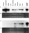
6Ckine is expressed in both wild-type and plt mice. Total RNA (20 μg/lane) from either BALB/c (A) or plt (C) mouse tissues was hybridized with a 32P-labeled 6Ckine probe and subjected to autoradiography. BALB/c mice showed 6Ckine expression in various lymphoid and nonlymphoid tissues, whereas plt mice showed expression only in nonlymphoid tissue. Total spleen RNA (20 μg) from BALB/c mice was included as a positive control in each blot. Ethidium bromide–stained gel of BALB/c and plt RNA is shown in B and D, respectively.
Similar articles
- Mice lacking expression of the chemokines CCL21-ser and CCL19 (plt mice) demonstrate delayed but enhanced T cell immune responses.
Mori S, Nakano H, Aritomi K, Wang CR, Gunn MD, Kakiuchi T. Mori S, et al. J Exp Med. 2001 Jan 15;193(2):207-18. doi: 10.1084/jem.193.2.207. J Exp Med. 2001. PMID: 11148224 Free PMC article. - Mice lacking expression of secondary lymphoid organ chemokine have defects in lymphocyte homing and dendritic cell localization.
Gunn MD, Kyuwa S, Tam C, Kakiuchi T, Matsuzawa A, Williams LT, Nakano H. Gunn MD, et al. J Exp Med. 1999 Feb 1;189(3):451-60. doi: 10.1084/jem.189.3.451. J Exp Med. 1999. PMID: 9927507 Free PMC article. - The CC chemokine thymus-derived chemotactic agent 4 (TCA-4, secondary lymphoid tissue chemokine, 6Ckine, exodus-2) triggers lymphocyte function-associated antigen 1-mediated arrest of rolling T lymphocytes in peripheral lymph node high endothelial venules.
Stein JV, Rot A, Luo Y, Narasimhaswamy M, Nakano H, Gunn MD, Matsuzawa A, Quackenbush EJ, Dorf ME, von Andrian UH. Stein JV, et al. J Exp Med. 2000 Jan 3;191(1):61-76. doi: 10.1084/jem.191.1.61. J Exp Med. 2000. PMID: 10620605 Free PMC article. - The CC chemokine 6Ckine binds the CXC chemokine receptor CXCR3.
Soto H, Wang W, Strieter RM, Copeland NG, Gilbert DJ, Jenkins NA, Hedrick J, Zlotnik A. Soto H, et al. Proc Natl Acad Sci U S A. 1998 Jul 7;95(14):8205-10. doi: 10.1073/pnas.95.14.8205. Proc Natl Acad Sci U S A. 1998. PMID: 9653165 Free PMC article.
Cited by
- Graft Site Microenvironment Determines Dendritic Cell Trafficking Through the CCR7-CCL19/21 Axis.
Hua J, Stevenson W, Dohlman TH, Inomata T, Tahvildari M, Calcagno N, Pirmadjid N, Sadrai Z, Chauhan SK, Dana R. Hua J, et al. Invest Ophthalmol Vis Sci. 2016 Mar;57(3):1457-67. doi: 10.1167/iovs.15-17551. Invest Ophthalmol Vis Sci. 2016. PMID: 27031839 Free PMC article. - Structure and Immune Function of Afferent Lymphatics and Their Mechanistic Contribution to Dendritic Cell and T Cell Trafficking.
Arasa J, Collado-Diaz V, Halin C. Arasa J, et al. Cells. 2021 May 20;10(5):1269. doi: 10.3390/cells10051269. Cells. 2021. PMID: 34065513 Free PMC article. Review. - The CCR7 ligand elc (CCL19) is transcytosed in high endothelial venules and mediates T cell recruitment.
Baekkevold ES, Yamanaka T, Palframan RT, Carlsen HS, Reinholt FP, von Andrian UH, Brandtzaeg P, Haraldsen G. Baekkevold ES, et al. J Exp Med. 2001 May 7;193(9):1105-12. doi: 10.1084/jem.193.9.1105. J Exp Med. 2001. PMID: 11342595 Free PMC article. - Lymphatic endothelial cells - key players in regulation of tolerance and immunity.
Tewalt EF, Cohen JN, Rouhani SJ, Engelhard VH. Tewalt EF, et al. Front Immunol. 2012 Sep 28;3:305. doi: 10.3389/fimmu.2012.00305. eCollection 2012. Front Immunol. 2012. PMID: 23060883 Free PMC article. - Increased number of mature dendritic cells in Crohn's disease: evidence for a chemokine mediated retention mechanism.
Middel P, Raddatz D, Gunawan B, Haller F, Radzun HJ. Middel P, et al. Gut. 2006 Feb;55(2):220-7. doi: 10.1136/gut.2004.063008. Epub 2005 Aug 23. Gut. 2006. PMID: 16118351 Free PMC article.
References
- Nagira M., Imai T., Hieshima K., Kusuda J., Ridanpaa M., Takagi S., Nishimura M., Kakizaki M., Nomiyama H., Yoshie O. Molecular cloning of a novel human CC chemokine secondary lymphoid-tissue chemokine that is a potent chemoattractant for lymphocytes and mapped to chromosome 9p13. J. Biol. Chem. 1997;272:19518–19524. - PubMed
- Hromas R., Kim C.H., Klemsz M., Krathwohl M., Fife K., Cooper S., Schnizlein-Bick C., Broxmeyer H.E. Isolation and characterization of Exodus-2, a novel C-C chemokine with a unique 37-amino acid carboxyl-terminal extension. J. Immunol. 1997;159:2554–2558. - PubMed
- Tanabe S., Lu Z., Luo Y., Quackenbush E., Berman M., Collins-Racie L., Mi S., Reilly C., Lo D., Jacobs K. Identification of a new mouse β-chemokine, thymus-derived chemotactic agent 4, with activity on T lymphocytes and mesangial cells. J. Immunol. 1997;159:5671–5679. - PubMed
- Hedrick J.A., Zlotnik A. Identification and characterization of a novel beta chemokine containing six conserved cysteines. J. Immunol. 1997;159:1589–1593. - PubMed
- Willimann K., Legler D.F., Loetscher M., Roos R.S., Delgado M.B., Clark-Lewis I., Baggiolini M., Moser B. The chemokine SLC is expressed in T cell areas of lymph nodes and mucosal lymphoid tissues and attracts activated T cells via CCR7. Eur. J. Immunol. 1998;28:2025–2034. - PubMed


