Running enhances neurogenesis, learning, and long-term potentiation in mice - PubMed (original) (raw)
Running enhances neurogenesis, learning, and long-term potentiation in mice
H van Praag et al. Proc Natl Acad Sci U S A. 1999.
Abstract
Running increases neurogenesis in the dentate gyrus of the hippocampus, a brain structure that is important for memory function. Consequently, spatial learning and long-term potentiation (LTP) were tested in groups of mice housed either with a running wheel (runners) or under standard conditions (controls). Mice were injected with bromodeoxyuridine to label dividing cells and trained in the Morris water maze. LTP was studied in the dentate gyrus and area CA1 in hippocampal slices from these mice. Running improved water maze performance, increased bromodeoxyuridine-positive cell numbers, and selectively enhanced dentate gyrus LTP. Our results indicate that physical activity can regulate hippocampal neurogenesis, synaptic plasticity, and learning.
Figures
Figure 1
Flowchart of the experiment. Controls were housed in standard 30- by 18-cm cages, whereas runners had 48- by 26-cm housing with free access to a running wheel (1). Mice in both conditions received BrdU (50 μg/g per day) injections for the first 10 days after their housing assignment. After 1 month in their respective environments, mice were tested in the water maze (2) between days 30 and 36 or between days 43 and 49. Mice were anesthetized and decapitated between days 54 and 118; one hemisphere was used for electrophysiology; the other was used for immunocytochemistry (3).
Figure 2
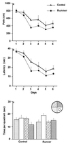
Water maze learning in controls and runners trained with two trials per day (four-trial data are not shown here, but see description in Results). Mice were trained over 6 days to find the hidden platform in the Morris water maze. A significant difference developed between the groups (P < 0.04) in path length (a) and (P < 0.047) in latency (b). Results of probe test 4 hr after the last trial on day 6 (c).
Figure 3
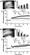
LTP in dentate gyrus (a) and area CA1 (b). (a1, b1) Digital images of hippocampal slices showing the position of the stimulation and recording electrodes. (a2, b2) Paired-pulse facilitation at 50-, 100-, 200-, and 500-ms interpulse intervals. EPSP, excitatory postsynaptic potential. There was no difference between slices from controls and runners (P > 0.91). (a3, b3) Time course of LTP in slices from controls (▵) and runners (●) . In addition, representative examples are shown of evoked responses immediately before (Pre) and 30 min after (Post) induction of LTP. Example waveforms are the average of 20 responses recorded over a 5-min period. Population spikes were apparent in some animals in each group after LTP induction. (Scale bars under a1 and b1 indicate 250 μm.)
Figure 4
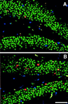
Confocal images of BrdU-positive cells in control (A) and runner coronal sections (B). Sections were immunofluorescent triple-labeled for BrdU (red), NeuN, indicating neuronal phenotype (green), and S100β, selective for glial phenotype (blue). (Scale bar indicates 50 μm.)
Similar articles
- Effects of voluntary exercise on synaptic plasticity and gene expression in the dentate gyrus of adult male Sprague-Dawley rats in vivo.
Farmer J, Zhao X, van Praag H, Wodtke K, Gage FH, Christie BR. Farmer J, et al. Neuroscience. 2004;124(1):71-9. doi: 10.1016/j.neuroscience.2003.09.029. Neuroscience. 2004. PMID: 14960340 - Functional analysis of neurovascular adaptations to exercise in the dentate gyrus of young adult mice associated with cognitive gain.
Clark PJ, Brzezinska WJ, Puchalski EK, Krone DA, Rhodes JS. Clark PJ, et al. Hippocampus. 2009 Oct;19(10):937-50. doi: 10.1002/hipo.20543. Hippocampus. 2009. PMID: 19132736 Free PMC article. - Lack of JWA Enhances Neurogenesis and Long-Term Potentiation in Hippocampal Dentate Gyrus Leading to Spatial Cognitive Potentiation.
Sha S, Xu J, Lu ZH, Hong J, Qu WJ, Zhou JW, Chen L. Sha S, et al. Mol Neurobiol. 2016 Jan;53(1):355-368. doi: 10.1007/s12035-014-9010-4. Epub 2014 Nov 30. Mol Neurobiol. 2016. PMID: 25432888 - Synaptic plasticity and learning and memory: LTP and beyond.
Hölscher C. Hölscher C. J Neurosci Res. 1999 Oct 1;58(1):62-75. J Neurosci Res. 1999. PMID: 10491572 Review. - Stress modulation of hippocampal activity--spotlight on the dentate gyrus.
Fa M, Xia L, Anunu R, Kehat O, Kriebel M, Volkmer H, Richter-Levin G. Fa M, et al. Neurobiol Learn Mem. 2014 Jul;112:53-60. doi: 10.1016/j.nlm.2014.04.008. Epub 2014 Apr 18. Neurobiol Learn Mem. 2014. PMID: 24747273 Review.
Cited by
- Exercise, memory, and the hippocampus: Uncovering modifiable lifestyle reserve factors in refractory epilepsy.
Stasenko A, Kaestner E, Schadler A, Brady E, Rodriguez J, Roth RW, Gleichgerrcht E, Helm JL, Drane DL, McDonald CR. Stasenko A, et al. Epilepsy Behav Rep. 2024 Oct 28;28:100721. doi: 10.1016/j.ebr.2024.100721. eCollection 2024. Epilepsy Behav Rep. 2024. PMID: 39555495 Free PMC article. - Running in mice increases the expression of brain hemoglobin-related genes interacting with the GH/IGF-1 system.
Walser M, Karlsson L, Motalleb R, Isgaard J, Kuhn HG, Svensson J, Åberg ND. Walser M, et al. Sci Rep. 2024 Oct 26;14(1):25464. doi: 10.1038/s41598-024-77489-1. Sci Rep. 2024. PMID: 39462081 Free PMC article. - Entorhinal cortex-hippocampal circuit connectivity in health and disease.
Hernández-Frausto M, Vivar C. Hernández-Frausto M, et al. Front Hum Neurosci. 2024 Sep 20;18:1448791. doi: 10.3389/fnhum.2024.1448791. eCollection 2024. Front Hum Neurosci. 2024. PMID: 39372192 Free PMC article. Review. - The intricate interplay between microglia and adult neurogenesis in Alzheimer's disease.
Früholz I, Meyer-Luehmann M. Früholz I, et al. Front Cell Neurosci. 2024 Sep 18;18:1456253. doi: 10.3389/fncel.2024.1456253. eCollection 2024. Front Cell Neurosci. 2024. PMID: 39360265 Free PMC article. Review. - Effect of Aerobic Exercise versus Non-Invasive Brain Stimulation on Cognitive Function in Multiple Sclerosis: A Systematic Review and Meta-Analysis.
Elkhooly M, Di Stadio A, Bernitsas E. Elkhooly M, et al. Brain Sci. 2024 Jul 30;14(8):771. doi: 10.3390/brainsci14080771. Brain Sci. 2024. PMID: 39199465 Free PMC article. Review.
References
- Gage F H, Kempermann G, Palmer T D, Peterson D A, Ray J. J Neurobiol. 1998;36:249–266. - PubMed
- Goldman S A, Luskin M B. Trends Neurosci. 1998;21:107–114. - PubMed
- Kempermann G, Kuhn H G, Gage F H. Nature (London) 1997;386:493–495. - PubMed
- van Praag H, Kempermann G, Gage F H. Nat Neurosci. 1999;2:266–270. - PubMed
Publication types
MeSH terms
LinkOut - more resources
Full Text Sources
Other Literature Sources
Molecular Biology Databases
Miscellaneous


