Nerve growth factor is an autocrine factor essential for the survival of macrophages infected with HIV - PubMed (original) (raw)
Nerve growth factor is an autocrine factor essential for the survival of macrophages infected with HIV
E Garaci et al. Proc Natl Acad Sci U S A. 1999.
Abstract
Nerve growth factor (NGF) is a neurotrophin with the ability to exert specific effects on cells of the immune system. Human monocytes/macrophages (M/M) infected in vitro with HIV type 1 (HIV-1) are able to produce substantial levels of NGF that are associated with enhanced expression of the high-affinity NGF receptor (p140 trkA) on the M/M surface. Treatment of HIV-infected human M/M with anti-NGF Ab blocking the biological activity of NGF leads to a marked decrease of the expression of p140 trkA high-affinity receptor, a concomitant increased expression of p75(NTR) low-affinity receptor for NGF, and the occurrence of apoptotic death of M/M. Taken together, these findings suggest a role for NGF as an autocrine survival factor that rescues human M/M from the cytopathic effect caused by HIV infection.
Figures
Figure 1
NGF production by macrophages infected with HIV. NGF production was assessed in the supernatants of M/M immediately before virus challenge (A, small dots) and 5 days after mock infection (big dots) or HIV infection (hatched bar). Immunocytochemical analysis was performed on M/M (B) at the same time points, that is, immediately before virus challenge (A) and 5 days after mock infection (B) or HIV infection (C). NGF production in the supernatants of HIV-infected M/M is statistically greater than that found in the supernatants of mock-infected M/M (either before or after mock infection) [ANOVA:F(2, 6) = 96.448; P = 0.001]. Values represent mean ± SE of an experiment representative of three.
Figure 2
Expression of high-affinity receptors on macrophages infected by HIV. Treatment of HIV-infected M/M with anti-NGF Ab (M/M + HIV + anti-NGF), but not with an IgG isotypic Ab (M/M + HIV + IgG), modifies the cellular expression of p140 trkA high-affinity receptor. Quantitative analysis of p140 trkA-immunoreactive cells carried out with a computerized image-analysis system (Zeiss Axiophot 2 microscope equipped with a Vidas Kontron system) shows that the increase of p140 trkA immunopositive cells in HIV-infected human M/M (M/M + HIV) was statistically significant (ANOVA: F(4, 85) = 116.017;P < 0.01) compared with mock-infected human M/M (M/M). Treatment of HIV-infected M/M with anti-NGF Ab (M/M + HIV + anti-NGF) yielded a statistically significant decrease (P < 0.01) of p140 trkA immunoreactivity in comparison to HIV-infected M/M (M/M + HIV), mock-infected human M/M (M/M + anti-NGF), or HIV-infected M/M with an irrelevant IgG Ab (M/M + HIV + IgG).
Figure 3
Expression of low-affinity receptors in macrophages infected with HIV. A dramatic increase of p75NTR receptor expression was consistently obtained by treating HIV-infected M/M with anti-NGF Ab. An IgG isotypic Ab was totally ineffective. A quantitative analysis of p75NTR immunoreactive cells (carried out with a computerized image-analysis system) shows a statistically significant increase of p75NTR immunopositive cells in HIV-infected human M/M treated with anti-NGF Ab compared with all other samples, either infected or not (ANOVA: F(4, 85) = 289.354;P = 0.001).
Figure 4
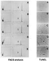
Programmed cell death in HIV-infected macrophages exposed to anti-NGF Ab. (Left) FACS analysis. Apoptosis was detected by DNA labeling with propidium iodide, a fluorescent intercalating dye that allows DNA quantification. Apoptotic nuclei appeared as a broad hypodiploid DNA peak (black arrow in each panel) easily discriminable from the narrow peak of nuclei with normal diploid DNA counted in the red fluorescence channel. DNA fragmentation has been detected in 5% of mock-infected human M/M (A). Results in the same range were observed in HIV-infected M/M (9.1% of propidium-positive cells; B), in mock-infected M/M exposed to anti-NGF Ab (6.9%; C), or in HIV-infected M/M treated with the IgG isotypic Ab (8.7%; E). By contrast, exposure of HIV-infected cells to anti-NGF Ab (D) induces DNA fragmentation in 40.3% of M/M in this experiment representative of five different tests. (Right) TUNEL. Immunocytochemical studies performed by TUNEL show nuclei with round condensed chromatin in HIV-infected M/M exposed to anti-NGF Ab (D) far more than in any of the other M/M samples tested (A, B,C, and E). The figure presents data from a typical experiment of three.
Figure 5
Effect of NGF starvation upon HIV production in macrophages. A dramatic difference of virus production was detected between HIV-infected M/M exposed to anti-NGF Ab (■), HIV-infected M/M (●), or HIV-infected M/M exposed to an IgG-isotypic irrelevant Ab (▴). HIV p24 gag antigen production was evaluated by commercially available ELISA at days 2, 5, 8, 11, and 14 after virus infection. The figure represents a typical experiment of three.
Similar articles
- NGF ligand alters NGF signaling via p75(NTR) and trkA.
Niederhauser O, Mangold M, Schubenel R, Kusznir EA, Schmidt D, Hertel C. Niederhauser O, et al. J Neurosci Res. 2000 Aug 1;61(3):263-72. doi: 10.1002/1097-4547(20000801)61:3<263::AID-JNR4>3.0.CO;2-M. J Neurosci Res. 2000. PMID: 10900073 - Nerve growth factor (NGF) influences differentiation and proliferation of myogenic cells in vitro via TrKA.
Rende M, Brizi E, Conner J, Treves S, Censier K, Provenzano C, Taglialatela G, Sanna PP, Donato R. Rende M, et al. Int J Dev Neurosci. 2000 Dec;18(8):869-85. doi: 10.1016/s0736-5748(00)00041-1. Int J Dev Neurosci. 2000. PMID: 11154856 - Distinction between differentiation, cell cycle, and apoptosis signals in PC12 cells by the nerve growth factor mutant delta9/13, which is selective for the p75 neurotrophin receptor.
Hughes AL, Messineo-Jones D, Lad SP, Neet KE. Hughes AL, et al. J Neurosci Res. 2001 Jan 1;63(1):10-9. doi: 10.1002/1097-4547(20010101)63:1<10::AID-JNR2>3.0.CO;2-R. J Neurosci Res. 2001. PMID: 11169609 - Autocrine nerve growth factor in human keratinocytes.
Pincelli C, Marconi A. Pincelli C, et al. J Dermatol Sci. 2000 Feb;22(2):71-9. doi: 10.1016/s0923-1811(99)00065-1. J Dermatol Sci. 2000. PMID: 10674819 Review. - Manipulation of the nerve growth factor network in prostate cancer.
Papatsoris AG, Liolitsa D, Deliveliotis C. Papatsoris AG, et al. Expert Opin Investig Drugs. 2007 Mar;16(3):303-9. doi: 10.1517/13543784.16.3.303. Expert Opin Investig Drugs. 2007. PMID: 17302525 Review.
Cited by
- Nerve growth factor: a neuroimmune crosstalk mediator for all seasons.
Skaper SD. Skaper SD. Immunology. 2017 May;151(1):1-15. doi: 10.1111/imm.12717. Epub 2017 Feb 21. Immunology. 2017. PMID: 28112808 Free PMC article. Review. - Neurotrophins and the immune system.
Vega JA, García-Suárez O, Hannestad J, Pérez-Pérez M, Germanà A. Vega JA, et al. J Anat. 2003 Jul;203(1):1-19. doi: 10.1046/j.1469-7580.2003.00203.x. J Anat. 2003. PMID: 12892403 Free PMC article. Review. - HIV Persistence, Latency, and Cure Approaches: Where Are We Now?
Chou TC, Maggirwar NS, Marsden MD. Chou TC, et al. Viruses. 2024 Jul 19;16(7):1163. doi: 10.3390/v16071163. Viruses. 2024. PMID: 39066325 Free PMC article. Review. - The genome of fowlpox virus.
Afonso CL, Tulman ER, Lu Z, Zsak L, Kutish GF, Rock DL. Afonso CL, et al. J Virol. 2000 Apr;74(8):3815-31. doi: 10.1128/jvi.74.8.3815-3831.2000. J Virol. 2000. PMID: 10729156 Free PMC article. - Nerve growth factor stimulation promotes CXCL-12 attraction of monocytes but decreases human immunodeficiency virus replication in attracted population.
Samah B, Porcheray F, Dereuddre-Bosquet N, Gras G. Samah B, et al. J Neurovirol. 2009 Jan;15(1):71-80. doi: 10.1080/13550280802482575. Epub 2008 Nov 19. J Neurovirol. 2009. PMID: 19023688
References
- Meltzer M S, Nakamura M, Hansen B D, Turpin J A, Kalter D C, Gendelman H E. AIDS Res Hum Retroviruses. 1990;6:967–971. - PubMed
- Popovic M, Gartner S. Lancet. 1987;ii:916. - PubMed
- Gartner S, Markovits P, Markovitz D M, Kaplan M H, Gallo R C, Popovic M. Science. 1986;233:215–219. - PubMed
Publication types
MeSH terms
Substances
LinkOut - more resources
Full Text Sources
Other Literature Sources
Research Materials


