Tissue-specific insulin resistance in mice with mutations in the insulin receptor, IRS-1, and IRS-2 - PubMed (original) (raw)
Tissue-specific insulin resistance in mice with mutations in the insulin receptor, IRS-1, and IRS-2
Y Kido et al. J Clin Invest. 2000 Jan.
Abstract
Type 2 diabetes is characterized by abnormalities of insulin action in muscle, adipose tissue, and liver and by altered beta-cell function. To analyze the role of the insulin signaling pathway in these processes, we have generated mice with combined heterozygous null mutations in insulin receptor (ir), insulin receptor substrate (irs-1), and/or irs-2. Diabetes developed in 40% of ir/irs-1/irs-2(+/-), 20% of ir/irs-1(+/-), 17% of ir/irs-2(+/-), and 5% of ir(+/-) mice. Although combined heterozygosity for ir/irs-1(+/-) and ir/irs-2(+/-) results in a similar number of diabetic mice, there are significant differences in the underlying metabolic abnormalities. ir/irs-1(+/-) mice develop severe insulin resistance in skeletal muscle and liver, with compensatory beta-cell hyperplasia. In contrast, ir/irs-2(+/-) mice develop severe insulin resistance in liver, mild insulin resistance in skeletal muscle, and modest beta-cell hyperplasia. Triple heterozygotes develop severe insulin resistance in skeletal muscle and liver and marked beta-cell hyperplasia. These data indicate tissue-specific differences in the roles of IRSs to mediate insulin action, with irs-1 playing a prominent role in skeletal muscle and irs-2 in liver. They also provide a practical demonstration of the polygenic and genetically heterogeneous interactions underlying the inheritance of type 2 diabetes.
Figures
Figure 1
Growth curves of mutant mice. Mice were weighed at 2, 4, and 6 months. Each point represents the mean body weight of at least 25 mice for each genotype. The SEM is not shown and was less than 3% at each time point for each genotype. P < 0.05 for ir/irs-1+/– or ir/irs-1/irs-2+/– versus wild-type, ir+/– and ir/irs-2+/–. P = NS for ir/irs-1+/– versus ir/irs-1/irs-2+/–.
Figure 2
Effect of combined ir and _irs_-1 and/or _irs_-2 mutations on glucose and insulin levels. (a) Whole blood glucose levels in animals of the indicated genotype. (b) Plasma insulin levels as measured by RIA. Values were determined in fed mice between 0900 and 1100 hours at 2 and 6 months of age. The results represent means of at least 30 wild-type, ir+/–, ir/irs-1+/–, or ir/irs-2+/– mice and 24 ir/irs-1/irs-2+/– mice.
Figure 3
Correlation between insulin and glucose values. (a) Scattergram representation of plasma insulin and glucose values in ir/irs-1+/– (filled circles), ir/irs-2+/– (filled triangles), and ir/irs-1/irs-2+/– mice (open circles). All mice were studied at 6 months of age. (b) Serum insulin values in nondiabetic (N) and diabetic (D) animals of the indicated genotype at 6 months of age.
Figure 4
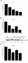
Insulin-stimulated PI3-kinase activity in liver and muscle. Liver (a) and hindlimb muscle (b) extracts from 8- to 12-week-old animals were immunoprecipitated with antiphosphotyrosine and subjected to PI3-kinase assay as described in Methods. The results are expressed as percentage of PI3-kinase activity in insulin-treated wild-type mice. The data represent mean ± SEM from five independent experiments. (c and d) Association between p85 and phosphotyrosine-containing proteins in epididymal fat tissues. Epididymal fat pads were isolated from 8- to 12-week-old mice after insulin stimulation and solubilized as indicated. Triton-soluble proteins were immunoprecipitated with antiphosphotyrosine antibodies followed by immunoblotting with anti-p85 antibody. A representative blot is shown (c). and quantitation of the results from 3 independent experiments is shown in the bar graphs (d). The intensity of the autoradiographic bands was quantitated using the NIH image software. The results are expressed as percentage of p85 associated with phosphotyrosine-containing proteins in insulin-stimulated wild-type mice. The data represent mean ± SEM.
Figure 5
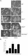
Islet morphology and analysis of β-cell mass in mutant mice. (a) Hematoxylin and eosin staining. Representative β-cells from diabetic and nondiabetic animals of each genotype were shown. (b) Quantitation of β-cell area in animals of the indicated genotype was performed using the Openlab image analysis software. Results are expressed as the percentage of the total surveyed area containing cells positive for insulin. Both diabetic and nondiabetic animals were studied.
Similar articles
- Analysis of compensatory beta-cell response in mice with combined mutations of Insr and Irs2.
Kim JJ, Kido Y, Scherer PE, White MF, Accili D. Kim JJ, et al. Am J Physiol Endocrinol Metab. 2007 Jun;292(6):E1694-701. doi: 10.1152/ajpendo.00430.2006. Epub 2007 Feb 13. Am J Physiol Endocrinol Metab. 2007. PMID: 17299086 - N-3 polyunsaturated fatty acids prevent the defect of insulin receptor signaling in muscle.
Taouis M, Dagou C, Ster C, Durand G, Pinault M, Delarue J. Taouis M, et al. Am J Physiol Endocrinol Metab. 2002 Mar;282(3):E664-71. doi: 10.1152/ajpendo.00320.2001. Am J Physiol Endocrinol Metab. 2002. PMID: 11832371 - Irs-2 coordinates Igf-1 receptor-mediated beta-cell development and peripheral insulin signalling.
Withers DJ, Burks DJ, Towery HH, Altamuro SL, Flint CL, White MF. Withers DJ, et al. Nat Genet. 1999 Sep;23(1):32-40. doi: 10.1038/12631. Nat Genet. 1999. PMID: 10471495 - Tissue-specificity of insulin action and resistance.
Benito M. Benito M. Arch Physiol Biochem. 2011 Jul;117(3):96-104. doi: 10.3109/13813455.2011.563748. Epub 2011 Apr 20. Arch Physiol Biochem. 2011. PMID: 21506723 Review. - [Insulin resistance. Receptor and post-receptor abnormalities].
Liguori M, Urso R, Fatigante G. Liguori M, et al. Minerva Endocrinol. 1998 Jun;23(2):37-52. Minerva Endocrinol. 1998. PMID: 9844354 Review. Italian.
Cited by
- The double-stranded RNA-dependent protein kinase differentially regulates insulin receptor substrates 1 and 2 in HepG2 cells.
Yang X, Nath A, Opperman MJ, Chan C. Yang X, et al. Mol Biol Cell. 2010 Oct 1;21(19):3449-58. doi: 10.1091/mbc.E10-06-0481. Epub 2010 Aug 4. Mol Biol Cell. 2010. PMID: 20685959 Free PMC article. - Klotho induces insulin resistance possibly through interference with GLUT4 translocation and activation of Akt, GSK3β, and PFKfβ3 in 3T3-L1 adipocyte cells.
Hasannejad M, Samsamshariat SZ, Esmaili A, Jahanian-Najafabadi A. Hasannejad M, et al. Res Pharm Sci. 2019 Aug;14(4):369-377. doi: 10.4103/1735-5362.263627. Res Pharm Sci. 2019. PMID: 31516514 Free PMC article. - Myeloid cell-restricted insulin receptor deficiency protects against obesity-induced inflammation and systemic insulin resistance.
Mauer J, Chaurasia B, Plum L, Quast T, Hampel B, Blüher M, Kolanus W, Kahn CR, Brüning JC. Mauer J, et al. PLoS Genet. 2010 May 6;6(5):e1000938. doi: 10.1371/journal.pgen.1000938. PLoS Genet. 2010. PMID: 20463885 Free PMC article. - Fulminant type 1 diabetes mellitus observed in insulin receptor substrate 2 deficient mice.
Arai T, Hashimoto H, Kawai K, Mori A, Ohnishi Y, Hioki K, Ito M, Saito M, Ueyama Y, Ohsugi M, Suzuki R, Kubota N, Yamauchi T, Tobe K, Kadowaki T, Kosaka K. Arai T, et al. Clin Exp Med. 2008 Jun;8(2):93-9. doi: 10.1007/s10238-008-0163-1. Epub 2008 Jul 11. Clin Exp Med. 2008. PMID: 18618219 - Diabetes in mice with selective impairment of insulin action in Glut4-expressing tissues.
Lin HV, Ren H, Samuel VT, Lee HY, Lu TY, Shulman GI, Accili D. Lin HV, et al. Diabetes. 2011 Mar;60(3):700-9. doi: 10.2337/db10-1056. Epub 2011 Jan 24. Diabetes. 2011. PMID: 21266328 Free PMC article.
References
- Kahn CR. New concepts in the pathogenesis of diabetes mellitus. Adv Intern Med. 1996;41:285–321. - PubMed
- Taylor SI. Deconstructing type 2 diabetes. Cell. 1999;97:9–12. - PubMed
- Polonsky KS, Sturis J, Bell GI. Non-insulin-dependent diabetes mellitus: a genetically programmed failure of the beta cell to compensate for insulin resistance. N Engl J Med. 1996;334:777–783. - PubMed
- Lauro D, et al. Impaired glucose tolerance in mice with a targeted impairment of insulin action in muscle and adipose tissue. Nat Genet. 1998;20:294–298. - PubMed
- Bruning JC, et al. A muscle-specific insulin receptor knockout exhibits features of the metabolic syndrome of NIDDM without altering glucose tolerance. Mol Cell. 1998;2:559–569. - PubMed
Publication types
MeSH terms
Substances
LinkOut - more resources
Full Text Sources
Other Literature Sources
Medical
Molecular Biology Databases


