Progression of meiotic DNA replication is modulated by interchromosomal interaction proteins, negatively by Spo11p and positively by Rec8p - PubMed (original) (raw)
. 2000 Feb 15;14(4):493-503.
Affiliations
- PMID: 10691741
- PMCID: PMC316381
Progression of meiotic DNA replication is modulated by interchromosomal interaction proteins, negatively by Spo11p and positively by Rec8p
R S Cha et al. Genes Dev. 2000.
Abstract
Spo11p is a key mediator of interhomolog interactions during meiosis. Deletion of the SPO11 gene decreases the length of S phase by approximately 25%. Rec8p is a key coordinator of meiotic interhomolog and intersister interactions. Deletion of the REC8 gene increases S-phase length, by approximately 10% in wild-type and approximately 30% in a spo11Delta background. Thus, the progression of DNA replication is modulated by interchromosomal interaction proteins. The spo11-Y135F DSB (double strand break) catalysis-defective mutant is normal for S-phase modulation and DSB-independent homolog pairing but is defective for later events, formation of DSBs, and synaptonemal complexes. Thus, earlier and later functions of Spo11 are defined. We propose that meiotic S-phase progression is linked directly to development of specific chromosomal features required for meiotic interhomolog interactions and that this feedback process is built upon a more fundamental mechanism, common to all cell types, by which S-phase progression is coupled to development of nascent intersister connections and/or related aspects of chromosome morphogenesis. Roles for Rec8 and/or Spo11 in progression through other stages of meiosis are also revealed.
Figures
Figure 1
Measurement of meiotic and mitotic S-phase lengths. (A) Progression of DNA replication was monitored by FACS analysis of samples collected at specified time points during meiotic (SPM) and mitotic (YPD) time courses. Cells progress from an unreplicated state indicated by their presence in a single peak of fluorescence (2C), to a state in which bulk DNA replication is complete, as indicated by movement into a second peak of twice the fluorescence intensity of the first peak (4C). (B) The fraction of cells undergoing DNA replication at each time point was evaluated from the corresponding FACS profile. The fractional area under the indicated rectangle was taken to represent the fraction of cells in S phase; the fractional area under the indicated triangle, doubled, was taken to represent the fraction of cells in G2/M. The appropriateness of these approximations is discussed in Materials and Methods. (C) Fraction of cells undergoing DNA replication at each time point in A (SPM and YPD + Noc cultures) is estimated as in Materials and Methods and plotted as a function of time. The lengths of mitotic (17.0 min) and meiotic (60.9 min) S phases were calculated as described in the text (also see Materials and Methods). (D) Cumulative curves for mitotic and meiotic DNA replication. The fraction of cells that have entered (solid line) and exited (broken line) S phase at each time point is calculated (Materials and Methods) as a function of time. The times at which 50% of active cells have entered mitotic (75 min) and meiotic (165 min) S phases are indicated. The lengths of mitotic (17 min) and meiotic (60.9 min) S phases is the time distance between the Entering S and Exiting S lines as depicted.
Figure 2
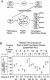
Experimental system. (A) NKY3000 is a self-diploidized spore clone of NKY730. A panel of homothallic heterozygous strains were generated by standard yeast transformation procedures. Each heterozygotic derivative of NKY3000 was sporulated and the four haploid spore clones from the same ascus were allowed to undergo self-diploidization. The resulting four diploid strains, two wild type and two mutant, were analyzed simultaneously during parallel synchronous meiotic time courses. (B) Distribution of wild-type meiotic S-phase lengths. (Left) The distribution of S-phase lengths observed for pairs of wild-type spore clones derived as described in A. The mean and
s.d.
, 76.7 ± 18.9 min, is of 34 measurements from 17 different experiments. (Right) The variation between the two sibling cultures examined in parallel in a single experiment. Average length of S phase for each pair of wild-type cultures from a single experiment is summarized below.
Figure 3
Meiotic S-phase is shorter in spo11Δ. (A) Progression of meiotic DNA replication in two wild-type (○,□) and two spo11Δ (●,█) strains derived from NKY3002. (B) Lengths of S phases in wild type and spo11Δ. (see Fig. 1 and text for details). (C) The fraction of cells that have entered (solid line) and exited (broken line) S phase is calculated for the four cultures in A (Materials and Methods). Arrows indicate the time at which 50% of the active cells have entered S phase in each culture. (D) Lengths of meiotic S phase in wild-type and spo11Δ cultures from three different experiments. Open and solid symbols represent the calculated lengths of meiotic S phase in wild type and spo11Δ, respectively. Results from A are summarized in experiment 1. NKY3002 was analyzed for experiments 1 and 2, and NKY3196 for experiment 3. For experiment 2, one of the two wild-type and spo11Δ spore clones was analyzed in duplicate giving rise to a total of six cultures analyzed. Also shown are the average lengths of meiotic S phase for wild-type and mutant spore clones in each experiment. (E) The relative lengths of meiotic S phase in spo11Δ cultures (see text for detail). (Left) Each shaded oval corresponds to a specific ● and █ in D. (Right) Σ1–3 is the distribution of normalized S-phase length in seven spo11Δ cultures. The mean and standard deviation are used in a one-sample _t_-test; the confidence level that the mean value for spo11Δ (0.771) is different from that of the wild type (1.0) is >99.5%. (F) Kinetics of entry into meiotic S phase is comparable in SPO11 and spo11Δ strains. The time at which 50% of active cells have entered S-phase in each SPO11 and spo11Δ culture of the above-mentioned three independent experiments are summarized. Mean values of seven SPO11 and spo11Δ cultures are shown (right) (Σ1–3).
Figure 4
Relative lengths of meiotic S-phase in various mutants. (A–D) Lengths of meiotic S-phase in various mutants relative to wild type (defined as 1.0) are calculated as described in Fig. 3E. The number of circles in each strain represents the number of cultures analyzed. (A) (spo11Δ) NKY3002 and NKY3196; (spo11–Y135F) NKY3001. Analysis of NKY3197 indicated that HA-epitope tagging of Spo11p did not affect the timely occurrence of major events during meiosis, including meiotic S phase (data not shown). (B) (rec102Δ) NKY3135; (red1Δ) NKY3190; (hop1Δ) NKY3191. (C) (rec8Δ) NKY3157; (rec8Δ spo11Δ) NKY3194.
Figure 5
DSB-independent homolog pairing in SPO11, spo11Δ, and spo11–Y135F. Synchronous meiotic cultures for NKY1992 (SPO11; A), BWY168A (spo11Δ; B), and BWY119 (spo11–Y135F; C) were set up as described in Materials and Methods. Samples were harvested at specified time points and the extent of homolog pairing is measured by FISH analysis (Weiner and Kleckner 1994). Two homologs are defined as paired if the distance between them, visualized by a fluorescence labeled probe, is ≤0.7 μm (Weiner and Kleckner 1994). The kinetics of loss and re-establishment of pairing, (○) as well as the entry into (solid line) and exit from meiotic (broken line) S-phase are summarized.
Similar articles
- Multiple roles of Spo11 in meiotic chromosome behavior.
Celerin M, Merino ST, Stone JE, Menzie AM, Zolan ME. Celerin M, et al. EMBO J. 2000 Jun 1;19(11):2739-50. doi: 10.1093/emboj/19.11.2739. EMBO J. 2000. PMID: 10835371 Free PMC article. - Wild-type levels of Spo11-induced DSBs are required for normal single-strand resection during meiosis.
Neale MJ, Ramachandran M, Trelles-Sticken E, Scherthan H, Goldman AS. Neale MJ, et al. Mol Cell. 2002 Apr;9(4):835-46. doi: 10.1016/s1097-2765(02)00498-7. Mol Cell. 2002. PMID: 11983174 - Functional interactions between SPO11 and REC102 during initiation of meiotic recombination in Saccharomyces cerevisiae.
Kee K, Keeney S. Kee K, et al. Genetics. 2002 Jan;160(1):111-22. doi: 10.1093/genetics/160.1.111. Genetics. 2002. PMID: 11805049 Free PMC article. - [SPO11: an activity that promotes DNA breaks required for meiosis].
Baudat F, de Massy B. Baudat F, et al. Med Sci (Paris). 2004 Feb;20(2):213-8. doi: 10.1051/medsci/2004202213. Med Sci (Paris). 2004. PMID: 14997442 Review. French. - [The role of pre-meiotic S phase].
Watanabe Y. Watanabe Y. Tanpakushitsu Kakusan Koso. 2002 Jan;47(1):45-50. Tanpakushitsu Kakusan Koso. 2002. PMID: 11808194 Review. Japanese. No abstract available.
Cited by
- How to halve ploidy: lessons from budding yeast meiosis.
Kerr GW, Sarkar S, Arumugam P. Kerr GW, et al. Cell Mol Life Sci. 2012 Sep;69(18):3037-51. doi: 10.1007/s00018-012-0974-9. Epub 2012 Apr 6. Cell Mol Life Sci. 2012. PMID: 22481439 Free PMC article. Review. - Identification of residues in yeast Spo11p critical for meiotic DNA double-strand break formation.
Diaz RL, Alcid AD, Berger JM, Keeney S. Diaz RL, et al. Mol Cell Biol. 2002 Feb;22(4):1106-15. doi: 10.1128/MCB.22.4.1106-1115.2002. Mol Cell Biol. 2002. PMID: 11809802 Free PMC article. - Recruitment of Rec8, Pds5 and Rad61/Wapl to meiotic homolog pairing, recombination, axis formation and S-phase.
Hong S, Joo JH, Yun H, Kleckner N, Kim KP. Hong S, et al. Nucleic Acids Res. 2019 Dec 16;47(22):11691-11708. doi: 10.1093/nar/gkz903. Nucleic Acids Res. 2019. PMID: 31617566 Free PMC article. - The Nucleoporin Nup2 Contains a Meiotic-Autonomous Region that Promotes the Dynamic Chromosome Events of Meiosis.
Chu DB, Gromova T, Newman TAC, Burgess SM. Chu DB, et al. Genetics. 2017 Jul;206(3):1319-1337. doi: 10.1534/genetics.116.194555. Epub 2017 Apr 28. Genetics. 2017. PMID: 28455351 Free PMC article. - Homologous pairing preceding SPO11-mediated double-strand breaks in mice.
Boateng KA, Bellani MA, Gregoretti IV, Pratto F, Camerini-Otero RD. Boateng KA, et al. Dev Cell. 2013 Jan 28;24(2):196-205. doi: 10.1016/j.devcel.2012.12.002. Epub 2013 Jan 11. Dev Cell. 2013. PMID: 23318132 Free PMC article.
References
- Bennett M, Smith J. The effect of polyploidy on meiotic duration and pollen development in cereal anthers. Proc R Soc Lond B. 1972;181:81–107.
- Bergerat A, de Massy B, Gadelle D, Varoutas P-C, Nicolas A, Forterre P. An atypical topoisomerase II from archaea with implications for meiotic recombination. Nature. 1997;386:414–417. - PubMed
Publication types
MeSH terms
Substances
Grants and funding
- R01 GM044794/GM/NIGMS NIH HHS/United States
- R37 GM025326/GM/NIGMS NIH HHS/United States
- R37-GM25326/GM/NIGMS NIH HHS/United States
- R01-GM44794/GM/NIGMS NIH HHS/United States
LinkOut - more resources
Full Text Sources
Other Literature Sources
Molecular Biology Databases


