Differential viral protein expression in Kaposi's sarcoma-associated herpesvirus-infected diseases: Kaposi's sarcoma, primary effusion lymphoma, and multicentric Castleman's disease - PubMed (original) (raw)
Differential viral protein expression in Kaposi's sarcoma-associated herpesvirus-infected diseases: Kaposi's sarcoma, primary effusion lymphoma, and multicentric Castleman's disease
C Parravicini et al. Am J Pathol. 2000 Mar.
Abstract
Kaposi's sarcoma (KS)-associated herpesvirus (KSHV) is linked to KS, primary effusion lymphomas (PEL), and a subset of multicentric Castleman's disease (MCD). Transcript mapping studies using PEL cell lines have allowed preliminary classification of viral gene expression into constitutive (class I) and inducible (class II/III) categories. To determine whether viral gene expression differs in vivo, we examined tissue sections of KSHV-infected disorders, using specific antibodies against proteins that are representative of the different expression classes of KSHV genes. ORF73/LANA appears to be a surrogate marker for KSHV infection because it is constitutively expressed in vitro and in vivo in all KSHV-infected cells. Expression of vIRF1, vIL6, and PF-8 proteins in the infected B cells of MCD lymph nodes reproduces the expression pattern observed in TPA-stimulated KSHV-infected B-cell lines. In contrast, the protein expression of the inducible viral genes that we tested in KS and PEL biopsies is restricted to PF-8 and vIL6, respectively. The tightly restricted expression of KSHV proteins in vivo differs from the dysregulated expression of inducible KSHV genes in vitro and suggests that viral gene expression in KSHV-infected cell lines does not accurately reflect what occurs in diseased tissues. These differences may be related to either cell-specific or immune restriction of viral replication.
Figures
Figure 1.
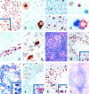
A: LANA is constitutively expressed in nearly all BCBL-1 cells as a speckled nuclear pattern (inset). Similar expression is seen also in BC-1 and BCP-1 cells (DAB/hematoxylin counterstain). B: vIRF1 in BCBL-1 cells is localized to the cytoplasm in one cell (right) and in the nucleus of another (left), consistent with nuclear translocation. C: Only 1% of BCBL-1 cells express vIRF1 without TPA (upper panel) but up to >20% (lower panel) after 48 hours of TPA stimulation (DAB/hematoxylin counterstain). D: With TPA stimulation (upper panel) BCBL-1 cells may express nuclear PF-8 alone (brown), cytoplasmic vIRF1 alone (blue), or may coexpress both proteins. In some cells double stained for PF-8 and vIRF1, the latter shows a pseudolinear intracytoplasmic pattern reminiscent of rough endoplasmic reticulum positivity (lower panel) (AEC/FastBlue; no counterstain). E: Section of myocardium with infiltrating PEL. All neoplastic cells express nuclear LANA (AEC/hematoxylin counterstain). F: Peritoneal biopsy with infiltrating PEL. Only a few tumor cells are immunopositive for vIL6 (DAB/hematoxylin counterstain). G: Hyalinization of germinal centers, targetoid mantle zone, and increased vascularity in a lymph node biopsy from plasma cell variant, KSHV-related multicentric Castleman’s disease (H&E stain). H: KSHV LANA protein expression is restricted to a subpopulation of mantle zone lymphocytes that show a speckled nuclear staining pattern (inset) (DAB/hematoxylin counterstain). I: Double-stained section of an MCD lymph node showing that nearly all of the KSHV-infected mantle zone lymphocytes expressing LANA (light brown) are negative for CD79 (blue), with only a few double-positive cells (inset). Similar findings are obtained by double staining with other B-cell antigens expressed by normal mantle zone lymphocytes. (DAB/FastBlue; no counterstain). J: MCD lymph node section with expression of vIL6 (brown) restricted to a subpopulation of mantle zone cells having immunoblastic morphology (inset) (DAB/hematoxylin counterstain). K: In double-stained sections, vIRF1 (blue) in mantle zone cells is also positive for LANA (inset). Double-stained lymphocytes, however, account for only 10% to 30% of the LANA-positive cells (DAB/FastBlue; no counterstain). L: MCD frozen section, same case as in C, shows that only rare cells at the border between mantle zone and germinal center express PF-8 (DAB/hematoxylin counterstain).
Figure 2.
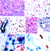
A: Spindle cell area with slit-like vascular spaces in nodular KS (H&E). B: Serial section of the case in A, stained for LANA, showing a speckled/granular nuclear reactivity in KS spindle cells (AEC/hematoxylin counterstain). C: In double-stained sections, LANA is detectable in the nuclei of KS cells (red) but not in the endothelial cells nor in the actin-positive smooth muscle cells (blue) of normal vessels entrapped within the tumor (AEC/FastBlue; no counterstain). D: Angiomatous area of a patch KS lesion containing CD34-positive endothelial cells (blue) lining KS vascular spaces, which also express LANA (brown) (H&E, left panel; DAB/FastBlue without counterstain, right panel). E: In double-stained sections, none of the KS cells expressing LANA are positive for CD68 (blue) or other leukocytic antigens, but rare LANA-positive KS spindle cells are positive for CD68 (inset). (DAB/FastBlue; no counterstain). F: Only rare cells are positive in KS frozen section stained for PF-8; CD68-positive monocytes (blue) are consistently negative (DAB/FastBlue; no counterstain).
Similar articles
- Distinct expression of Kaposi's sarcoma-associated herpesvirus-encoded proteins in Kaposi's sarcoma and multicentric Castleman's disease.
Abe Y, Matsubara D, Gatanaga H, Oka S, Kimura S, Sasao Y, Saitoh K, Fujii T, Sato Y, Sata T, Katano H. Abe Y, et al. Pathol Int. 2006 Oct;56(10):617-24. doi: 10.1111/j.1440-1827.2006.02017.x. Pathol Int. 2006. PMID: 16984619 - Expression of a virus-derived cytokine, KSHV vIL-6, in HIV-seronegative Castleman's disease.
Parravicini C, Corbellino M, Paulli M, Magrini U, Lazzarino M, Moore PS, Chang Y. Parravicini C, et al. Am J Pathol. 1997 Dec;151(6):1517-22. Am J Pathol. 1997. PMID: 9403701 Free PMC article. - The pleiotropic effects of Kaposi's sarcoma herpesvirus.
Schulz TF. Schulz TF. J Pathol. 2006 Jan;208(2):187-98. doi: 10.1002/path.1904. J Pathol. 2006. PMID: 16362980 Review. - Expression and localization of human herpesvirus 8-encoded proteins in primary effusion lymphoma, Kaposi's sarcoma, and multicentric Castleman's disease.
Katano H, Sato Y, Kurata T, Mori S, Sata T. Katano H, et al. Virology. 2000 Apr 10;269(2):335-44. doi: 10.1006/viro.2000.0196. Virology. 2000. PMID: 10753712
Cited by
- Cell Cycle Regulatory Functions of the KSHV Oncoprotein LANA.
Wei F, Gan J, Wang C, Zhu C, Cai Q. Wei F, et al. Front Microbiol. 2016 Mar 30;7:334. doi: 10.3389/fmicb.2016.00334. eCollection 2016. Front Microbiol. 2016. PMID: 27065950 Free PMC article. Review. - Systemic expression of Kaposi sarcoma herpesvirus (KSHV) Vflip in endothelial cells leads to a profound proinflammatory phenotype and myeloid lineage remodeling in vivo.
Ballon G, Akar G, Cesarman E. Ballon G, et al. PLoS Pathog. 2015 Jan 21;11(1):e1004581. doi: 10.1371/journal.ppat.1004581. eCollection 2015 Jan. PLoS Pathog. 2015. PMID: 25607954 Free PMC article. - The Kaposi's sarcoma-associated herpesvirus LANA protein stabilizes and activates c-Myc.
Liu J, Martin HJ, Liao G, Hayward SD. Liu J, et al. J Virol. 2007 Oct;81(19):10451-9. doi: 10.1128/JVI.00804-07. Epub 2007 Jul 18. J Virol. 2007. PMID: 17634226 Free PMC article. - Kaposi's sarcoma-associated herpesvirus immunoevasion and tumorigenesis: two sides of the same coin?
Moore PS, Chang Y. Moore PS, et al. Annu Rev Microbiol. 2003;57:609-39. doi: 10.1146/annurev.micro.57.030502.090824. Annu Rev Microbiol. 2003. PMID: 14527293 Free PMC article. Review. - Quantitative RNAseq analysis of Ugandan KS tumors reveals KSHV gene expression dominated by transcription from the LTd downstream latency promoter.
Rose TM, Bruce AG, Barcy S, Fitzgibbon M, Matsumoto LR, Ikoma M, Casper C, Orem J, Phipps W. Rose TM, et al. PLoS Pathog. 2018 Dec 17;14(12):e1007441. doi: 10.1371/journal.ppat.1007441. eCollection 2018 Dec. PLoS Pathog. 2018. PMID: 30557332 Free PMC article.
References
- Chang Y, Cesarman E, Pessin MS, Lee F, Culpepper J, Knowles DM, Moore PS: Identification of herpesvirus-like DNA sequences in AIDS-associated Kaposi’s sarcoma. Science 1994, 266:1865-1869 - PubMed
- Cesarman E, Chang Y, Moore PS, Said JW, Knowles DM: Kaposi’s sarcoma-associated herpesvirus-like DNA sequences in AIDS- related body-cavity-based lymphomas. N Engl J Med 1995, 332:1186-1191 - PubMed
- Soulier J, Grollet L, Oksenhendler E, Cacoub P, Cazals HD, Babinet P, d’Agay MF, Clauvel JP, Raphael M, Degos L, et al: Kaposi’s sarcoma-associated herpesvirus-like DNA sequences in multicentric Castleman’s disease. Blood 1995, 86:1276-1280 - PubMed
- Cesarman E, Moore PS, Rao RH, Inghirami G, Knowles DM, Chang Y: In vitro establishment and characterization of two acquired immunodeficiency syndrome-related lymphoma cell lines (BC-1 and BC-2) containing Kaposi’s sarcoma-associated herpesvirus-like (KSHV) DNA sequences. Blood 1995, 86:2708-2714 - PubMed
- Boshoff C, Gao SJ, Healy LE, Matthews S, Thomas AJ, Coignet L, Warnke RA, Strauchen JA, Matutes E, Kamel OW, Moore PS, Weiss RA, Chang Y: Establishing a KSHV+ cell line (BCP-1) from peripheral blood and characterizing its growth in Nod/SCID mice. Blood 1998, 91:1671-1679 - PubMed
Publication types
MeSH terms
Substances
Grants and funding
- CA82056/CA/NCI NIH HHS/United States
- R01 CA075911/CA/NCI NIH HHS/United States
- R01 CA067391/CA/NCI NIH HHS/United States
- CA67391/CA/NCI NIH HHS/United States
- CA75911/CA/NCI NIH HHS/United States
LinkOut - more resources
Full Text Sources
Other Literature Sources
Medical

