Production of hepatocyte growth factor during acute myocardial infarction - PubMed (original) (raw)
Production of hepatocyte growth factor during acute myocardial infarction
Y Zhu et al. Heart. 2000 Apr.
Abstract
Objective: To investigate the clinical significance of circulating hepatocyte growth factor (HGF) and the role of peripheral blood mononuclear cells (monocytes), which are a possible source of HGF, in patients with acute myocardial infarction.
Design and patients: 37 patients with acute myocardial infarction and 13 normal control subjects were recruited. Peripheral venous blood samples were drawn from the infarct patients 1, 7, 14, and 21 days after onset. Monocytes were isolated from peripheral blood at those times. HGF concentrations in serum and in a culture medium of monocytes after incubation for 24 hours (monocyte HGF levels) were measured by enzyme linked immunosorbent assay.
Results: Serum HGF and monocyte HGF values within seven days after onset of myocardial infarction were significantly higher than those of control subjects and decreased by day 14. There were significant positive correlations between serum HGF and monocyte HGF levels on day 7; between maximum plasma creatine phosphokinase levels and serum HGF levels on day 1; between maximum plasma C reactive protein and serum HGF levels; and between maximum C reactive protein and monocyte HGF levels. Monocyte HGF levels were raised in the patients with progression of ventricular enlargement in the course of acute myocardial infarction.
Conclusions: Early serum HGF concentrations reflect the extent of myocardial damage in acute myocardial infarction patients. Inflammation after acute myocardial infarction is supposed to be involved in enhanced HGF production. Monocytes may play an important role in ventricular remodelling after acute myocardial infarction by releasing the cardiovascular protective mitogen HGF.
Figures
Figure 1
Time course of serum hepatocyte growth factor (HGF) concentrations in patients with acute myocardial infarction. HGF was determined by enzyme linked immunosorbent assay as described in Methods. Serum HGF reached a peak on day 1 and decreased by day 21. Serum HGF in the patients was significantly higher than in the normal controls on day 1 (0.54 (0.11) v 0.12 (0.03) ng/ml, p < 0.05), and remained significantly higher on day 7. Filled circles, patients with acute myocardial infarction; empty circles, control subjects. Error bars = SEM. *p < 0.05 v control subjects.
Figure 2
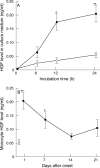
(A) Hepatocyte growth factor (HGF) concentrations in the supernatant of cultured peripheral blood mononuclear cells (monocytes) in patients with acute myocardial infarction and in control subjects. Monocytes were isolated and cultured as described in Methods. HGF levels in the monocyte culture medium increased time dependently until 24 hours after incubation. (B) Changes in HGF production by monocytes in the course of acute myocardial infarction. HGF levels in the monocyte culture medium after incubation for 24 hours are called "monocyte HGF levels" and used as a marker of the HGF producing ability of monocytes. Monocyte HGF levels were significantly higher than those of control subjects on day 1 but decreased by day 14. Filled circles, patients with acute myocardial infarction; empty circles, control subjects. Error bars = SEM. *p < 0.05; **p < 0.01 v control subjects.
Figure 3
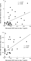
Correlations between serum hepatocyte growth factor (HGF) levels and monocyte HGF levels in patients with acute myocardial infarction: a weak correlation was found between serum HGF and monocyte HGF on day 1 after onset of (panel A, r = +0.47, p < 0.05), and a positive correlation was found between these two variables on day 7 after onset (panel B, r = +0.54, p < 0.01).
Figure 4
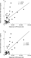
Correlations between maximum creatine phosphokinase (CPK) and hepatocyte growth factor (HGF): there was a significant positive correlation between maximum plasma CPK and both maximum serum HGF (panel A, r = +0.54, p < 0.01) and serum HGF on day 1 (panel B, r = +0.61, p < 0.001).
Figure 5
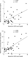
Correlations between maximum C reactive protein levels and hepatocyte growth factor (HGF) levels: there was a significant positive correlation between maximum plasma C reactive protein and maximum serum HGF (panel A, r = +0.55, p < 0.01), and between maximum C reactive protein and maximum monocyte HGF (panel B, r = +0.65, p < 0.001).
Figure 6
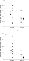
Hepatocyte growth factor (HGF) levels in the patients with or without left ventricular dilatation in the course of acute myocardial infarction. We divided the patients into two groups according to the changes in left ventricular end diastolic volume index (LVEDVI) (group A: eight patients with increases in LVEDVI during the course of acute myocardial infarction; group B: eight patients without increases in LVEDVI). (A) Mean serum HGF on day 7 in group A was higher than in group B, but the difference was not significant (0.47 (0.10) v 0.29 (0.05) ng/ml, p = 0.13). (B) Monocyte HGF levels on day 7 in group A were significantly higher than in group B (0.30 (0.07) v 0.08 (0.03) ng/ml, p < 0.05). Error bars = SEM. *p < 0.05.
Similar articles
- Hepatocyte growth factor production may be related to the inflammatory response in patients with acute myocardial infarction.
Shimada Y, Yoshiyama M, Jissho S, Kamimori K, Nakamura Y, Iida H, Takeuchi K, Yoshikawa J. Shimada Y, et al. Circ J. 2002 Mar;66(3):253-6. doi: 10.1253/circj.66.253. Circ J. 2002. PMID: 11922273 - Increased circulating hepatocyte growth factor in the early stage of acute myocardial infarction.
Matsumori A, Furukawa Y, Hashimoto T, Ono K, Shioi T, Okada M, Iwasaki A, Nishio R, Sasayama S. Matsumori A, et al. Biochem Biophys Res Commun. 1996 Apr 16;221(2):391-5. doi: 10.1006/bbrc.1996.0606. Biochem Biophys Res Commun. 1996. PMID: 8619866 - Circulating levels of hepatocyte growth factor and left ventricular remodelling after acute myocardial infarction (from the REVE-2 study).
Lamblin N, Bauters A, Fertin M, de Groote P, Pinet F, Bauters C. Lamblin N, et al. Eur J Heart Fail. 2011 Dec;13(12):1314-22. doi: 10.1093/eurjhf/hfr137. Epub 2011 Oct 13. Eur J Heart Fail. 2011. PMID: 21996026 - Serial changes in serum VEGF and HGF in patients with acute myocardial infarction.
Soeki T, Tamura Y, Shinohara H, Tanaka H, Bando K, Fukuda N. Soeki T, et al. Cardiology. 2000;93(3):168-74. doi: 10.1159/000007022. Cardiology. 2000. PMID: 10965088 - Hepatocyte growth factor(HGF): a new biochemical marker for acute myocardial infarction.
Sato T, Yoshinouchi T, Sakamoto T, Fujieda H, Murao S, Sato H, Kobayashi H, Ohe T. Sato T, et al. Heart Vessels. 1997;12(5):241-6. doi: 10.1007/BF02766790. Heart Vessels. 1997. PMID: 9846810
Cited by
- Monocyte activation state regulates monocyte-induced endothelial proliferation through Met signaling.
Schubert SY, Benarroch A, Monter-Solans J, Edelman ER. Schubert SY, et al. Blood. 2010 Apr 22;115(16):3407-12. doi: 10.1182/blood-2009-02-207340. Epub 2010 Feb 26. Blood. 2010. PMID: 20190195 Free PMC article. - In vitro hepatic trans-differentiation of human mesenchymal stem cells using sera from congestive/ischemic liver during cardiac failure.
Bishi DK, Mathapati S, Cherian KM, Guhathakurta S, Verma RS. Bishi DK, et al. PLoS One. 2014 Mar 18;9(3):e92397. doi: 10.1371/journal.pone.0092397. eCollection 2014. PLoS One. 2014. PMID: 24642599 Free PMC article. - Activated platelets interfere with recruitment of mesenchymal stem cells to apoptotic cardiac cells via high mobility group box 1/Toll-like receptor 4-mediated down-regulation of hepatocyte growth factor receptor MET.
Vogel S, Chatterjee M, Metzger K, Borst O, Geisler T, Seizer P, Müller I, Mack A, Schumann S, Bühring HJ, Lang F, Sorg RV, Langer H, Gawaz M. Vogel S, et al. J Biol Chem. 2014 Apr 18;289(16):11068-11082. doi: 10.1074/jbc.M113.530287. Epub 2014 Feb 24. J Biol Chem. 2014. PMID: 24567328 Free PMC article. - Intestinal hormones and growth factors: effects on the small intestine.
Drozdowski L, Thomson AB. Drozdowski L, et al. World J Gastroenterol. 2009 Jan 28;15(4):385-406. doi: 10.3748/wjg.15.385. World J Gastroenterol. 2009. PMID: 19152442 Free PMC article. Review. - Hepatocyte growth factor is associated with greater risk of extracoronary calcification: results from the multiethnic study of atherosclerosis.
Ogunmoroti O, Osibogun O, Ferraro RA, Ndunda PM, Larson NB, Decker PA, Bielinski SJ, Blumenthal RS, Budoff MJ, Michos ED. Ogunmoroti O, et al. Open Heart. 2022 May;9(1):e001971. doi: 10.1136/openhrt-2022-001971. Open Heart. 2022. PMID: 35641100 Free PMC article.
References
- J Cell Biol. 1994 Dec;127(6 Pt 2):1783-7 - PubMed
- Lancet. 1995 Feb 4;345(8945):293-5 - PubMed
- Biochem Biophys Res Commun. 1995 Oct 13;215(2):483-8 - PubMed
- Biochem Biophys Res Commun. 1996 Apr 16;221(2):391-5 - PubMed
- Hypertension. 1996 Sep;28(3):409-13 - PubMed
MeSH terms
Substances
LinkOut - more resources
Full Text Sources
Medical
Research Materials

