Role of Gab1 in heart, placenta, and skin development and growth factor- and cytokine-induced extracellular signal-regulated kinase mitogen-activated protein kinase activation - PubMed (original) (raw)
Role of Gab1 in heart, placenta, and skin development and growth factor- and cytokine-induced extracellular signal-regulated kinase mitogen-activated protein kinase activation
M Itoh et al. Mol Cell Biol. 2000 May.
Abstract
Gab1 is a member of the Gab/DOS (Daughter of Sevenless) family of adapter molecules, which contain a pleckstrin homology (PH) domain and potential binding sites for SH2 and SH3 domains. Gab1 is tyrosine phosphorylated upon stimulation of various cytokines, growth factors, and antigen receptors in cell lines and interacts with signaling molecules, such as SHP-2 and phosphatidylinositol 3-kinase, although its biological roles have not yet been established. To reveal the functions of Gab1 in vivo, we generated mice lacking Gab1 by gene targeting. Gab1-deficient embryos died in utero and displayed developmental defects in the heart, placenta, and skin, which were similar to phenotypes observed in mice lacking signals of the hepatocyte growth factor/scatter factor, platelet-derived growth factor, and epidermal growth factor pathways. Consistent with these observations, extracellular signal-regulated kinase mitogen-activated protein (ERK MAP) kinases were activated at much lower levels in cells from Gab1-deficient embryos in response to these growth factors or to stimulation of the cytokine receptor gp130. These results indicate that Gab1 is a common player in a broad range of growth factor and cytokine signaling pathways linking ERK MAP kinase activation.
Figures
FIG. 1
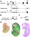
Targeting disruption of the Gab1 locus. (A) Restriction map of the Gab1 locus and targeting vector. The deletion region contains an exon encoding a part of the PH domain. This region was replaced by an en-2 splice acceptor-IRES–β-geo pA cassette. RI, _Eco_RI; Xb, _Xba_I; B, _Bam_HI. (B) Southern blot analysis for genotyping embryos. _Eco_RI-digested DNA from wild-type (+/+), heterozygous (+/−), and homozygous (−/−) embryos was hybridized with the 5′ probe shown in panel A. TK, thymidine kinase. (C) Immunoblotting analysis of Gab1. Gab1 immunoprecipitates from Gab1−/−, Gab1+/−, and Gab1+/+ embryonic fibroblasts were analyzed by immunoblotting with the anti-Gab1 antibody. (D) β-Galactosidase staining. Heterozygous embryos were fixed at E11.5 (left) and E13.5 (middle) and stained with X-Gal. Sagittal sections of stained E13.5 embryos are also shown (right).
FIG. 2
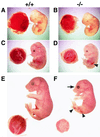
External appearance and placentas of wild-type and Gab1−/− embryos. (A and B) E11.5; (C and D) E13.5; (E and F) E17.5. Homozygous embryos were retarded in growth compared with wild-type littermates after E13.5. Mutant placentas were pale and smaller than the wild type. The arrowheads and arrow indicate hemorrhage and an open eye with no lid, respectively.
FIG. 3
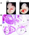
Developmental defects in heart of Gab1−/− embryos. (A) External appearance of E11.5 wild-type (+/+) and homozygous (−/−) embryos. Gab1−/− embryos contained blood in the pericardial cavity, indicated by the arrowhead. (B and C) Heart sections from wild-type (left) and Gab1−/− (right) embryos at E13.5 (B) E15.5 (C). Note the hypoblastic ventricle in the heart of Gab1−/− embryos (B and C). a, atrium; v, ventricle; s, septum; vw, ventricular wall.
FIG. 4
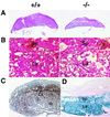
Underdeveloped placentas in Gab1−/− embryos. Shown are sections of wild-type (+/+) and Gab1−/− placentas of E13.5 embryos at low (A) and high (B) magnifications. There were fewer trophoblast cells in the labyrinth region of Gab1−/− placentas. Sp, spongiotrophoblast; La, labyrinthine trophoblast. Gab1 expression was detected by immunohistochemistry with anti-Gab1 antibody (C) and by LacZ expression (D), with counterstaining with hematoxylin (C) and eosin (D). Expression of Gab1 was detected in the spongiotrophoblast and labyrinthine trophoblast.
FIG. 5
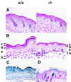
Developmental defects in skin of Gab1−/− embryos. Shown are sections of epidermis in Gab1+/+ and Gab1−/− embryos at low (A) and high (B) magnifications. The epidermal layer was thinner and hair follicles were underdeveloped. c, stratum corneum; g, stratum granulosum; s, stratum spinosum; b, basal layer. Gab1 expression was detected by immunohistochemistry with anti-Gab1 antibody (C) and by LacZ expression (D), with counterstaining with hematoxylin (C) and eosin (D). Expression of Gab1 was detected in the epidermis and hair follicles.
FIG. 6
Reduction in gp130-dependent ERK MAP kinase activation in Gab1−/− cells. (A) gp130-mediated ERK activation. Gab1+/+ and Gab1−/− embryonic fibroblasts were stimulated with IL-6 and sIL-6Rα for the indicated periods or with tetradecanoyl phorbol acetate (TPA) for 30 min. Lysates were immunoblotted with anti-diphospho-ERKs or anti-ERK2 antibodies. Kinase activities were determined by an immunoprecipitation (IP) kinase assay using the anti-ERK2 antibody and MBP. The amount of incorporated 32P in MBP was determined and is indicated as relative activity (versus activity in unstimulated Gab1+/+ cells) (RA). Tyrosine phosphorylation and expression of SHP-2 and STAT3 are also indicated. TCL, total cell lysate. (B) Transfection of Gab1 rescued the ERK activation in Gab1−/− cells. Gab1+/+ or Gab1−/− fibroblasts were transfected with an expression vector for human Gab1 and Flag-tagged ERK2 and stimulated with IL-6 and sIL-6Rα for 15 min. Expression of Flag-tagged ERK2 and Gab1 is indicated. Flag-tagged ERK2 was immunoprecipitated with anti-Flag antibody and was subjected to an in vitro kinase assay. The results are shown by autoradiography and indicated as relative activities. IB, immunoblot.
FIG. 7
Reduced activation of ERK MAP kinase in response to EGF, PDGF, and HGF. (A) EGF-induced ERK activation and tyrosine phosphorylation of Shc. Gab1+/+ and Gab−/− embryonic fibroblasts were stimulated with EGF for the indicated periods. The indicated isoform of Shc is p52. PY, phosphotyrosine. (B) PDGF-induced ERK activation and tyrosine phosphorylation of SHP-2 and Shc. (C) c-Met-induced ERK activation. Embryonic fibroblasts were transfected with expression vectors for Flag-tagged ERK2 and c-Met. At 20 h after transfection, cells were stimulated with HGF for 10 min (+) or left unstimulated (−). ERK activities were determined by the in vitro kinase assay, using anti-Flag immunoprecipitates (IP). Similar amounts of c-Met were expressed in Gab1+/+ and Gab1−/− cells (data not shown). IB, immunoblot.
Similar articles
- Gab1 acts as an adapter molecule linking the cytokine receptor gp130 to ERK mitogen-activated protein kinase.
Takahashi-Tezuka M, Yoshida Y, Fukada T, Ohtani T, Yamanaka Y, Nishida K, Nakajima K, Hibi M, Hirano T. Takahashi-Tezuka M, et al. Mol Cell Biol. 1998 Jul;18(7):4109-17. doi: 10.1128/MCB.18.7.4109. Mol Cell Biol. 1998. PMID: 9632795 Free PMC article. - Activation of gp130 transduces hypertrophic signal through interaction of scaffolding/docking protein Gab1 with tyrosine phosphatase SHP2 in cardiomyocytes.
Nakaoka Y, Nishida K, Fujio Y, Izumi M, Terai K, Oshima Y, Sugiyama S, Matsuda S, Koyasu S, Yamauchi-Takihara K, Hirano T, Kawase I, Hirota H. Nakaoka Y, et al. Circ Res. 2003 Aug 8;93(3):221-9. doi: 10.1161/01.RES.0000085562.48906.4A. Epub 2003 Jul 10. Circ Res. 2003. PMID: 12855672 - Gab-family adapter proteins act downstream of cytokine and growth factor receptors and T- and B-cell antigen receptors.
Nishida K, Yoshida Y, Itoh M, Fukada T, Ohtani T, Shirogane T, Atsumi T, Takahashi-Tezuka M, Ishihara K, Hibi M, Hirano T. Nishida K, et al. Blood. 1999 Mar 15;93(6):1809-16. Blood. 1999. PMID: 10068651 - Gab-family adapter molecules in signal transduction of cytokine and growth factor receptors, and T and B cell antigen receptors.
Hibi M, Hirano T. Hibi M, et al. Leuk Lymphoma. 2000 Apr;37(3-4):299-307. doi: 10.3109/10428190009089430. Leuk Lymphoma. 2000. PMID: 10752981 Review. - The role of Gab family scaffolding adapter proteins in the signal transduction of cytokine and growth factor receptors.
Nishida K, Hirano T. Nishida K, et al. Cancer Sci. 2003 Dec;94(12):1029-33. doi: 10.1111/j.1349-7006.2003.tb01396.x. Cancer Sci. 2003. PMID: 14662016 Free PMC article. Review.
Cited by
- Grb-2-associated binder 1 (Gab1) regulates postnatal ischemic and VEGF-induced angiogenesis through the protein kinase A-endothelial NOS pathway.
Lu Y, Xiong Y, Huo Y, Han J, Yang X, Zhang R, Zhu DS, Klein-Hessling S, Li J, Zhang X, Han X, Li Y, Shen B, He Y, Shibuya M, Feng GS, Luo J. Lu Y, et al. Proc Natl Acad Sci U S A. 2011 Feb 15;108(7):2957-62. doi: 10.1073/pnas.1009395108. Epub 2011 Jan 31. Proc Natl Acad Sci U S A. 2011. PMID: 21282639 Free PMC article. - Loss of H3K27me3 imprinting in the Sfmbt2 miRNA cluster causes enlargement of cloned mouse placentas.
Inoue K, Ogonuki N, Kamimura S, Inoue H, Matoba S, Hirose M, Honda A, Miura K, Hada M, Hasegawa A, Watanabe N, Dodo Y, Mochida K, Ogura A. Inoue K, et al. Nat Commun. 2020 May 1;11(1):2150. doi: 10.1038/s41467-020-16044-8. Nat Commun. 2020. PMID: 32358519 Free PMC article. - Allelic H3K27me3 to allelic DNA methylation switch maintains noncanonical imprinting in extraembryonic cells.
Chen Z, Yin Q, Inoue A, Zhang C, Zhang Y. Chen Z, et al. Sci Adv. 2019 Dec 20;5(12):eaay7246. doi: 10.1126/sciadv.aay7246. eCollection 2019 Dec. Sci Adv. 2019. PMID: 32064321 Free PMC article. - Essential roles of Gab1 tyrosine phosphorylation in growth factor-mediated signaling and angiogenesis.
Wang W, Xu S, Yin M, Jin ZG. Wang W, et al. Int J Cardiol. 2015 Feb 15;181:180-4. doi: 10.1016/j.ijcard.2014.10.148. Epub 2014 Oct 24. Int J Cardiol. 2015. PMID: 25528308 Free PMC article. Review. - Gene expression analysis of mammary tissue during fetal bud formation and growth in two pig breeds--indications of prenatal initiation of postnatal phenotypic differences.
Chomwisarutkun K, Murani E, Ponsuksili S, Wimmers K. Chomwisarutkun K, et al. BMC Dev Biol. 2012 Apr 26;12:13. doi: 10.1186/1471-213X-12-13. BMC Dev Biol. 2012. PMID: 22537077 Free PMC article.
References
- Baker J, Liu J P, Robertson E J, Efstratiadis A. Role of insulin-like growth factors in embryonic and postnatal growth. Cell. 1993;75:73–82. - PubMed
- Bladt F, Riethmacher D, Isenmann S, Aguzzi A, Birchmeier C. Essential role for the c-met receptor in the migration of myogenic precursor cells into the limb bud. Nature. 1995;376:768–771. - PubMed
- Cheng A M, Saxton T M, Sakai R, Kulkarni S, Mbamalu G, Vogel W, Tortorice C G, Cardiff R D, Cross J C, Muller W J, Pawson T. Mammalian Grb2 regulates multiple steps in embryonic development and malignant transformation. Cell. 1998;95:793–803. - PubMed
Publication types
MeSH terms
Substances
LinkOut - more resources
Full Text Sources
Other Literature Sources
Molecular Biology Databases
Miscellaneous
