Changes in bacterial community structure in the colon of pigs fed different experimental diets and after infection with Brachyspira hyodysenteriae - PubMed (original) (raw)
Changes in bacterial community structure in the colon of pigs fed different experimental diets and after infection with Brachyspira hyodysenteriae
T D Leser et al. Appl Environ Microbiol. 2000 Aug.
Abstract
Bacterial communities in the large intestines of pigs were compared using terminal restriction fragment length polymorphism (T-RFLP) analysis targeting the 16S ribosomal DNA. The pigs were fed different experimental diets based on either modified standard feed or cooked rice supplemented with dietary fibers. After feeding of the animals with the experimental diets for 2 weeks, differences in the bacterial community structure in the spiral colon were detected in the form of different profiles of terminal restriction fragments (T-RFs). Some of the T-RFs were universally distributed, i.e., they were found in all samples, while others varied in distribution and were related to specific diets. The reproducibility of the T-RFLP profiles between individual animals within the diet groups was high. In the control group, the profiles remained unchanged throughout the experiment and were similar between two independent but identical experiments. When the animals were experimentally infected with Brachyspira hyodysenteriae, causing swine dysentery, many of the T-RFs fluctuated, suggesting a destabilization of the microbial community.
Figures
FIG. 1
T-RFLP profiles from the colon lumenal contents of two pigs in the control group from experiment 1. K-31 was sampled after the animals had been fed the standard diet for 2 weeks; K-19 was sampled 4 weeks later. Thirty-nine peaks were detected in the K-31 sample (solid fill); 41 peaks were detected in the K-19 sample. The Dice coefficient of the two samples was 0.975.
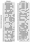
FIG. 2
T-RFs included in the analysis. The sizes of the fragments are in base pairs. Refer to Table 1 for an explanation of the diets (K to G). Solid squares show fragments that were present in samples from the diet groups after 2 weeks of feeding the experimental diets to the animals and before the infection with B. hyodysenteriae. Arrows pointing downward in the “−” column indicate that the fragment disappeared in one or more diet groups after infection. Arrows pointing upward in the “+” column indicate fragments that were first detected in a new diet group after the infection.
Similar articles
- Protection of pigs from swine dysentery by vaccination with recombinant BmpB, a 29.7 kDa outer-membrane lipoprotein of Brachyspira hyodysenteriae.
La T, Phillips ND, Reichel MP, Hampson DJ. La T, et al. Vet Microbiol. 2004 Aug 19;102(1-2):97-109. doi: 10.1016/j.vetmic.2004.06.004. Vet Microbiol. 2004. PMID: 15288932 - A novel method for isolation of Brachyspira (Serpulina) hyodysenteriae from pigs with swine dysentery in Italy.
Calderaro A, Merialdi G, Perini S, Ragni P, Guégan R, Dettori G, Chezzi C. Calderaro A, et al. Vet Microbiol. 2001 May 3;80(1):47-52. doi: 10.1016/s0378-1135(00)00374-6. Vet Microbiol. 2001. PMID: 11278122 - Serologic detection of Brachyspira (Serpulina) hyodysenteriae infections.
La T, Hampson DJ. La T, et al. Anim Health Res Rev. 2001 Jun;2(1):45-52. Anim Health Res Rev. 2001. PMID: 11708746 Review. - Swine dysentery: aetiology, pathogenicity, determinants of transmission and the fight against the disease.
Alvarez-Ordóñez A, Martínez-Lobo FJ, Arguello H, Carvajal A, Rubio P. Alvarez-Ordóñez A, et al. Int J Environ Res Public Health. 2013 May 10;10(5):1927-47. doi: 10.3390/ijerph10051927. Int J Environ Res Public Health. 2013. PMID: 23665849 Free PMC article. Review.
Cited by
- Changes in the Swine Gut Microbiota in Response to Porcine Epidemic Diarrhea Infection.
Koh HW, Kim MS, Lee JS, Kim H, Park SJ. Koh HW, et al. Microbes Environ. 2015;30(3):284-7. doi: 10.1264/jsme2.ME15046. Epub 2015 Jul 25. Microbes Environ. 2015. PMID: 26212519 Free PMC article. - From structure to function: the ecology of host-associated microbial communities.
Robinson CJ, Bohannan BJ, Young VB. Robinson CJ, et al. Microbiol Mol Biol Rev. 2010 Sep;74(3):453-76. doi: 10.1128/MMBR.00014-10. Microbiol Mol Biol Rev. 2010. PMID: 20805407 Free PMC article. Review. - Escherichia coli populations in Great Lakes waterfowl exhibit spatial stability and temporal shifting.
Hansen DL, Ishii S, Sadowsky MJ, Hicks RE. Hansen DL, et al. Appl Environ Microbiol. 2009 Mar;75(6):1546-51. doi: 10.1128/AEM.00444-08. Epub 2009 Jan 9. Appl Environ Microbiol. 2009. PMID: 19139226 Free PMC article. - Characterization of the bacterial fecal microbiota composition of pigs preceding the clinical signs of swine dysentery.
Barbosa JA, Aguirre JCP, Nosach R, Harding JCS, Cantarelli VS, Costa MO. Barbosa JA, et al. PLoS One. 2023 Nov 10;18(11):e0294273. doi: 10.1371/journal.pone.0294273. eCollection 2023. PLoS One. 2023. PMID: 37948383 Free PMC article. - Dietary Fiber Ameliorates Lipopolysaccharide-Induced Intestinal Barrier Function Damage in Piglets by Modulation of Intestinal Microbiome.
Sun X, Cui Y, Su Y, Gao Z, Diao X, Li J, Zhu X, Li D, Li Z, Wang C, Shi Y. Sun X, et al. mSystems. 2021 Apr 6;6(2):e01374-20. doi: 10.1128/mSystems.01374-20. mSystems. 2021. PMID: 33824201 Free PMC article.
References
- Ausubel F M, Brent R, Kingston R E, Moore D D, Smith J A, Seidman J G, Struhl K, editors. Current protocols in molecular biology. New York, N.Y: John Wiley & Sons, Inc.; 1988. pp. 2.4.1–2.4.5.
- Avaniss-Aghajani E, Jones K, Chapman D, Brunk C. A molecular technique for identification of bacteria using small subunit ribosomal RNA sequences. BioTechniques. 1994;17:144–149. - PubMed
- Bourlioux P. What is currently known about the molecular mechanisms of colonisation resistance? Anaerobe. 1997;3:179–184. - PubMed
Publication types
MeSH terms
Substances
LinkOut - more resources
Full Text Sources
Other Literature Sources

