The weaver mutation reverses the function of dopamine and GABA in mouse dopaminergic neurons - PubMed (original) (raw)
The weaver mutation reverses the function of dopamine and GABA in mouse dopaminergic neurons
E Guatteo et al. J Neurosci. 2000.
Abstract
In the present study, we characterized the intrinsic electrophysiological properties and the membrane currents activated by dopamine (DA) D(2) and GABA(B) receptors in midbrain dopaminergic neurons, maintained in vitro in a slice preparation, from wild-type and homozygous weaver (wv/wv) mice. By using patch-clamp techniques, we found that membrane potential, apparent input resistance, and spontaneous firing of wv/wv dopaminergic neurons were similar to those of dopamine-containing cells recorded from nonaffected (+/+) animals. More interestingly, the wv/wv neurons were excited rather than inhibited by dopamine and the GABA(B) agonist baclofen. This neurotransmitter-mediated excitation was attributable to the activation of a G-protein-gated inward current that reversed polarity at a membrane potential of approximately -30 mV. We suggest that the altered behavior of the receptor-operated wv G-protein-gated inwardly rectifying K(+) channel 2 (GIRK2) might be related to the selective degeneration of the dopaminergic neurons. In addition, the wv GIRK2 would not only suppress the autoreceptor-mediated feedback inhibition of DA release but could also establish a feedforward mechanism of DA release in the terminal fields.
Figures
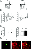
Fig. 1.
Electrophysiological and immunohistochemical identification of midbrain dopaminergic neurons in +/+ and_wv/wv_ mice. A, Hyperpolarizing voltage steps from −60 to −120 mV (10 mV increments,_V_h of −40 mV) activated the mixed cation current (I_h) in both genotypes. Calibration: 250 msec, 500 pA. B, The corresponding current-clamp recording showed that hyperpolarizing pulses (−0.5 and −1 nA) activated the I_h current, thus producing a typical sag potential in both cells taken form the two genotypes. A depolarizing current pulse (0.5 nA) elicited a pacemaker-like sequence of action potentials in both neurons. Action potential amplitudes are clipped because of low sampling rate of the digital interface. Time bar: 100 msec. C,Left, The white square indicates the mean resting membrane potential (RMP) of +/+ dopaminergic cells (−47.6 ± 1.5 mV, n = 17), and the_black square indicates the mean resting membrane potential of wv/wv cells (−47.1 ± 1.3 mV,n = 18); values were not significantly different (p = 0.81). Right, The_columns indicate the mean spontaneous firing recorded in cell-attached configuration in +/+ (white) (2.1 ± 0.12 Hz, n = 7) and wv/wv(black) (2.4 ± 0.2 Hz, n = 15) neurons; values were not significantly different (p = 0.35). D, Confocal laser scanning microscope image of a wv/wv neuron loaded with biocytin (5 m
m
) through the patch pipette showing typical features of a dopaminergic neuron (magnification, 20×):a, biocytin staining as revealed by FITC fluorescence;b, TH immunostaining as revealed by TRITC fluorescence (note that many neurons, including the recorded one, resulted in being TH-positive, within the SNc); c, merged image of the two fluorescent stainings.
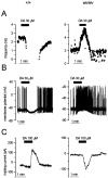
Fig. 2.
DA mediates inhibition of +/+ and excitation of_wv/wv_ neurons. A, Cell-attached recordings from a +/+ (left) and wv/wv(right) cell showing the changes of spontaneous firing during the extracellular application of DA (30 μ
m
). DA clearly inhibited the firing of the +/+ neuron, whereas it increased the activity of the wv/wv neuron. B, Whole-cell current-clamp recordings of two dopaminergic cells in which DA caused membrane hyperpolarization–inhibition (+/+) and depolarization–excitation (wv/wv). C, Voltage-clamp recordings (at _V_h of −40 mV) showing the activation of outward (+/+) and inward (wv/wv) currents caused by DA (100 μ
m
) in +/+ and wv/wv dopaminergic cells, respectively.
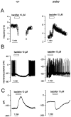
Fig. 3.
The GABAB agonist baclofen mediates inhibition of +/+ and excitation of wv/wv neurons.A, Cell-attached recordings from a +/+ (left) and a wv/wv (right) neuron showing the modification of the spontaneous firing caused by the GABAB receptor agonist baclofen (10 μ
m
). Note the increase in firing frequency induced by this compound in the_wv/wv_ neuron. B, Whole-cell current-clamp recordings showing the modification in membrane potential and firing activity in +/+ and wv/wv neurons. C, Corresponding voltage-clamp recordings (at_V_h of −40 mV) showing the changes in membrane current caused by baclofen in a +/+ versus a_wv/wv_ neuron. Note that, like dopamine, baclofen activated an inward rather than outward current in _wv/wv_neurons.
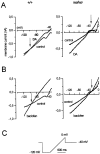
Fig. 4.
Properties of the DA- and baclofen-induced currents in wv/wv neurons. A, Voltage ramps (from −120 to 0 mV) delivered in control condition and in the presence of DA revealed that the DA-induced (100 μ
m
) current reversed at −86 ± 6 mV (n = 4) in +/+ neurons (one cell is shown in the left), whereas it reversed at −27 ± 3.2 mV (n = 6) in_wv/wv_ neurons (one cell is shown in the_right_). B, The baclofen-activated (10 μ
m
) current reversed at negative potentials (−87 ± 3 mV, n = 4) in +/+ neurons (one cell is shown in the left), whereas it reversed at −34.1 ± 3.2 mV (n = 6) in wv/wv neurons (one cell is shown in the right). The current traces of the_wv/wv_ neurons were recorded in the presence of TTX (0.5 μ
m
), tetraethylammonium chloride (5 m
m
), and nifedipine (10 μ
m
) to reduce voltage-dependent sodium, potassium, and calcium conductances. C shows the protocol used to induce the slow depolarizing ramps.
Fig. 5.
The cation channel blockers QX-314 and ZD 7288 inhibit the DA- and baclofen-induced inward currents in_wv/wv_ neurons. A, B, The DA-induced (100 μ
m
) current was strongly inhibited by QX-314 (100 μ
m
) and ZD 7288 (50 μ
m
).C, D, QX-314 (100 μ
m
) and ZD 7288 (50 μ
m
) also inhibited the current induced by baclofen (10 μ
m
). Note that the protocol of the voltage ramps in this and the following figures is the same shown in Figure4_C_.
Fig. 6.
D2 and GABAB receptor antagonists reduce the DA- and baclofen-induced inward currents.A, The inward current induced by both agonists in_wv/wv_ dopaminergic neurons was inhibited by the presence of sulpiride (10 μ
m
; B,top) and CGP 55845A (250 n
m
;B, bottom). C, The plots show the mean values of DA-induced (top, black columns) and baclofen-induced (bottom,white columns) inward currents at the points indicated by asterisks in A (−120 mV). The DA-induced inward current was 207 ± 33 pA (_n_= 7; left bar) and was significantly reduced by QX-314 (100 μ
m
) to 111 ± 13 pA (paired _t_test, p < 0.05; middle bar) and by sulpiride (10 μ
m
) to 117 ± 17 pA (paired_t_ test, p < 0.05; right bar). The baclofen-induced inward current of 198 ± 28 pA (n = 6; left bar) was significantly reduced by QX-314 (100 μ
m
) to 63 ± 15 (paired_t_ test, p < 0.01; middle bar) and by CGP 55845A (250 n
m
) to 21 ± 8 pA (paired t test, p < 0.01;right bar).
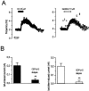
Fig. 7.
D2 and GABABreceptors couple to wv GIRK2 in a G-protein-dependent manner. A, Recordings of the spontaneous firing of two_wv/wv_ neurons in cell-attached configuration with a pipette solution containing the GTP analog GTP-γ-S (0.6 m
m
). Note that both DA (left) and baclofen (right) induced an increase of the spontaneous firing before the rupture of the membrane patches. B, The_black columns_ show the amplitude of the DA-induced current (at −120 mV) (204 ± 30 pA, _n_= 10) in control conditions and during the dialysis with GTP-γ-S (38 ± 14 pA, n = 5) (p < 0.05, unpaired data). The white columns show the amplitude of the baclofen-induced current (at −120 mV) (205 ± 40 pA, n = 6) in control condition and during the intracellular dialysis with GTP-γ-S (57 ± 10, n = 5) (p < 0.05, unpaired data). C, GTP-γ-S induced a tonic inward current that was reversibly blocked by QX-314 and irreversibly inhibited by ZD 7288. Note that, under this condition, the response to DA application was not observed.
Fig. 8.
GDP-β-S prevents the DA- and baclofen-induced inward currents. A, Cell-attached recordings of two_wv/wv_ neurons with a pipette containing the GDP analog GDP-β-S (0.6 m
m
) showing the increase of the spontaneous firing caused by DA and baclofen. B,Left, black columns, In whole-cell configuration, the DA-induced current (at −120 mV) was 204 ± 30 pA (n = 10) and was significantly reduced by GDP-β-S to 39 ± 15 pA (n = 5,p < 0.01, unpaired data). Right,white columns, The baclofen-induced current (at −120 mV) was 205 ± 40 pA (n = 6) and was significantly reduced by GDP-β-S to 21 ± 19 pA (n = 5, p < 0.01, unpaired data).
Similar articles
- Voltage-gated calcium channels mediate intracellular calcium increase in weaver dopaminergic neurons during stimulation of D2 and GABAB receptors.
Guatteo E, Bengtson CP, Bernardi G, Mercuri NB. Guatteo E, et al. J Neurophysiol. 2004 Dec;92(6):3368-74. doi: 10.1152/jn.00602.2004. Epub 2004 Jul 7. J Neurophysiol. 2004. PMID: 15240766 - The weaver mouse gain-of-function phenotype of dopaminergic midbrain neurons is determined by coactivation of wvGirk2 and K-ATP channels.
Liss B, Neu A, Roeper J. Liss B, et al. J Neurosci. 1999 Oct 15;19(20):8839-48. doi: 10.1523/JNEUROSCI.19-20-08839.1999. J Neurosci. 1999. PMID: 10516303 Free PMC article. - Defective gamma-aminobutyric acid type B receptor-activated inwardly rectifying K+ currents in cerebellar granule cells isolated from weaver and Girk2 null mutant mice.
Slesinger PA, Stoffel M, Jan YN, Jan LY. Slesinger PA, et al. Proc Natl Acad Sci U S A. 1997 Oct 28;94(22):12210-7. doi: 10.1073/pnas.94.22.12210. Proc Natl Acad Sci U S A. 1997. PMID: 9342388 Free PMC article. - Cell death in weaver mouse cerebellum.
Harkins AB, Fox AP. Harkins AB, et al. Cerebellum. 2002 Jul;1(3):201-6. doi: 10.1080/14734220260418420. Cerebellum. 2002. PMID: 12879981 Review. - Rapid effects of estrogen on G protein-coupled receptor activation of potassium channels in the central nervous system (CNS).
Kelly MJ, Qiu J, Wagner EJ, Rønnekleiv OK. Kelly MJ, et al. J Steroid Biochem Mol Biol. 2002 Dec;83(1-5):187-93. doi: 10.1016/s0960-0760(02)00249-2. J Steroid Biochem Mol Biol. 2002. PMID: 12650715 Review.
Cited by
- I(h) channels contribute to the different functional properties of identified dopaminergic subpopulations in the midbrain.
Neuhoff H, Neu A, Liss B, Roeper J. Neuhoff H, et al. J Neurosci. 2002 Feb 15;22(4):1290-302. doi: 10.1523/JNEUROSCI.22-04-01290.2002. J Neurosci. 2002. PMID: 11850457 Free PMC article. - Cell type analysis of functional fetal dopamine cell suspension transplants in the striatum and substantia nigra of patients with Parkinson's disease.
Mendez I, Sanchez-Pernaute R, Cooper O, Viñuela A, Ferrari D, Björklund L, Dagher A, Isacson O. Mendez I, et al. Brain. 2005 Jul;128(Pt 7):1498-510. doi: 10.1093/brain/awh510. Epub 2005 May 4. Brain. 2005. PMID: 15872020 Free PMC article.
References
- Bertolino M, Llinas RR. The central role of voltage-activated and receptor-operated calcium channels in neuronal cells. Annu Rev Pharmacol Toxicol. 1992;32:399–421. - PubMed
- Choi DW. Calcium: still center-stage in hypoxic-ischemic neuronal death. Trends Neurosci. 1995;18:58–60. - PubMed
- Gupta AM, Felten DL, Ghetti B. Selective loss of monoaminergic neurones in the weaver mutant mice: an immunocytochemical study. Brain Res. 1987;402:379–382. - PubMed
Publication types
MeSH terms
Substances
LinkOut - more resources
Full Text Sources
Molecular Biology Databases
