Early alterations of the receptor-binding properties of H1, H2, and H3 avian influenza virus hemagglutinins after their introduction into mammals - PubMed (original) (raw)
Early alterations of the receptor-binding properties of H1, H2, and H3 avian influenza virus hemagglutinins after their introduction into mammals
M Matrosovich et al. J Virol. 2000 Sep.
Abstract
Interspecies transmission of influenza A viruses circulating in wild aquatic birds occasionally results in influenza outbreaks in mammals, including humans. To identify early changes in the receptor binding properties of the avian virus hemagglutinin (HA) after interspecies transmission and to determine the amino acid substitutions responsible for these alterations, we studied the HAs of the initial isolates from the human pandemics of 1957 (H2N2) and 1968 (H3N2), the European swine epizootic of 1979 (H1N1), and the seal epizootic of 1992 (H3N3), all of which were caused by the introduction of avian virus HAs into these species. The viruses were assayed for their ability to bind the synthetic sialylglycopolymers 3'SL-PAA and 6'SLN-PAA, which contained, respectively, 3'-sialyllactose (the receptor determinant preferentially recognized by avian influenza viruses) and 6'-sialyl(N-acetyllactosamine) (the receptor determinant for human viruses). Avian and seal viruses bound 6'SLN-PAA very weakly, whereas the earliest available human and swine epidemic viruses bound this polymer with a higher affinity. For the H2 and H3 strains, a single mutation, 226Q-->L, increased binding to 6'SLN-PAA, while among H1 swine viruses, the 190E-->D and 225G-->E mutations in the HA appeared important for the increased affinity of the viruses for 6'SLN-PAA. Amino acid substitutions at positions 190 and 225 with respect to the avian virus consensus sequence are also present in H1 human viruses, including those that circulated in 1918, suggesting that substitutions at these positions are important for the generation of H1 human pandemic strains. These results show that the receptor-binding specificity of the HA is altered early after the transmission of an avian virus to humans and pigs and, therefore, may be a prerequisite for the highly effective replication and spread which characterize epidemic strains.
Figures
FIG. 1
Binding of sialylglycopolymers 3′SL-PAA (solid lines) and 6′SLN-PAA (dotted lines) by H3 subtype influenza virus strains A/duck/Hokkaido/33/80 and A/Los Angeles/2/87. The binding assay is described in Materials and Methods. The upper panels represent the primary data (dependence of absorbancy in the wells, _A_490, versus concentration of the polymer); the lower panels show the corresponding Scatchard plots.
FIG. 2
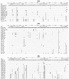
Partial HA amino acid sequences (HA1 positions 130 to 230) of influenza viruses that were tested for their binding to sialylglycopolymers. Full names of the virus strains are listed in Tables 1 to 3. Differences with respect to the top sequence are shown. The H3 numbering system is used; the position in the H1 HA that is absent from the H3 HA is indicated by an underscore. The RBS line shows the position of the amino acid with respect to the HA receptor-binding site. The “R” designates that the amino acid residue contacts either sialic acid or penultimate galactose in the X31 virus HA complex with 3′-sialyllactose (1HGG structure; Brookhaven Protein Database). The star indicates that an amino acid is within 15 Å of the C2 carbon atom of the sialic acid in the 1HGG structure. The figure was generated with GeneDoc 2.3 software (25).
FIG. 3
Positions of amino acids in the HA receptor-binding site that differ between early epidemic human and swine viruses and their closely related avian counterparts (stereo view). The figure is based on the crystallographic model of the X31 HA complex with the Neu5Acα2-6Gal-containing receptor analog LSTc (Neu5Acα2-6Galβ1-4GlcNAcβ1-3Galβ1-4Glc) (10). The solvent-accessible molecular surface of the protein is shown. The sialic acid residue and penultimate galactose ring of LSTc are displayed as thick stick bonds; the rest of the molecule is shown as a thin white line. The gray transparent sphere (C6) in close proximity to amino acid 226 represents the van der Waals surface of the C6′-carbon atom of Gal. The figure was generated with WebLab ViewerPro 3.10 (Molecular Simulations, Inc., San Diego, Calif.).
FIG. 4
Phylogenetic tree for H1 subtype influenza viruses from different hosts, as determined based on the amino acid sequences of the HA1. The tree was constructed as described in Materials and Methods using the HA sequences of viruses listed in Fig. 2 and the following sequences of classical swine and human viruses from GenBank: sw/Iowa/15/30, WSN/33, PR/8/34 (Cambridge strain), FM/1/47, USSR/90/77, Taiwan/1/86, and Finland/168/91. The figure was generated with the TREEVIEW 1.5.2 program (27).
Similar articles
- Avian influenza A viruses differ from human viruses by recognition of sialyloligosaccharides and gangliosides and by a higher conservation of the HA receptor-binding site.
Matrosovich MN, Gambaryan AS, Teneberg S, Piskarev VE, Yamnikova SS, Lvov DK, Robertson JS, Karlsson KA. Matrosovich MN, et al. Virology. 1997 Jun 23;233(1):224-34. doi: 10.1006/viro.1997.8580. Virology. 1997. PMID: 9201232 - 6-sulfo sialyl Lewis X is the common receptor determinant recognized by H5, H6, H7 and H9 influenza viruses of terrestrial poultry.
Gambaryan AS, Tuzikov AB, Pazynina GV, Desheva JA, Bovin NV, Matrosovich MN, Klimov AI. Gambaryan AS, et al. Virol J. 2008 Jul 24;5:85. doi: 10.1186/1743-422X-5-85. Virol J. 2008. PMID: 18652681 Free PMC article. - Glycan microarray analysis of the hemagglutinins from modern and pandemic influenza viruses reveals different receptor specificities.
Stevens J, Blixt O, Glaser L, Taubenberger JK, Palese P, Paulson JC, Wilson IA. Stevens J, et al. J Mol Biol. 2006 Feb 3;355(5):1143-55. doi: 10.1016/j.jmb.2005.11.002. Epub 2005 Nov 18. J Mol Biol. 2006. PMID: 16343533 - Adaptation of influenza viruses to human airway receptors.
Thompson AJ, Paulson JC. Thompson AJ, et al. J Biol Chem. 2021 Jan-Jun;296:100017. doi: 10.1074/jbc.REV120.013309. Epub 2020 Nov 22. J Biol Chem. 2021. PMID: 33144323 Free PMC article. Review. - Recent zoonoses caused by influenza A viruses.
Alexander DJ, Brown IH. Alexander DJ, et al. Rev Sci Tech. 2000 Apr;19(1):197-225. doi: 10.20506/rst.19.1.1220. Rev Sci Tech. 2000. PMID: 11189716 Review.
Cited by
- Effects of different HA and NA gene combinations on the growth characteristics of the H3N8 influenza candidate vaccine virus.
Liu L, Li Z, Liu J, Li X, Zhou J, Xiao N, Yang L, Wang D. Liu L, et al. Vaccine X. 2024 Jul 18;19:100531. doi: 10.1016/j.jvacx.2024.100531. eCollection 2024 Aug. Vaccine X. 2024. PMID: 39157684 Free PMC article. - Exploring Potential Intermediates in the Cross-Species Transmission of Influenza A Virus to Humans.
Lee CY. Lee CY. Viruses. 2024 Jul 14;16(7):1129. doi: 10.3390/v16071129. Viruses. 2024. PMID: 39066291 Free PMC article. Review. - Age-dependent heterogeneity in the antigenic effects of mutations to influenza hemagglutinin.
Welsh FC, Eguia RT, Lee JM, Haddox HK, Galloway J, Van Vinh Chau N, Loes AN, Huddleston J, Yu TC, Quynh Le M, Nhat NTD, Thi Le Thanh N, Greninger AL, Chu HY, Englund JA, Bedford T, Matsen FA 4th, Boni MF, Bloom JD. Welsh FC, et al. Cell Host Microbe. 2024 Aug 14;32(8):1397-1411.e11. doi: 10.1016/j.chom.2024.06.015. Epub 2024 Jul 19. Cell Host Microbe. 2024. PMID: 39032493 - Molecular Markers and Mechanisms of Influenza A Virus Cross-Species Transmission and New Host Adaptation.
Guo X, Zhou Y, Yan H, An Q, Liang C, Liu L, Qian J. Guo X, et al. Viruses. 2024 May 30;16(6):883. doi: 10.3390/v16060883. Viruses. 2024. PMID: 38932174 Free PMC article. Review. - R229I substitution from oseltamivir induction in HA1 region significantly increased the fitness of a H7N9 virus bearing NA 292K.
Tang J, Zou SM, Zhou JF, Gao RB, Xin L, Zeng XX, Huang WJ, Li XY, Cheng YH, Liu LQ, Xiao N, Wang DY. Tang J, et al. Emerg Microbes Infect. 2024 Dec;13(1):2373314. doi: 10.1080/22221751.2024.2373314. Epub 2024 Jul 16. Emerg Microbes Infect. 2024. PMID: 38922326 Free PMC article.
References
- Baum L G, Paulson J C. Sialyloligosaccharides of the respiratory epithelium in the selection of human influenza virus receptor specificity. Acta Histochem Suppl Band. 1990;XL:35–38. - PubMed
- Bovin N V, Korchagina E Y, Zemlyanukhina T V, Byramova N E, Galanina O E, Zemlyakov A E, Ivanov A E, Zubov V P, Mochalova L V. Synthesis of polymeric neoglycoconjugates based on N-substituted polyacrylamides. Glycoconj J. 1993;10:142–151. - PubMed
- Brown I H, Ludwig S, Olsen C W, Hannoun C, Scholtissek C, Hinshaw V S, Harris P A, McCauley J W, Strong I, Alexander D J. Antigenic and genetic analyses of H1N1 influenza A viruses from European pigs. J Gen Virol. 1997;78:553–562. - PubMed
- Callan R J, Early G, Kida H, Hinshaw V S. The appearance of H3 influenza viruses in seals. J Gen Virol. 1995;76:199–203. - PubMed
Publication types
MeSH terms
Substances
LinkOut - more resources
Full Text Sources
Other Literature Sources


