Diagnostic ultrasound activation of contrast agent gas bodies induces capillary rupture in mice - PubMed (original) (raw)
Diagnostic ultrasound activation of contrast agent gas bodies induces capillary rupture in mice
D L Miller et al. Proc Natl Acad Sci U S A. 2000.
Abstract
Interaction of diagnostic ultrasound with gas bodies produces a useful contrast effect in medical images, but the same interaction also represents a mechanism for bioeffects. Anesthetized hairless mice were scanned by using a 2.5-MHz transducer (610-ns pulses with 3.6-kHz repetition frequency and 61-Hz frame rate) after injection of Optison and Evans blue dye. Petechial hemorrhages (PHs) in intestine and abdominal muscle were counted 15 min after exposure to characterize capillary rupture, and Evans blue extravasation was evaluated in samples of muscle tissue. For 5 ml small middle dotkg(-1) contrast agent and exposure to 10 alternating 10-s on and off periods, PH counts in muscle were approximately proportional to the square of peak negative pressure amplitude and were statistically significant above 0.64 MPa. PH counts in intestine and Evans blue extravasation into muscle tissue were significant above 1. 0 MPa. The PH effect in muscle was proportional to contrast dose and was statistically significant for the lowest dose of 0.05 ml small middle dotkg(-1). The effects decreased nearly to sham levels if the exposure was delayed 5 min. The PH effect in abdominal muscle was significant and statistically indistinguishable for uninterrupted 100-s exposure, 10-s exposure, 100 scans repeated at 1 Hz, and even for a single scan. The results confirms a previous report of PH induction by diagnostic ultrasound with contrast agent in mammalian skeletal muscle [Skyba, D. M., Price, R. J., Linka, A. Z., Skalak, T. C. & Kaul, S. (1998) Circulation 98, 290-293].
Figures
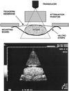
Figure 1
(Upper) The experimental setup. (Lower) The ultrasound image obtained during exposure with the attenuation phantom.
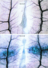
Figure 2
Examples of the effects found in the abdominal muscle and fat. (Lower) PHs and Evans blue extravasation with 5 ml⋅kg-1 contrast agent and 10 × 10s exposure at 2.8 MPa. (Upper) No effect with the same exposure but without injected contrast-agent gas bodies. (Scale bars: 1 mm.)
Figure 3
Mean PH counts with standard error bars in muscle and intestine for 5 ml⋅kg-1 contrast agent at the indicated pressure amplitudes. The curve plotted with the muscle results was fitted to the data means by linear regression against the square of the amplitude (_r_2 = 0.995).
Figure 4
The results for Evans blue extravasation for 5 ml⋅kg-1 contrast agent at the indicated pressure amplitudes, obtained from the same samples used for the points in Fig. 3.
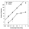
Figure 5
Mean PH counts with standard error bars in muscle and intestine for 10 × 10s exposure at 2.8 MPa with the indicated contrast agent doses. The line indicates linear dependence of the muscle counts on dose (except the maximum dose) (_r_2 = 0.988).
Figure 6
The results for Evans blue extravasation for the indicated contrast agent doses, obtained from the same samples used for the points in Fig. 5.
Figure 7
A comparison of muscle PH counts after different exposure timing sequences at 2.8 MPa with 5 ml⋅kg-1 contrast agent.
Similar articles
- Overview of experimental studies of biological effects of medical ultrasound caused by gas body activation and inertial cavitation.
Miller DL. Miller DL. Prog Biophys Mol Biol. 2007 Jan-Apr;93(1-3):314-30. doi: 10.1016/j.pbiomolbio.2006.07.027. Epub 2006 Aug 22. Prog Biophys Mol Biol. 2007. PMID: 16989895 Review. - Capillary Hemorrhage Induced by Contrast-Enhanced Diagnostic Ultrasound in Rat Intestine.
Lu X, Dou C, Fabiilli ML, Miller DL. Lu X, et al. Ultrasound Med Biol. 2019 Aug;45(8):2133-2139. doi: 10.1016/j.ultrasmedbio.2019.04.012. Epub 2019 May 14. Ultrasound Med Biol. 2019. PMID: 31101449 Free PMC article. - The influence of ultrasound frequency and gas-body composition on the contrast agent-mediated enhancement of vascular bioeffects in mouse intestine.
Miller DL, Gies RA. Miller DL, et al. Ultrasound Med Biol. 2000 Feb;26(2):307-13. doi: 10.1016/s0301-5629(99)00138-6. Ultrasound Med Biol. 2000. PMID: 10722920 - Gas-body-based contrast agent enhances vascular bioeffects of 1.09 MHz ultrasound on mouse intestine.
Miller DL, Gies RA. Miller DL, et al. Ultrasound Med Biol. 1998 Oct;24(8):1201-8. doi: 10.1016/s0301-5629(98)00063-5. Ultrasound Med Biol. 1998. PMID: 9833589 - Bioeffects considerations for diagnostic ultrasound contrast agents.
Miller DL, Averkiou MA, Brayman AA, Everbach EC, Holland CK, Wible JH Jr, Wu J. Miller DL, et al. J Ultrasound Med. 2008 Apr;27(4):611-32; quiz 633-6. doi: 10.7863/jum.2008.27.4.611. J Ultrasound Med. 2008. PMID: 18359911 Review.
Cited by
- Research methodology for in vivo measurements of resting energy expenditure, daily body temperature, metabolic heat and non-viral tissue-specific gene therapy in baboons.
Frost PA, Chen S, Rodriguez-Ayala E, Laviada-Molina HA, Vaquera Z, Gaytan-Saucedo JF, Li WH, Haack K, Grayburn PA, Sayers K, Cole SA, Bastarrachea RA. Frost PA, et al. Res Vet Sci. 2020 Dec;133:136-145. doi: 10.1016/j.rvsc.2020.09.020. Epub 2020 Sep 20. Res Vet Sci. 2020. PMID: 32979746 Free PMC article. - Controlled ultrasound-induced blood-brain barrier disruption using passive acoustic emissions monitoring.
Arvanitis CD, Livingstone MS, Vykhodtseva N, McDannold N. Arvanitis CD, et al. PLoS One. 2012;7(9):e45783. doi: 10.1371/journal.pone.0045783. Epub 2012 Sep 24. PLoS One. 2012. PMID: 23029240 Free PMC article. - Microbubbles improve the ablation efficiency of extracorporeal high intensity focused ultrasound against kidney tissues.
Yu T, Hu D, Xu C. Yu T, et al. World J Urol. 2008 Dec;26(6):631-6. doi: 10.1007/s00345-008-0290-z. Epub 2008 Jul 2. World J Urol. 2008. PMID: 18594828 - Therapeutic Ultrasound Parameter Optimization for Drug Delivery Applied to a Murine Model of Hepatocellular Carcinoma.
Telichko AV, Wang H, Bachawal S, Kumar SU, Bose JC, Paulmurugan R, Dahl JJ. Telichko AV, et al. Ultrasound Med Biol. 2021 Feb;47(2):309-322. doi: 10.1016/j.ultrasmedbio.2020.09.009. Epub 2020 Nov 3. Ultrasound Med Biol. 2021. PMID: 33153807 Free PMC article. - Nephron injury induced by diagnostic ultrasound imaging at high mechanical index with gas body contrast agent.
Williams AR, Wiggins RC, Wharram BL, Goyal M, Dou C, Johnson KJ, Miller DL. Williams AR, et al. Ultrasound Med Biol. 2007 Aug;33(8):1336-44. doi: 10.1016/j.ultrasmedbio.2007.03.002. Epub 2007 May 16. Ultrasound Med Biol. 2007. PMID: 17507144 Free PMC article.
References
- Golberg B B, Liu J B, Forsberg F. Ultrasound Med Biol. 1994;20:319–333. - PubMed
- Burns P N. In: Diagnostic Ultrasound. Rumack C, Wilson S, Charboneau J, editors. Vol. 1. New York: Mosby; 1998. pp. 57–84.
- Killam A, Dittrich H. In: Ultrasound Contrast Agents. Goldberg B, editor. London: Martin Dunitz; 1997. pp. 43–55.
- Cotter B, Mahmud E, Kwan O L, DeMaria A N. In: Ultrasound Contrast Agents. Goldberg B, editor. London: Martin Dunitz; 1997. pp. 31–42.
- Food and Drug Administration. Information for Manufacturers Seeking Clearance of Diagnostic Ultrasound Systems and Transducers. U.S. Food and Drug Administration, Rockville, MD: Center for Devices and Radiological Health; 1997.
Publication types
MeSH terms
Substances
LinkOut - more resources
Full Text Sources
Other Literature Sources
Miscellaneous


