Blockade of T lymphocyte costimulation with cytotoxic T lymphocyte-associated antigen 4-immunoglobulin (CTLA4Ig) reverses the cellular pathology of psoriatic plaques, including the activation of keratinocytes, dendritic cells, and endothelial cells - PubMed (original) (raw)
Blockade of T lymphocyte costimulation with cytotoxic T lymphocyte-associated antigen 4-immunoglobulin (CTLA4Ig) reverses the cellular pathology of psoriatic plaques, including the activation of keratinocytes, dendritic cells, and endothelial cells
J R Abrams et al. J Exp Med. 2000.
Abstract
Efficient T cell activation is dependent on the intimate contact between antigen-presenting cells (APCs) and T cells. The engagement of the B7 family of molecules on APCs with CD28 and CD152 (cytotoxic T lymphocyte-associated antigen 4 [CTLA-4]) receptors on T cells delivers costimulatory signal(s) important in T cell activation. We investigated the dependence of pathologic cellular activation in psoriatic plaques on B7-mediated T cell costimulation. Patients with psoriasis vulgaris received four intravenous infusions of the soluble chimeric protein CTLA4Ig (BMS-188667) in a 26-wk, phase I, open label dose escalation study. Clinical improvement was associated with reduced cellular activation of lesional T cells, keratinocytes, dendritic cells (DCs), and vascular endothelium. Expression of CD40, CD54, and major histocompatibility complex (MHC) class II HLA-DR antigens by lesional keratinocytes was markedly reduced in serial biopsy specimens. Concurrent reductions in B7-1 (CD80), B7-2 (CD86), CD40, MHC class II, CD83, DC-lysosomal-associated membrane glycoprotein (DC-LAMP), and CD11c expression were detected on lesional DCs, which also decreased in number within lesional biopsies. Skin explant experiments suggested that these alterations in activated or mature DCs were not the result of direct toxicity of CTLA4Ig for DCs. Decreased lesional vascular ectasia and tortuosity were also observed and were accompanied by reduced presence of E-selectin, P-selectin, and CD54 on vascular endothelium. This study highlights the critical and proximal role of T cell activation through the B7-CD28/CD152 costimulatory pathway in maintaining the pathology of psoriasis, including the newly recognized accumulation of mature DCs in the epidermis.
Figures
Figure 1
Decrease in psoriatic lesional leukocytic infiltrates after administration of CTLA4Ig. Representative immunohistochemical findings in serial biopsies at day 1 (top), day 36 (middle), and day 78 (bottom) from prospectively identified lesions in 9 of the 11 patients accrued to the CTLA4Ig 25 and 50 mg/kg dose levels. Illustrated in A–L are data from a patient who received CTLA4Ig 25 mg/kg/dose. T cells present within the lesion were detected by immunostaining with mAbs to CD3 (A–C), CD25 (D–F), or CD8 (G–I). Serial reductions in T cell infiltrates were observed after initiation of CTLA4Ig infusions (day 1) and were associated with progressive epidermal thinning. Neutrophilic infiltrates detected by immunostaining with mAbs to elastase (J–L) were also decreased in serial biopsies. At day 36 (K), neutrophils were largely confined to capillaries in the papillary dermis (arrows). Original magnifications: ×200.
Figure 2
Changes in psoriatic lesional T cell numbers and keratinocyte hyperplasia across the 11 patients accrued to the CTLA4Ig 25 and 50 mg/kg dose groups. (A) Mean percent decrease in values for epidermal thickness (black squares) and numbers of T cells within the epidermis (red diamonds) or dermis (blue circles) compared with baseline (day 1) are illustrated over the first 78 d of the study period. Asterisks indicate statistical significance (*P < 0.05; **P < 0.001). P values were based on a two-sided t test for no change at the indicated study day compared with day 1. (B) Correlation plots of the percent change in infiltrating epidermal or dermal T cells versus percent change in epidermal thickness for each patient at all sampling time points. Individual data points are an average derived from triplicate analyses. Epidermal thickness was calculated by quantitating the cross-sectional surface area beneath a 1-mm linear region of a representative histological section using computer-assisted image analysis. Positive correlations between the change in T (CD3+) cell numbers and epidermal thickness were evident (epidermal CD3+: r = 0.73; dermal CD3+: r = 0.61). Discordant responses characterized by reductions in epidermal thickness that were not accompanied by similar reductions in T cell numbers within a specific compartment are indicated (open red squares).
Figure 4
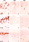
Increased CD1a reactivity and reduced CD80 and CD86 expression in psoriatic lesional biopsies after administration of CTLA4Ig. Serial biopsies were obtained from the perimeter of a single lesion in a patient accrued to the CTLA4Ig 25 mg/kg dose level, and are representative of the histological findings in the clinical responders within the 25 and 50 mg/kg dosing cohorts. An increasing density of CD1a+ epidermal LC staining was observed after initiation of treatment (A–C). Positive CD80 and CD86 immunoreactivity was seen in both the epidermis and dermis at day 1 (D and G). Diminished staining for CD80 and CD86 is seen at day 36 (E and H). At day 78, CD80 expression was no longer detectable within the epidermis, and minimal CD80+ staining was present in the dermis (F). A low level of staining for CD86, predominantly within the dermis, was present at day 78 (I). Original magnifications: ×400.
Figure 3
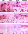
Diminished immunohistochemical detection of CD40, ICAM-1, HLA-DR, and FXIIIa in psoriatic lesional skin after administration of CTLA4Ig. Keratinocytes within the suprapapillary regions (arrows) and DCs displayed marked upregulation of expression of CD40, ICAM-1 (CD54), and HLA-DR at day 1 (top), which serially decreased at study days 36 (middle) and 78 (bottom). Superficial capillaries in untreated psoriatic lesions also exhibited increased ICAM-1 and CD40 staining (arrowheads; A and D), which was reduced at days 36 (B and E) and 78 (C and F). Phenotypic alterations of lesional DCs stained with mAbs to HLA-DR and FXIIIa are observed at days 36 and 78 after the initial infusion (G–L). Higher magnification views of the boxed regions are shown in the inset areas in G–L, and demonstrate a progressive decrease in the number and length of branching processes radiating from the cell body (dendricity) of the stained cells. Results shown are from serial biopsies of a single lesion in a patient administered CTLA4Ig 25 mg/kg and are representative of the nine patients achieving a 50% or greater improvement in clinical scores. Original magnifications: (A–F) ×400; (G–L) ×200; (G–L, insets) ×800.
Figure 5
Alterations in immunohistochemical markers of DC maturational status in psoriatic lesional biopsies after CTLA4Ig administration. Biopsy material obtained at day 1 (top) and day 78 (bottom) from a patient accrued to the CTLA4Ig 25 mg/kg dose level are illustrated and were reflective of the changes observed in all clinical responders. The DC-restricted markers CD83 and DC-LAMP, expressed at high levels on mature DCs, demonstrated an increased magnitude of staining in both the epidermis and dermis at day 1 (A, C, and Day 1 Dermis DC-LAMP). Marked reductions in the density of staining for both CD83 and DC-LAMP were observed; little residual staining was evident at day 78 (B, D, and Day 78 Dermis DC-LAMP). Immunoreactivity for the leukocyte integrin CD11c, abundantly expressed on DCs, was also decreased within the epidermal and dermal compartments at day 78 compared with day 1 (E and F). Residual CD11c reactivity was restricted to the dermis at day 78. The population of immature DCs characterized by MMR positivity was distributed exclusively within the dermal compartment and displayed small decreases in immunoreactivity at day 78 compared with baseline examination (G and H). Original magnifications: (A, B, E–H) ×400; (C and D) ×800.
Figure 6
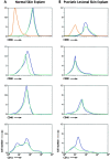
Four-color flow cytometry analysis of emigrating leukocytes from normal and psoriatic lesional skin explants. DCs (CD3−DR+CD45+) emigrating from normal skin (A) or psoriatic lesional skin explants (B) after 3 d of culture were analyzed for expression of CD80, CD86, CD40, or CD1a, and the results were plotted as histograms of cell number versus log10 fluorescence. Isotype controls for the DC population gates (CD3−DR+CD45+) used in the two series of analyses are illustrated in A and B (red lines). Cultures were performed with media alone (blue lines) or in the presence of CTLA4Ig (100 μg/ml; green lines). The data in A are representative of four independent experiments with normal skin explants; data in B are comparable to three independent experiments with psoriatic lesional skin. The results from all experiments are summarized in Table .
Figure 7
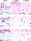
Alterations in psoriatic lesional vascular immunohistology in serial cryostat sections after administration of CTLA4Ig. Illustrated in A–I are representative immunohistochemical findings in biopsies obtained at day 1 (top), day 36 (middle), and day 78 (bottom) from the perimeter of a single lesion in clinically responding subjects accrued to the CTLA4Ig 25 and 50 mg/kg dose levels; data shown are from one subject accrued to the 50 mg/kg dose level. Immunostaining with mAbs to laminin (A–C) present in the basement membrane of blood vessels (and epidermis) illustrates the progressive decrease in lesional vascular ectasia and tortuosity after CTLA4Ig administration. Increased E-selectin (CD62E) and P-selectin (CD62P) reactivity was noted in superficial (arrows) and deep dermal vessels (arrowheads) within chronically inflamed psoriatic skin at day 1 (D and G). E-selectin staining decreases serially in both the superficial and deep dermal vascular beds after administration of CTLA4Ig (E and F). Expression of P-selectin is selectively decreased in expression in the superficial capillaries at days 36 and 78. Original magnifications: ×200.
Similar articles
- CTLA4Ig-mediated blockade of T-cell costimulation in patients with psoriasis vulgaris.
Abrams JR, Lebwohl MG, Guzzo CA, Jegasothy BV, Goldfarb MT, Goffe BS, Menter A, Lowe NJ, Krueger G, Brown MJ, Weiner RS, Birkhofer MJ, Warner GL, Berry KK, Linsley PS, Krueger JG, Ochs HD, Kelley SL, Kang S. Abrams JR, et al. J Clin Invest. 1999 May;103(9):1243-52. doi: 10.1172/JCI5857. J Clin Invest. 1999. PMID: 10225967 Free PMC article. Clinical Trial. - Costimulation blockade promotes the apoptotic death of graft-infiltrating T cells and prolongs survival of hepatic allografts from FLT3L-treated donors.
Li W, Lu L, Wang Z, Wang L, Fung JJ, Thomson AW, Qian S. Li W, et al. Transplantation. 2001 Oct 27;72(8):1423-32. doi: 10.1097/00007890-200110270-00016. Transplantation. 2001. PMID: 11685115 - Costimulation of superantigen-activated T lymphocytes by autologous dendritic cells is dependent on B7.
Nestle FO, Thompson C, Shimizu Y, Turka LA, Nickoloff BJ. Nestle FO, et al. Cell Immunol. 1994 Jun;156(1):220-9. doi: 10.1006/cimm.1994.1166. Cell Immunol. 1994. PMID: 7515331 - [Regulation of T-cell activation by CD28 and CTLA-4].
Nagel T, Kalden JR, Manger B. Nagel T, et al. Med Klin (Munich). 1998 Oct 15;93(10):592-7. doi: 10.1007/BF03042674. Med Klin (Munich). 1998. PMID: 9849050 Review. German. - CTLA4-Ig: a novel immunosuppressive agent.
Najafian N, Sayegh MH. Najafian N, et al. Expert Opin Investig Drugs. 2000 Sep;9(9):2147-57. doi: 10.1517/13543784.9.9.2147. Expert Opin Investig Drugs. 2000. PMID: 11060799 Review.
Cited by
- Psoriatic arthritis: pathogenesis and novel immunomodulatory approaches to treatment.
Cassell S, Kavanaugh A. Cassell S, et al. J Immune Based Ther Vaccines. 2005 Sep 2;3:6. doi: 10.1186/1476-8518-3-6. J Immune Based Ther Vaccines. 2005. PMID: 16138929 Free PMC article. - Antagonistic and agonistic anti-canine CD28 monoclonal antibodies: tools for allogeneic transplantation.
Graves SS, Stone DM, Loretz C, Peterson LJ, Lesnikova M, Hwang B, Georges GE, Nash R, Storb R. Graves SS, et al. Transplantation. 2011 Apr 27;91(8):833-40. doi: 10.1097/TP.0b013e31820f07ff. Transplantation. 2011. PMID: 21343872 Free PMC article. - Impaired synthesis of erythropoietin, glutamine synthetase and metallothionein in the skin of NOD/SCID/gamma(c)(null) and Foxn1 nu/nu mice with misbalanced production of MHC class II complex.
Danielyan L, Verleysdonk S, Buadze M, Gleiter CH, Buniatian GH. Danielyan L, et al. Neurochem Res. 2010 Jun;35(6):899-908. doi: 10.1007/s11064-009-0074-x. Epub 2009 Oct 14. Neurochem Res. 2010. PMID: 19826948 - Novel immunobiologics for psoriasis.
Ghosh N, Singh PN, Kumar V. Ghosh N, et al. Indian J Pharmacol. 2008 Jun;40(3):95-102. doi: 10.4103/0253-7613.42300. Indian J Pharmacol. 2008. PMID: 20040934 Free PMC article. - Avoiding horror autotoxicus: the importance of dendritic cells in peripheral T cell tolerance.
Steinman RM, Nussenzweig MC. Steinman RM, et al. Proc Natl Acad Sci U S A. 2002 Jan 8;99(1):351-8. doi: 10.1073/pnas.231606698. Epub 2002 Jan 2. Proc Natl Acad Sci U S A. 2002. PMID: 11773639 Free PMC article. Review.
References
- Paukkonen K., Naukkarinen A., Horsmanheimo M. The development of manifest psoriatic lesions is linked with the invasion of CD8+ T cells and CD11c+ macrophages into the epidermis. Arch. Dermatol. Res. 1992;284:375–379. - PubMed
- Demiden A., Taylor J.R., Grammer S.F., Streilein J.W. T-lymphocyte-activating properties of epidermal antigen-presenting cells from normal and psoriatic skinevidence that psoriatic epidermal antigen-presenting cells resemble cultured normal Langerhans cells. J. Invest. Dermatol. 1991;97:454–460. - PubMed
- Banchereau J., Steinman R.M. Dendritic cells and the control of immunity. Nature. 1998;392:245–252. - PubMed
- Cerio R., Griffiths C.E.M., Cooper K.D., Nickoloff B.J., Headington J.T. Characterization of factor XIIIa positive dermal dendritic cells in normal and inflamed skin. Br. J. Dermatol. 1989;121:421–431. - PubMed
MeSH terms
Substances
LinkOut - more resources
Full Text Sources
Other Literature Sources
Medical
Research Materials
Miscellaneous


