Cell cycle-regulated phosphorylation of p220(NPAT) by cyclin E/Cdk2 in Cajal bodies promotes histone gene transcription - PubMed (original) (raw)
Cell cycle-regulated phosphorylation of p220(NPAT) by cyclin E/Cdk2 in Cajal bodies promotes histone gene transcription
T Ma et al. Genes Dev. 2000.
Abstract
Cyclin E/Cdk2 acts at the G1/S-phase transition to promote the E2F transcriptional program and the initiation of DNA synthesis. To explore further how cyclin E/Cdk2 controls S-phase events, we examined the subcellular localization of the cyclin E/Cdk2 interacting protein p220(NPAT) and its regulation by phosphorylation. p220 is localized to discrete nuclear foci. Diploid fibroblasts in Go and G1 contain two p220 foci, whereas S- and G2-phase cells contain primarily four p220 foci. Cells in metaphase and telophase have no detectable focus. p220 foci contain cyclin E and are coincident with Cajal bodies (CBs), subnuclear organelles that associate with histone gene clusters on chromosomes 1 and 6. Interestingly, p220 foci associate with chromosome 6 throughout the cell cycle and with chromosome 1 during S phase. Five cyclin E/Cdk2 phosphorylation sites in p220 were identified. Phospho-specific antibodies against two of these sites react with p220 within CBs in a cell cycle-specific manner. The timing of p220 phosphorylation correlates with the appearance of cyclin E in CBs at the G1/S boundary, and this phosphorylation is maintained until prophase. Expression of p220 activates transcription of the histone H2B promoter. Importantly, mutation of Cdk2 phosphorylation sites to alanine abrogates the ability of p220 to activate the histone H2B promoter. Collectively, these results strongly suggest that p220(NPAT) links cyclical cyclin E/Cdk2 kinase activity to replication-dependent histone gene transcription.
Figures
Figure 1
p220 is located in cell cycle–regulated nuclear foci. (a) Affinity-purified polyclonal antibodies against p220 immunoprecipitate a closely spaced doublet of proteins 220 kD in molecular mass from tissue culture cells. For a large-scale immunoprecipitation, nuclear extracts from 293T cells (44 mg in 9 mL) were immunoprecipitated with 20 μg of anti-p220 antibodies or pre-immune IgG bound to 80 μL of protein A–Sepharose. Washed immunoprecipitates were separated using SDS–PAGE, and the gel was stained with Coomassie blue (top). A small fraction of this immune complex was immunoblotted with anti-p220 antibodies (bottom). (M) Molecular mass markers with masses indicated at left; (NRS) normal rabbit sera; (IP) immunoprecipitate. (b) p220 is localized in discrete nuclear foci. WI38 fibroblasts were subjected to indirect immunofluorescence using anti-p220 antibodies in the presence (right) or absence (left) of 0.5 μg of antigen. (red) p220; (blue) nuclei stained with 4′,6-diamidino-2-phenylindole (DAPI). (c) Cells with four p220 foci accumulate during S phase. Asynchronous bjTERT fibroblasts were pulse-labeled with BrdU for 60 min and then stained for p220 and BrdU. The number of p220 foci in BrdU-positive and BrdU-negative cells was determined from a minimum of 100 cells. (d) An example of BrdU-positive (green) cells displaying three or four p220 foci (red), whereas a BrdU-negative cell had two p220 foci. DAPI staining of nuclei is in blue.
Figure 2
S-phase entry from quiescence is accompanied by the generation of four p220 foci in normal diploid fibroblasts. Quantification of nuclear DNA contents in human dermal fibroblasts containing different numbers of foci as detected with antibodies against p220 (a) or with an antibody against the phosphopeptide-spanning T1270 (b). Two hundred cells were analyzed for each experiment. (c) Quiescent normal dermal fibroblasts were stimulated to enter the cell cycle by serum addition. Cells were pulsed-labeled with BrdU at the indicated times before immunofluorescence to detect p220 and BrdU. The number of p220 foci was determined in 200 cells per time point, 100 each for BrdU-positive and BrdU-negative cells. (DAPI) 4′,6-diamidino-2-phenylindole.
Figure 3
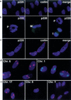
Association of p220 foci with Cajal bodies (CBs) and with domains of chromosomes 1 and 6. In all panels, 4′,6-diamidino-2-phenylindole (DAPI) stained nuclear DNA blue. (a) Normal dermal fibroblasts were subjected to immunofluorescence by using anti-p220 (green) and anti-p80coilin monoclonal antibodies (red) known to stain CBs. Co-localization is demonstrated in the merged image. (b) p220 foci are present in prophase (left; right, cell on top), but are no longer detectable in metaphase (middle) and telophase (right, cell at bottom). Prophase cells with four p220 foci were also observed (not shown). (c) HeLa cells contain CBs devoid of p220 foci. In a–c, p220 is green and DAPI is blue; in a and c, coilin is red. (d) and (e) p220 foci are associated with chromosomes 1 and 6 but not with other chromosomes. Normal dermal fibroblasts were stained for p220 and for the indicated chromosomal domains by using chromosome paints. For chromosomes 6, 17, 5, and Y, p220 is green and chromosome paint is red. For chromosome 1, p220 is red and chromosome paint green. (Chr.) Chromosome; (M) metaphase; (P) prophase; (T) telophase.
Figure 4
p220 preferentially associates with and is phosphorylated by cyclin E/Cdk2 in vitro. Immobilized cyclin/Cdk complexes were incubated with control insect cell lysates or insect cell lysates containing Flag-p220, as described in Materials and Methods. Complexes were washed with lysis buffer followed by 10 mM MgCl2 and 20 mM Tris-HCl. Some samples were supplemented with γ-[32P]ATP for 20 min before SDS–PAGE and visualization of proteins by immunoblotting or autoradiography. Flag-p220 was detected by anti-flag antibodies. The quantities of GST–cyclin/Cdk complexes were similar, as determined by immunoblotting with GST antibodies. Cdk2 complexes associated with an insect cell protein migrating slightly faster than p220 that was also a substrate for the kinase (indicated by an asterisk). An anti-flag immunoprecipitate of Flag-p220 (lane 19) was included as a control.
Figure 5
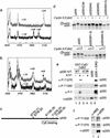
Identification of phosphorylation sites in p220. (a,b) A portion of the matrix-assisted laser desorption/ionization mass spectrometry (MALDI/TOF) mass spectra before (upper spectra) and after (lower spectra) treatment with calf intestinal phosphatase (CIP), showing the doubly phosphorylated peptide encompassing the sequence of 742–788 from recombinant (a) and endogenous (b) p220. To generate cyclin E/Cdk2–phosphorylated p220, 2 μg of Flag-p220 immobilized on anti-flag agarose was allowed to associate with 1 μg of cyclin E/Cdk2 and washed complexes incubated with 1 mM ATP (50 min at 25°C). (c) Schematic diagram of p220 phosphorylation sites and the cyclin E binding domain inferred by expression cloning (Zhao et al. 1998; Ma et al. 1999). (d) Altered mobility of p220 in response to cyclin E/Cdk2–mediated phosphorylation requires S775 and S779. In vitro translation products were incubated in the presence or absence of 20 nM cyclin E/Cdk2 for 20 min at 30°C (top) or with 20 and 50 nM cyclin E/Cdk2 for 20 min at 30°C (bottom) before electrophoresis and autoradiography. (e) Specificity of phosphopeptide-specific antibodies. In vitro–translated p220 or p220ΔCdk (50 μL) was incubated with 1 μg of GST–cyclin E/Cdk2 immobilized on GSH-Sepharose, and washed complexes were incubated in the presence or absence of 1 mM ATP (lanes 1_–_4). Proteins were separated by SDS–PAGE and immunoblotted. The anti-phosphoThr antibody (New England Biolabs), which reacts with a large number of different phosphoThr-Pro-containing sequences, did not recognize p220ΔCdk. As a control, Flag-p220 from insect cells (∼100 ng) was incubated with or without 50 nM cyclin E/Cdk2 and 1 mM ATP before immunoblotting. (WT) Wild type. (f) Anti-p220 immune complexes from 293T cells were separated by SDS–PAGE and immunoblotted with the indicated antibodies. (NRS) Normal rabbit serum.
Figure 6
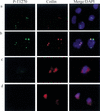
p220 in Cajal bodies (CBs) is phosphorylated on Cdk sites in a cell cycle–specific manner. Dual detection with anti-phospho-T1270 antibodies and p80coilin in dermal fibroblasts is shown. Focal co-localization was observed in S phase (a) and prophase (b), but antiphospho-T1270 did not detect any foci in metaphase (c) or telophase (d). a also contains three cells that display anti-coilin reactive foci but not anti-phospho-T1270 reactive foci. The DNA content of these cells is consistent with G1 phase (Fig. 2b). Only diffused coilin signals were observed in c and d. (Green) anti-phospho-T1270; (red) anti-coilin; (blue) 4′,6-diamidino-2-phenylindole (DAPI).
Figure 7
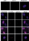
Co-localization of cyclin E with anti-phospho-T1270 reactive foci in a cell cycle–dependent manner. (a) Cyclin E (red) co-localizes with p220 (green) in a cell containing four p220 foci. Nuclei are in blue. (b_–_e) Growing diploid fibroblasts were labeled with BrdU for 1 h and subjected to immunofluorescence using anti-phospho-T1270 (red), anti-cyclin E (green), anti-BrdU (magenta), and 4′,6-diamidino-2-phenylindole (DAPI; blue) to visualize nuclei. (b) A BrdU-negative cell containing two phospho-T1270 foci that co-localize with cyclin E. This field also contains a BrdU-negative cell that is negative for both anti–cyclin E and anti-phospho-T1270 antibodies. (c) A BrdU-positive cell containing two phospho-T1270 foci that co-localize with cyclin E adjacent to a BrdU-negative cell that is negative for both anti–cyclin E and anti-phospho-T1270 antibodies. (d) BrdU-positive cells containing two or four phospho-T1270 foci that co-localize with cyclin E. (e) A BrdU-negative cell containing four phospho-T1270 foci that lacks cyclin E staining.
Figure 8
Cyclin E/Cdk2–mediated phosphorylation of p220 is important for optimal p220-induced histone H2B transcriptional activation. (a) pCMV, pCMV-p220, or pCMV-p220ΔCdk was transfected into 293T cells (3.5-cm dish), along with 50 ng of pCMV-β-galactosidase and 50 ng of pGL-H2B-luciferase reporter plasmid, and subsequently processes for luciferase and β-galactosidase activity as described in Materials and Methods. Luciferase activities were normalized relative to β-galactosidase activities. (b) A portion of extracts used in a was immunoblotted using anti-p220 antibodies. The wild-type p220 protein comigrates with the endogenous protein found at low levels, whereas p220ΔCdk migrates slightly faster as a result of the absence of phosphorylation. (c) The results of two independent experiments each performed using triplicate independent calcium phosphate precipitates derived from 10 μg of pCMV, pCMV-p220, or pCMV-p220ΔCdk are shown. (−) Lacking p220; (WT) wild type. (d) and (e) Expression levels for p220 and p220ΔCdk for experiments shown in c were similar as determined by immunoblotting (d) or immunofluorescence (e). (f) Expression of p57KIP2 blocks activation of the H2B promoter by p220 overexpression. 293T cells were transfected with limiting amounts of pCMV-p220 or p200ΔCdk expressing plasmids (5 μg) in the presence or absence of either 1 or 2 μg of pCMV-p57KIP2. At this level of expression, p220 leads to a twofold increase in reporter activity. p57KIP2 expression leads to a dramatic reduction in the level of p220-induced reporter activity in the presence or absence of p220 expression. The averages of duplicate independent transfections are shown.
Figure 9
S-phase entry in quiescent fibroblasts by cyclin E/Cdk2 expression is associated with the appearance of four p220 foci. WI38 fibroblasts were subjected to serum deprivation for 72 h before infection with Ad-cyclin E/Cdk2 and maintained in 0.1% serum. Twenty-four h after infection, cells were pulse-labeled with BrdU for 1 h before analysis of p220 staining by immunofluorescence. (a) An example of an S-phase cell from a cyclin E/Cdk2 infection containing four p220 foci adjacent to a non-S-phase cell containing two p220 foci. (b) Quantitation of p220 foci. Thirty to 100 nuclei of each class were counted.
Figure 10
Summary and proposed model of cell cycle–dependent transcriptional activation of histone genes on chromosomes 1 and 6 in association with Cajal bodies (CBs) as mediated by cyclin E/Cdk2 phosphorylation of p220. p220 foci are no longer detectable by metaphase and telophase but reappear in G1 phase. However, phosphorylation of p220 in CBs does not occur until cyclin E/Cdk2 is activated during late G1 phase/S phase.
References
- Ahn J, Gruen JR. The genomic organization of the histone clusters on human 6p21.3. Mamm Genome. 1999;10:768–770. - PubMed
- Brown NR, Noble ME, Endicott JA, Johnson LN. The structural basis for specificity of substrate and recruitment peptides for cyclin-dependent kinases. Nat Cell Biol. 1999;1:438–443. - PubMed
Publication types
MeSH terms
Substances
Grants and funding
- GM54137/GM/NIGMS NIH HHS/United States
- CA36200/CA/NCI NIH HHS/United States
- R01 CA036200/CA/NCI NIH HHS/United States
- DE/CA11910/DE/NIDCR NIH HHS/United States
- R01 GM054137/GM/NIGMS NIH HHS/United States
LinkOut - more resources
Full Text Sources
Other Literature Sources
Molecular Biology Databases


