Reactive oxygen species (ROS)-induced ROS release: a new phenomenon accompanying induction of the mitochondrial permeability transition in cardiac myocytes - PubMed (original) (raw)
Reactive oxygen species (ROS)-induced ROS release: a new phenomenon accompanying induction of the mitochondrial permeability transition in cardiac myocytes
D B Zorov et al. J Exp Med. 2000.
Abstract
We sought to understand the relationship between reactive oxygen species (ROS) and the mitochondrial permeability transition (MPT) in cardiac myocytes based on the observation of increased ROS production at sites of spontaneously deenergized mitochondria. We devised a new model enabling incremental ROS accumulation in individual mitochondria in isolated cardiac myocytes via photoactivation of tetramethylrhodamine derivatives, which also served to report the mitochondrial transmembrane potential, DeltaPsi. This ROS accumulation reproducibly triggered abrupt (and sometimes reversible) mitochondrial depolarization. This phenomenon was ascribed to MPT induction because (a) bongkrekic acid prevented it and (b) mitochondria became permeable for calcein ( approximately 620 daltons) concurrently with depolarization. These photodynamically produced "triggering" ROS caused the MPT induction, as the ROS scavenger Trolox prevented it. The time required for triggering ROS to induce the MPT was dependent on intrinsic cellular ROS-scavenging redox mechanisms, particularly glutathione. MPT induction caused by triggering ROS coincided with a burst of mitochondrial ROS generation, as measured by dichlorofluorescein fluorescence, which we have termed mitochondrial "ROS-induced ROS release" (RIRR). This MPT induction/RIRR phenomenon in cardiac myocytes often occurred synchronously and reversibly among long chains of adjacent mitochondria demonstrating apparent cooperativity. The observed link between MPT and RIRR could be a fundamental phenomenon in mitochondrial and cell biology.
Figures
Figure 1
Spatial organization and function of mitochondria in isolated adult rat cardiac myocytes. (A) Confocal plane of a myocyte loaded with TMRM (125 nM; bar = 5 μm). (B) z-section through A (1-μm resolution). Arrows in A and B denote mitochondria lacking TMRM sequestration. (C) Spontaneous ROS production at sites of low mitochondrial membrane potential: red, TMRM (125 nM) fluorescence; green, DCF (10 μM) fluorescence. (D) Exposure to antimycin A (25 μM) produces widespread numbers of depolarized mitochondria (loss of red TMRM fluorescence) together with increased ROS production (increased green DCF fluorescence). (E) Pretreatment with the ROS scavenger Trolox (2 mM) prevents the antimycin A–induced mitochondrial depolarization seen in D (cell labeled with TMRM and DCF as in D).
Figure 2
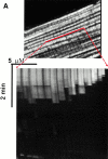
Experimental model: line scanning of mitochondrial arrays and photoexcitation ROS production. (A) Upper panel, confocal image of fluorescence in TMRM-loaded (125 nM) cardiac myocyte. Line drawn on image shows position scanned for experiment in bottom panel. Bottom panel shows 2 Hz line-scan image of TMRM fluorescence along mitochondrial row; time progresses from top (total scan 256 s). The dark regions between vertical columns are junctions between mitochondria. The sudden dissipation of TMRM fluorescence (white-to-black transitions) indicates mitochondrial ΔΨ loss. (B) Frequency distribution of ΔΨ transition times (time to 50% dissipation of ΔΨ) for the mitochondrial ensemble during line-scan imaging at 2 Hz as in A (n = 5 cells). (C) EPR spin-trapping, measured as the formation of DEPMPO/·OH and ·O2 − adducts during photoactivation of TMRM solutions. The system consisted of TMRM (100 μM) and DEPMPO (10 mM) in Hepes-buffered medium, pH 7.4, maintained at 23°C. While no signal was seen without light (a), a prominent spectrum of the DEPMPO/·OH (o symbol labeling central peaks) and ·O2 − (*central peaks) adducts was seen during illumination (b) and abolished partially (50–60%) by catalase (100 U/ml; c) or completely by SOD (1,000 U/ml; d). Trolox (2 mM) quenched these illumination-dependent signals by ∼60 ± 10% and produced a superimposed intense seven-line EPR signal of the Trolox-derived phenoxyl radical (e). Each spectrum is the sum of 24 1-min sequential acquisitions.
Figure 3
(A) Evidence of cooperativity and reversibility of ΔΨ (TMRM) dissipation during 2 Hz line-scan imaging. (B) The abrupt dissipation phase of TMRM fluorescence during line-scan imaging can be described by first-order kinetics. In this example, the dissipation of ΔΨ in a single mitochondrion occurs in step-wise fashion over several seconds. The inset shows all of the segments with negative slope (joined together; after background subtraction), fit by a single exponential model with τ1/2 = 0.54 s.
Figure 4
RIRR in single mitochondria. Representative cell that was dual-loaded with 125 nM TMRM (for ΔΨ) and 10 μM DCF (for ROS). (A) Typical pattern of ΔΨ dissipation at 10 Hz line-scan imaging. (B) Generation of ROS, as indicated by the increase in DCF fluorescence (acquired simultaneously with A). (C) Temporal relationship between ΔΨ and ROS production from the mitochondrial pair denoted by arrows in A and B. The trace at the bottom shows the hypothetical opening of the MPT pore. (D) Coordinated flickering of ΔΨ and RIRR in a single mitochondrion at 2 Hz line-scan imaging. (E) Relationship between ΔΨ and NAD(P) redox state during the MPT. ΔΨ and the MPT are assessed by changes in the TMRM (125 nM) fluorescence and the intrinsic autofluorescence excited at 351 nm (index of NAD[P] redox state), respectively, during 2 Hz line-scan imaging. (F) Inhibition of mitochondrial electron transport at Complex I prevents the mitochondrial ROS burst after induction of the MPT. Cell loading with TMRM and DCF and line-scan imaging protocol, as in D, except for the exposure to rotenone (0.1 and 1 μM) as indicated. Representative regions (encompassing groups of about six mitochondria over three sarcomeres) from the respective 2 Hz line-scan protocols are shown from each experimental group (top panel).
Figure 5
Triggering the RIRR process is not dependent on a sensitizing fluorophore. Cardiac myocyte dual-loaded with 400 nM MitoTracker Red CMXRos (ΔΨ, red signal) and 10 μM DCF (ROS, green signal); confocal line-scan imaging at 2 Hz.
Figure 6
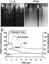
Demonstration of MPT induction by photoexcited trigger ROS. (A) Cells dual-loaded with TMRM (ΔΨ) and calcein-AM (the latter loaded under conditions that results in a cytosolic distribution initially in excess over that in mitochondria); line-scan imaging at 20 Hz. (B) 10 μM BA partially inhibits the MPT (ΔΨ; a and b) and RIRR (ROS; c and d) versus control. Cells were dual-loaded with TMRM and DCF; line-scan imaging at 2 Hz. (C) 100 μM BA completely inhibits the MPT versus control. Cells are loaded with 125 nM TMRM; line-scan at 2 Hz.
Figure 7
Scavenging the ROS trigger or exposure to exogenous NO inhibits the MPT. Cells loaded with 125 nM TMRM and confocal line-scan–imaged at 2 Hz. (A) Mean time to MPT induction in control versus pretreated cells as indicated: Trolox (2 mM); SNAP (100 μM); L-NAME (1 mM). Data represent the average from 8–10 cells in each group. (B) Evidence of endogenous production of NO by mitochondria after MPT induction and inhibition by L-NAME (4 mM). Myocytes were loaded with 125 nM TMRM (red) and 10 mM DAF-2 (green) and line scanned at 100 Hz.
Figure 9
Induction of Ca2+ sparks after the MPT. Cell is dual-loaded with 125 nM TMRM (ΔΨ) and fluo-3 (Ca2+) and line-scan imaged at 230 Hz. Representative example showing the dissipation of TMRM fluorescence from a single mitochondrion and a cluster of Ca2+ sparks in the immediate vicinity, within seconds of MPT induction. Inset: comparison of Ca2+ spark rate in proximity of MPT occurrence (i.e., within the sarcomere containing the involved mitochondria and within 3 s after MPT occurrence; n = 90 sparks) versus that at background (n = 150 sparks; P < 0.05).
Figure 8
Altered MPT characteristics after modulation of the redox state of soluble and protein thiols. (A) Comparison of MPT times (during 2 Hz line-scan imaging in TMRM-loaded cells) and cellular glutathione content in control versus 600 μM diethylmaleate-treated cells. (B) Development of apparent unstable MPT pore flickering during 2 Hz line-scan imaging in a representative TMRM-loaded (125 nM) cell exposed to 5 mM diethylmaleate. Arrows indicate movement artifacts.
Similar articles
- High fluence low-power laser irradiation induces mitochondrial permeability transition mediated by reactive oxygen species.
Wu S, Xing D, Gao X, Chen WR. Wu S, et al. J Cell Physiol. 2009 Mar;218(3):603-11. doi: 10.1002/jcp.21636. J Cell Physiol. 2009. PMID: 19006121 - Alteration in mitochondrial thiol enhances calcium ion dependent membrane permeability transition and dysfunction in vitro: a cross-talk between mtThiol, Ca(2+), and ROS.
Singh BK, Tripathi M, Pandey PK, Kakkar P. Singh BK, et al. Mol Cell Biochem. 2011 Nov;357(1-2):373-85. doi: 10.1007/s11010-011-0908-0. Epub 2011 Jul 12. Mol Cell Biochem. 2011. PMID: 21748338 - Mitochondrial ROS-induced ROS release: an update and review.
Zorov DB, Juhaszova M, Sollott SJ. Zorov DB, et al. Biochim Biophys Acta. 2006 May-Jun;1757(5-6):509-17. doi: 10.1016/j.bbabio.2006.04.029. Epub 2006 May 23. Biochim Biophys Acta. 2006. PMID: 16829228 Review. - Reactive oxygen species, but not Ca2+ overloading, trigger pH- and mitochondrial permeability transition-dependent death of adult rat myocytes after ischemia-reperfusion.
Kim JS, Jin Y, Lemasters JJ. Kim JS, et al. Am J Physiol Heart Circ Physiol. 2006 May;290(5):H2024-34. doi: 10.1152/ajpheart.00683.2005. Epub 2006 Jan 6. Am J Physiol Heart Circ Physiol. 2006. PMID: 16399872 - The mitochondrial permeability transition in cell death: a common mechanism in necrosis, apoptosis and autophagy.
Lemasters JJ, Nieminen AL, Qian T, Trost LC, Elmore SP, Nishimura Y, Crowe RA, Cascio WE, Bradham CA, Brenner DA, Herman B. Lemasters JJ, et al. Biochim Biophys Acta. 1998 Aug 10;1366(1-2):177-96. doi: 10.1016/s0005-2728(98)00112-1. Biochim Biophys Acta. 1998. PMID: 9714796 Review.
Cited by
- Monoamine Oxidases as Potential Contributors to Oxidative Stress in Diabetes: Time for a Study in Patients Undergoing Heart Surgery.
Duicu OM, Lighezan R, Sturza A, Ceausu RA, Borza C, Vaduva A, Noveanu L, Gaspar M, Ionac A, Feier H, Muntean DM, Mornos C. Duicu OM, et al. Biomed Res Int. 2015;2015:515437. doi: 10.1155/2015/515437. Epub 2015 May 25. Biomed Res Int. 2015. PMID: 26101773 Free PMC article. Review. - Mitochondrial reactive oxygen species (ROS) as signaling molecules of intracellular pathways triggered by the cardiac renin-angiotensin II-aldosterone system (RAAS).
De Giusti VC, Caldiz CI, Ennis IL, Pérez NG, Cingolani HE, Aiello EA. De Giusti VC, et al. Front Physiol. 2013 May 30;4:126. doi: 10.3389/fphys.2013.00126. eCollection 2013. Front Physiol. 2013. PMID: 23755021 Free PMC article. - Non-conventional mitochondrial permeability transition: Its regulation by mitochondrial dynamics.
Yoon Y, Lee H, Federico M, Sheu SS. Yoon Y, et al. Biochim Biophys Acta Bioenerg. 2023 Jan 1;1864(1):148914. doi: 10.1016/j.bbabio.2022.148914. Epub 2022 Sep 3. Biochim Biophys Acta Bioenerg. 2023. PMID: 36063902 Free PMC article. - Potential Role of Dipeptidyl Peptidase-4 in Regulating Mitochondria and Oxidative Stress in Cardiomyocytes.
Lee SY, Wu ST, Du CX, Ku HC. Lee SY, et al. Cardiovasc Toxicol. 2024 Oct;24(10):1090-1104. doi: 10.1007/s12012-024-09884-z. Epub 2024 Jul 2. Cardiovasc Toxicol. 2024. PMID: 38955919 - Cardiac mitochondrial network excitability: insights from computational analysis.
Zhou L, O'Rourke B. Zhou L, et al. Am J Physiol Heart Circ Physiol. 2012 Jun 1;302(11):H2178-89. doi: 10.1152/ajpheart.01073.2011. Epub 2012 Mar 16. Am J Physiol Heart Circ Physiol. 2012. PMID: 22427517 Free PMC article. Review.
References
- Williamson J.R. Mitochondrial function in heart. Annu. Rev. Physiol. 1979;41:485–506. - PubMed
- Gunter T.E., Pfeiffer D.R. Mechanisms by which mitochondria transport calcium. Am. J. Physiol. 1990;258:C755–786. - PubMed
- Green D.R., Reed J.C. Mitochondria and apoptosis. Science. 1998;281:1309–1312. - PubMed
- Zoratti M., Szabo I. The mitochondrial permeability transition. Biochim. Biophys. Acta. 1995;1241:139–176. - PubMed
Publication types
MeSH terms
Substances
Grants and funding
- HL63744/HL/NHLBI NIH HHS/United States
- R01 HL038324/HL/NHLBI NIH HHS/United States
- HL52315/HL/NHLBI NIH HHS/United States
- HL38324/HL/NHLBI NIH HHS/United States
- R01 HL063744/HL/NHLBI NIH HHS/United States
LinkOut - more resources
Full Text Sources
Other Literature Sources


