Gene transfer using recombinant rabbit hemorrhagic disease virus capsids with genetically modified DNA encapsidation capacity by addition of packaging sequences from the L1 or L2 protein of human papillomavirus type 16 - PubMed (original) (raw)
Gene transfer using recombinant rabbit hemorrhagic disease virus capsids with genetically modified DNA encapsidation capacity by addition of packaging sequences from the L1 or L2 protein of human papillomavirus type 16
S El Mehdaoui et al. J Virol. 2000 Nov.
Abstract
The aim of this study was to produce gene transfer vectors consisting of plasmid DNA packaged into virus-like particles (VLPs) with different cell tropisms. For this purpose, we have fused the N-terminally truncated VP60 capsid protein of the rabbit hemorrhagic disease virus (RHDV) with sequences which are expected to be sufficient to confer DNA packaging and gene transfer properties to the chimeric VLPs. Each of the two putative DNA-binding sequences of major L1 and minor L2 capsid proteins of human papillomavirus type 16 (HPV-16) were fused at the N terminus of the truncated VP60 protein. The two recombinant chimeric proteins expressed in insect cells self-assembled into VLPs similar in size and appearance to authentic RHDV virions. The chimeric proteins had acquired the ability to bind DNA. The two chimeric VLPs were therefore able to package plasmid DNA. However, only the chimeric VLPs containing the DNA packaging signal of the L1 protein were able efficiently to transfer genes into Cos-7 cells at a rate similar to that observed with papillomavirus L1 VLPs. It was possible to transfect only a very limited number of RK13 rabbit cells with the chimeric RHDV capsids containing the L2-binding sequence. The chimeric RHDV capsids containing the L1-binding sequence transfer genes into rabbit and hare cells at a higher rate than do HPV-16 L1 VLPs. However, no gene transfer was observed in human cell lines. The findings of this study demonstrate that the insertion of a DNA packaging sequence into a VLP which is not able to encapsidate DNA transforms this capsid into an artificial virus that could be used as a gene transfer vector. This possibility opens the way to designing new vectors with different cell tropisms by inserting such DNA packaging sequences into the major capsid proteins of other viruses.
Figures
FIG. 1
Schematic maps of VP60-L1BS and VP60-L2BS chimeric proteins.
FIG. 2
Western blot analysis of VP60Δ-L1BS and VP60Δ-L2BS protein expression. Recombinant capsid proteins were detected in the nuclear fraction of infected sf21 cells with an anti-RHDV monoclonal antibody. Lanes 1 and 2, VP60Δ-L1BS; lanes 4 and 5, VP60Δ-L2BS; lane 3, HPV-16 VLPs.
FIG. 3
Detection of DNA binding to VP60Δ-L1BS and VP60Δ-L2BS by Southwestern blotting using a digoxigenin-labeled plasmid probe. Lane 1, VP60Δ-L1BS; lane 2, VP60Δ; lane 3, VP60Δ-L2BS.
FIG. 4
Subcellular localization of VP60Δ (a and b) and VP60Δ-L1BS (c and d) VLPs by electron microscopy (bars, 200 nm [a and c] and 100 nm [b and d]).
FIG. 5
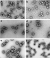
Identification of chimeric RHDV and HPV-16 VLPs by transmission electron microscopy. (a) VP60; (b) VP60Δ; (c) VP60Δ-L1BS; (d) VP60Δ-L2BS; (e) HPV-16 L1; (f) HPV-16 L1+L2. Bar, 100 nm.
FIG. 6
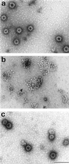
Purified VP60Δ-L2BS VLPs (a), capsomer-like structures obtained after treatment of VLPs with EGTA and dithiothreitol (b), VP60Δ-L2BS VLPs refolded after reassembly of the capsomers in the presence of CaCl2, DMSO and β-galactosidase plasmid (c). Bar, 100 nm.
FIG. 7
DNA protection assay using VP60Δ VLPs (lane 1), VP60Δ-L1BS VLPs (lane 2), VP60Δ-L2BS (lanes 3 and 4), and HPV-16 L1 VLPs (lane 5). M, molecular weight markers (λ phage digested with _Eco_RI and _Hin_dIII).
FIG. 8
Demonstration of gene expression of β-galactosidase in Cos-7 (a and b), R17 (c), HuH-7 (d), and CaCo2 (e and f) cells after gene transfer using VP60Δ-L1BS (a, c, d, and e) or HPV-16 L1 (b and f) capsids. Transfected cells expressing β-galactosidase were stained with X-Gal.
Similar articles
- Broad Cross-Protection Is Induced in Preclinical Models by a Human Papillomavirus Vaccine Composed of L1/L2 Chimeric Virus-Like Particles.
Boxus M, Fochesato M, Miseur A, Mertens E, Dendouga N, Brendle S, Balogh KK, Christensen ND, Giannini SL. Boxus M, et al. J Virol. 2016 Jun 24;90(14):6314-25. doi: 10.1128/JVI.00449-16. Print 2016 Jul 15. J Virol. 2016. PMID: 27147749 Free PMC article. - DNA packaging by L1 and L2 capsid proteins of bovine papillomavirus type 1.
Zhao KN, Sun XY, Frazer IH, Zhou J. Zhao KN, et al. Virology. 1998 Apr 10;243(2):482-91. doi: 10.1006/viro.1998.9091. Virology. 1998. PMID: 9568045 - Expression of human papillomavirus type 6 L1 and L2 isolated in China and self assembly of virus-like particles by the products.
Wang M, Wang LL, Chen LF, Han YH, Zou YH, Si JY, Song GX. Wang M, et al. Sheng Wu Hua Xue Yu Sheng Wu Wu Li Xue Bao (Shanghai). 2003 Jan;35(1):27-34. Sheng Wu Hua Xue Yu Sheng Wu Wu Li Xue Bao (Shanghai). 2003. PMID: 12518224 - L2, the minor capsid protein of papillomavirus.
Wang JW, Roden RB. Wang JW, et al. Virology. 2013 Oct;445(1-2):175-86. doi: 10.1016/j.virol.2013.04.017. Epub 2013 May 17. Virology. 2013. PMID: 23689062 Free PMC article. Review. - Papillomavirus virus-like particles as vehicles for the delivery of epitopes or genes.
Xu YF, Zhang YQ, Xu XM, Song GX. Xu YF, et al. Arch Virol. 2006 Nov;151(11):2133-48. doi: 10.1007/s00705-006-0798-8. Epub 2006 Jun 22. Arch Virol. 2006. PMID: 16791442 Review.
Cited by
- Recovery of infectious rabbit hemorrhagic disease virus from rabbits after direct inoculation with in vitro-transcribed RNA.
Liu G, Zhang Y, Ni Z, Yun T, Sheng Z, Liang H, Hua J, Li S, Du Q, Chen J. Liu G, et al. J Virol. 2006 Jul;80(13):6597-602. doi: 10.1128/JVI.02078-05. J Virol. 2006. PMID: 16775346 Free PMC article. - The epidemiology of conjunctival squamous cell carcinoma in Uganda.
Newton R, Ziegler J, Ateenyi-Agaba C, Bousarghin L, Casabonne D, Beral V, Mbidde E, Carpenter L, Reeves G, Parkin DM, Wabinga H, Mbulaiteye S, Jaffe H, Bourboulia D, Boshoff C, Touzé A, Coursaget P; Uganda Kaposi's Sarcoma Study Group. Newton R, et al. Br J Cancer. 2002 Jul 29;87(3):301-8. doi: 10.1038/sj.bjc.6600451. Br J Cancer. 2002. PMID: 12177799 Free PMC article. - Detection of neutralizing antibodies against human papillomaviruses (HPV) by inhibition of gene transfer mediated by HPV pseudovirions.
Bousarghin L, Combita-Rojas AL, Touzé A, El Mehdaoui S, Sizaret PY, Bravo MM, Coursaget P. Bousarghin L, et al. J Clin Microbiol. 2002 Mar;40(3):926-32. doi: 10.1128/JCM.40.3.926-932.2002. J Clin Microbiol. 2002. PMID: 11880418 Free PMC article. - Interactions between papillomavirus L1 and L2 capsid proteins.
Finnen RL, Erickson KD, Chen XS, Garcea RL. Finnen RL, et al. J Virol. 2003 Apr;77(8):4818-26. doi: 10.1128/jvi.77.8.4818-4826.2003. J Virol. 2003. PMID: 12663788 Free PMC article. - Identification of two cross-neutralizing linear epitopes within the L1 major capsid protein of human papillomaviruses.
Combita AL, Touzé A, Bousarghin L, Christensen ND, Coursaget P. Combita AL, et al. J Virol. 2002 Jul;76(13):6480-6. doi: 10.1128/jvi.76.13.6480-6486.2002. J Virol. 2002. PMID: 12050360 Free PMC article.
References
- Dmitriev I, Krasnykh V, Miller C R, Wang M, Kashentseva E, Mikheeva G, Belousova N, Curiel D T. An adenovirus vector with genetically modified fibers demonstrates expanded tropism via utilization of a coxsackievirus and adenovirus receptor-independent cell entry mechanism. J Virol. 1998;72:9706–9713. - PMC - PubMed
- Dooley S, Welter C, Blin N. Nonradioactive Southwestern analysis using chemiluminescent detection. Biotechniques. 1992;13:540–543. - PubMed
- Douglas J T, Miller C R, Kim M, Dmitriev I, Mikheeva G, Krasnykh V, Curiel D T. A system for the propagation of adenoviral vectors with genetically modified receptor specificities. Nat Biotechnol. 1999;17:470–475. - PubMed
Publication types
MeSH terms
Substances
LinkOut - more resources
Full Text Sources
Other Literature Sources
Miscellaneous


