Fc gamma R expression on macrophages is related to severity and chronicity of synovial inflammation and cartilage destruction during experimental immune-complex-mediated arthritis (ICA) - PubMed (original) (raw)
Fc gamma R expression on macrophages is related to severity and chronicity of synovial inflammation and cartilage destruction during experimental immune-complex-mediated arthritis (ICA)
A B Blom et al. Arthritis Res. 2000.
Abstract
STATEMENT OF FINDINGS: We investigated the role of Fc gamma receptors (Fc gamma Rs) on synovial macrophages in immune-complex-mediated arthritis (ICA). ICA elicited in knee joints of C57BL/6 mice caused a short-lasting, florid inflammation and reversible loss of proteoglycans (PGs), moderate chondrocyte death, and minor erosion of the cartilage. In contrast, when ICA was induced in knee joints of Fc receptor (FcR) gamma-chain(-/-) C57BL/6 mice, which lack functional Fc gamma RI and RIII, inflammation and cartilage destruction were prevented. When ICA was elicited in DBA/1 mice, a very severe, chronic inflammation was observed, and significantly more chondrocyte death and cartilage erosion than in arthritic C57BL/6 mice. The synovial lining and peritoneal macrophages of naïve DBA/1 mice expressed a significantly higher level of Fc gamma Rs than was seen in C57BL/6 mice. Moreover, elevated and prolonged expression of IL-1 was found after stimulation of these cells with immune complexes. Zymosan or streptococcal cell walls caused comparable inflammation and only mild cartilage destruction in all strains. We conclude that Fc gamma R expression on synovial macrophages may be related to the severity of synovial inflammation and cartilage destruction during ICA.
Figures
Figure 1
Inflammation in H&E-stained sections of mouse knee joints after induction of immune-mediated arthritis (ICA). (a) Section from FcR γ-chain-/- mouse 3 days after ICA induction. No inflammatory cells are visible in the joint space (js) or synovium (s). (b) Section from C57BL/6 (control) mouse 3 days after ICA induction. Florid inflammation is visible both in the joint space (exudate) and in the synovium (infiltrate) (orginal magnification 100×).
Figure 2
Semiquantitative mRNA measurements in synovial tissue of FcR γ-chain-/- and C57BL/6 (control) mice. Synovial mRNA levels of IL-1, IL-1Ra, MCP-1, and MIP-2, 6 and 24 h after induction of ICA. At certain points during the PCR reaction, samples were put on agarose gel and electrophoresis was performed. The number of the cycle in which the first band appeared was found: thus, a low cycle number means a higher mRNA content. Data were corrected for GAPDH signal. (Each group: n = 6.)
Figure 3
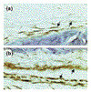
Expression of FcγRs in naïve knee joints of C57BL/6 and DBA/1 mice as detected by immunohistochemical staining using specific anti-FcγRII/III antibodies (mAb: 2.4G2) and subsequent development using di-aminobenzidine. (a) Naïve C57BL/6 mice. Very light staining of both synovial lining layer (arrows) and deeper layer. (b) Naïve DBA/1. Note the markedly higher staining intensity, especially of the cells of the synovial lining (arrows) (original magnification 200×).
Figure 4
Expression of FcγRs by resident peritoneal macrophages of C57BL/6 and DBA/1 mice as determined by fluorescence in FACS analysis. Almost twice as many receptors were expressed in DBA/1 as in C57BL/6 (control) mice. Cells were isolated from naïve mice using ice-cold medium and thereafter incubated with an anti-FcγR antibody (2.4G2) that recognises both FcγRII and RIII. Incubation with FITC-labeled secondary antibody followed.
Figure 5
IL-1 production by peritoneal macrophages. Production of bioactive IL-1 (pg/ml) by peritoneal macrophages of C57BL/6 and DBA/1 mice 24 and 48 h after stimulation with HAGGs (100 μg/ml). IL-1 production was measured using an IL-1-specific bioassay (NOB assay). The higher production by macrophages of DBA/1 than of C57BL/6 mice was significant (*P < 0.02).
Figure 6
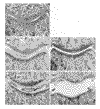
Cartilage damage during ICA. Safranin-O-stained knee-joint sections of FcR γ-chain-/-, C57BL/6, and DBA/1 mice 3 and 7 days after induction of ICA. Proteoglycan depletion was correlated with destaining of the superficial layer of the cartilage matrix, which is normally stained red. Both patellar (P) and femoral (F) cartilage are shown. (a) FcR γ-chain-/- 3 days after ICA induction. No proteoglycan depletion is seen. (b) C57BL/6 mouse 3 days after ICA induction. Marked destaining of the matrix is found. (c) C57BL/6 mouse 7 days after ICA induction. The absence of depleted areas suggests that the matrix has been completely restored. (d) DBA/1 mouse 3 days after ICA induction. The cartilage matrix is completely depleted, indicating considerable PG loss. (e) Same mouse strain (DBA/1) as in (d), 7 days after ICA induction. The cartilage matrix seems still devoid of PG, suggesting that no repair has taken place. Moreover, marked erosion of the matrix is visible, mainly of the femoral cartilage.
Similar articles
- Immune complexes, but not streptococcal cell walls or zymosan, cause chronic arthritis in mouse strains susceptible for collagen type II auto-immune arthritis.
Blom AB, van Lent PL, Holthuysen AE, van den Berg WB. Blom AB, et al. Cytokine. 1999 Dec;11(12):1046-56. doi: 10.1006/cyto.1999.0503. Cytokine. 1999. PMID: 10623430 - Fcgamma receptors directly mediate cartilage, but not bone, destruction in murine antigen-induced arthritis: uncoupling of cartilage damage from bone erosion and joint inflammation.
van Lent PL, Grevers L, Lubberts E, de Vries TJ, Nabbe KC, Verbeek S, Oppers B, Sloetjes A, Blom AB, van den Berg WB. van Lent PL, et al. Arthritis Rheum. 2006 Dec;54(12):3868-77. doi: 10.1002/art.22253. Arthritis Rheum. 2006. PMID: 17133594 - The role of macrophages in chronic arthritis.
van den Berg WB, van Lent PL. van den Berg WB, et al. Immunobiology. 1996 Oct;195(4-5):614-23. doi: 10.1016/S0171-2985(96)80026-X. Immunobiology. 1996. PMID: 8933161 Review. - Joint destruction in rheumatoid arthritis: biological bases.
Kingsley G, Panayi GS. Kingsley G, et al. Clin Exp Rheumatol. 1997 May-Jun;15 Suppl 17:S3-14. Clin Exp Rheumatol. 1997. PMID: 9266128 Review.
Cited by
- High production of proinflammatory and Th1 cytokines by dendritic cells from patients with rheumatoid arthritis, and down regulation upon FcgammaR triggering.
Radstake TR, van Lent PL, Pesman GJ, Blom AB, Sweep FG, Rönnelid J, Adema GJ, Barrera P, van den Berg WB. Radstake TR, et al. Ann Rheum Dis. 2004 Jun;63(6):696-702. doi: 10.1136/ard.2003.010033. Ann Rheum Dis. 2004. PMID: 15140777 Free PMC article. - Therapeutic blockade of CXCR2 rapidly clears inflammation in arthritis and atopic dermatitis models: demonstration with surrogate and humanized antibodies.
Alam MJ, Xie L, Ang C, Fahimi F, Willingham SB, Kueh AJ, Herold MJ, Mackay CR, Robert R. Alam MJ, et al. MAbs. 2020 Jan-Dec;12(1):1856460. doi: 10.1080/19420862.2020.1856460. MAbs. 2020. PMID: 33347356 Free PMC article. - Association of rheumatoid factor production with FcgammaRIIIa polymorphism in Taiwanese rheumatoid arthritis.
Chen JY, Wang CM, Wu JM, Ho HH, Luo SF. Chen JY, et al. Clin Exp Immunol. 2006 Apr;144(1):10-6. doi: 10.1111/j.1365-2249.2006.03021.x. Clin Exp Immunol. 2006. PMID: 16542359 Free PMC article. - Soluble FcgammaRIIa inhibits rheumatoid factor binding to immune complexes.
Wines BD, Gavin A, Powell MS, Steinitz M, Buchanan RR, Mark Hogarth P. Wines BD, et al. Immunology. 2003 Jun;109(2):246-54. doi: 10.1046/j.1365-2567.2003.01652.x. Immunology. 2003. PMID: 12757620 Free PMC article.
References
- Cooke TD, Richer S, Hurd E, Jasin HE. Localization of antigen-antibody complexes in intraarticular collagenous tissues. . Ann N Y Acad Sci. 1975;256:10–24. - PubMed
- Henson PM, Johnson HB, Spiegelberg HL. The release of granule enzymes from human neutrophils stimulated by aggregated immunoglobulins of different classes and subclasses. J Immunol. 1972;109:1182–1192. - PubMed
- Deo YM, Graziano RF, Repp R, van de Winkel JG. Clinical significance of IgG Fc receptors and Fc gamma R-directed immunotherapies. . Immunol Today. 1997;18:127–135. - PubMed
- Ravetch JV. Fc receptors. Curr Opin Immunol. 1997;9:121–125. - PubMed
MeSH terms
Substances
LinkOut - more resources
Full Text Sources
Other Literature Sources
Medical
Research Materials
Miscellaneous


