Sonic hedgehog promotes G(1) cyclin expression and sustained cell cycle progression in mammalian neuronal precursors - PubMed (original) (raw)
Sonic hedgehog promotes G(1) cyclin expression and sustained cell cycle progression in mammalian neuronal precursors
A M Kenney et al. Mol Cell Biol. 2000 Dec.
Abstract
Sonic hedgehog (Shh) signal transduction via the G-protein-coupled receptor, Smoothened, is required for proliferation of cerebellar granule neuron precursors (CGNPs) during development. Activating mutations in the Hedgehog pathway are also implicated in basal cell carcinoma and medulloblastoma, a tumor of the cerebellum in humans. However, Shh signaling interactions with cell cycle regulatory components in neural precursors are poorly understood, in part because appropriate immortalized cell lines are not available. We have utilized primary cultures from neonatal mouse cerebella in order to determine (i) whether Shh initiates or maintains cell cycle progression in CGNPs, (ii) if G(1) regulation by Shh resembles that of classical mitogens, and (iii) whether individual D-type cyclins are essential components of Shh proliferative signaling in CGNPs. Our results indicate that Shh can drive continued cycling in immature, proliferating CGNPs. Shh treatment resulted in sustained activity of the G(1) cyclin-Rb axis by regulating levels of cyclinD1, cyclinD2, and cyclinE mRNA transcripts and proteins. Analysis of CGNPs from cyclinD1(-/-) or cyclinD2(-/-) mice demonstrates that the Shh proliferative pathway does not require unique functions of cyclinD1 or cyclinD2 and that D-type cyclins overlap functionally in this regard. In contrast to many known mitogenic pathways, we show that Shh proliferative signaling is mitogen-activated protein kinase independent. Furthermore, protein synthesis is required for early effects on cyclin gene expression. Together, our results suggest that Shh proliferative signaling promotes synthesis of regulatory factor intermediates that upregulate or maintain cyclin gene expression and activity of the G(1) cyclin-Rb axis in proliferating granule neuron precursors.
Figures
FIG. 1
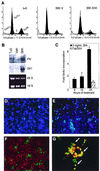
Characterization of proliferating cells in P4-5 neonatal mouse CGNP primary cultures. (A) Analysis of cell cycle phase distribution and Shh-signaling pathway activity in primary CGNP cultures. PI staining and flow cytometry were used to determine CGNP cell cycle distribution through the cell cycle after overnight culture in serum (t=0), 36 h of serum starvation (36h V), or 36 h of serum starvation in the presence of 3 μg of Shh per ml (36h Shh). Cell cycle phases associated with different levels of fluorescence are indicated. S-phase quantification is indicated below each plot (n indicates the number of separate cultures analyzed). (B) (Top) Northern blot analysis for expression of the Hedgehog transcriptional targets Patched (Ptc) and Gli1, after overnight culture in serum (T=0) and after a subsequent 24 h of treatment with vehicle (Veh) or 3 μg of Shh per ml. Note the strong upregulation of Ptc and Gli in Shh-treated cells. (Bottom) The ethidium-stained gel indicates RNA loading. (C) Quantification of BrdU incorporation levels (fold vehicle control) after various lengths of Shh treatment or after 24 h of Shh treatment in the presence of 10 μM forskolin. The asterisk indicates significant (P < 0.01) differences between vehicle- and Shh-treated samples. The data shown are average normalizations ± the SEM of approximately nine experiments per time point (see Materials and Methods). (D to G) Immunohistochemical analysis of proliferating P4-5 granule cell cultures. (D and E) Immunocytochemistry for BrdU-incorporating cells (Cy-2, green) in vehicle (D)- and Shh (E)-treated CGNP cultures. Note the increased number of cells undergoing DNA syntheses in the Shh-treated sample. DAPI was used to stain cell nuclei. (F and G) BrdU incorporation (Cy-2, green) segregated away from the Zic immunolabel (Cy-3, red), a marker of postmitotic granule cells (F), and colabeled with cells expressing Math-1 proteins (arrows, G). Note that some BrdU-positive, Math-1-negative cells (green) and BrdU-negative, Math-1-positive cells (red) were observed, possibly representing cells that have recently left the cell cycle and cells in early G1 phase of the cell cycle, respectively.
FIG. 2
Sonic hedgehog does not recruit quiescent CGNPs into the cell cycle. CGNPs were treated with Shh for 24 h after the indicated period of serum withdrawal, and then DNA synthesis levels were determined by measuring BrdU incorporation (solid bars). Alternatively, serum was replaced with Shh for 3 h; CGNPs were then serum and Shh starved for the indicated periods, after which Shh was added back for 24 h (striped bars). Note that Shh promoted significantly (approximately sixfold) increased levels of proliferation up to 6 h after serum withdrawal but not thereafter (solid bars). Similar results were obtained when an Shh pulse (3 h) was initially applied (striped bars). The infinity symbol (∞) indicates samples in which Shh added at t = 0, removed after 3 h, and measured for BrdU incorporation 24 h later (no Shh retreatment). Shh-induced BrdU incorporation is shown as the fold increase over baseline levels of BrdU incorporation in samples treated with vehicle alone. All data shown reflect results of three independent cerebellar preparations per time point treated with Shh versus vehicle alone. The asterisks indicate significant differences between Shh- and vehicle-treated CGNPs at P < 0.01 (∗∗) and P < 0.05 (∗) confidence levels.
FIG. 3
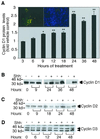
D-type cyclin protein regulation in Shh-treated versus vehicle-treated cultures. (A) Quantification of changes in cyclin D1 protein levels in the presence or absence of Shh for the indicated intervals. Western blots were quantified by densitometry, and values are expressed as the fold differences. The asterisks indicate significant differences between Shh- and vehicle-treated CGNPs at P < 0.01 (∗∗) and P < 0.05 (∗) confidence levels. (Inset, left) Immunohistochemical analysis of Shh-treated CGNPs showing overlap (yellow) of antibodies against cyclin D1 (Cy-2, green) and Math-1 (Cy-3, red). All cyclin D1-positive cells were also labeled with Math-1. The adjacent panel (inset, right) shows DAPI staining of the corresponding field of cells. (B to D) Representative time course of D-type cyclin protein level regulation by Shh. (B) Cyclin D1 appeared as a doublet with a relative mobility of 32 to 34 kDa. (C) Compared with controls, cyclin D2 levels did not appear to increase until after approximately 24 h. (D) Cyclin D3 levels were unaffected under the culture conditions tested.
FIG. 4
Analysis of Rb phosphorylation in Shh-treated granule cell cultures. Protein lysates (10 μg) were prepared from cells treated with vehicle alone, Shh (3 μg/ml), or Shh plus forskolin (10 μM) for the indicated periods of time and used for immunoblotting. Autoradiographs were analyzed by densitometry and signal intensities of P-pRb and pRb proteins were quantitated and expressed as a P-pRb/pRb ratio. The data shown are the average ratios ± the SEM from nine separate experiments. Significant differences (P ≤ 0.01) between Shh- and vehicle-treated samples are indicated by an asterisk.
FIG. 5
Proliferative effects of Shh signaling are independent of MAP kinase pathway activation. (A) Representative Western blot for phosphorylated p42-p44 ERK (top) in CGNPs treated with vehicle, BDNF (100 ng/ml), or Shh with or without MEK inhibitor for the indicated periods of time. The membrane was stripped and incubated with an antibody against total ERK (below) to demonstrate equivalent lane loading. (B) Quantification of DNA synthesis levels in CGNPs after 24 h of treatment with Shh, vehicle, or Shh plus the indicated concentration of MEK inhibitor. BrdU incorporation was measured by counting immunofluorescently labeled cells. Results shown are the averages of three separate cultures for each experiment + the SEM. The MEK inhibitor alone, or its solvent DMSO, had no effects on proliferation levels in cells treated with vehicle alone (not shown).
FIG. 6
Rapid upregulation of cyclin gene expression by Shh requires protein synthesis. A Northern blot analysis of cyclin gene expression in CGNPs treated for 3 h with vehicle or Shh, with or without 10 μg of cycloheximide per ml, in the absence of serum is shown. Note that treatment with Shh resulted in markedly increased levels of cyclinD1, cyclinD2, and cyclinE mRNA transcripts compared with controls. cyclinD3 expression was relatively unaffected. Addition of cycloheximide (Chx), however, abolished Shh effects on D- and E-type cyclin gene expression. GAPDH expression was used as a control for equivalent lane loading and transfer efficiency.
FIG. 7
Analysis of cyclin-Rb axis regulation and D-type cyclin function in _cyclinD1_-deficient CGNPs treated with Shh. A Western blot analysis of cell cycle regulatory protein response to Shh treatment in _cyclinD1_-deficient (−/−) mice and heterozygous (+/−) or wild-type (+/+) littermates is shown. (A) Western blot assay for hyperphosphorylated (PpRb) Rb protein, cyclin E, PCNA, and cyclin D3 after 24 h of Shh treatment. Note the equivalent responses of Rb hyperphosphorylation, cyclin E, and PCNA levels to Shh in all samples. (B) Intact D-type cyclin function in _cyclinD1_−/− CGNPs treated with Shh. CGNPs from cyclinD1 −/−, +/−, or +/+ littermates were treated with Shh for 15 h. Phosphorylation of Rb on serine-780 was analyzed by Western blotting (top). The membrane was stripped and reblotted for total Rb protein (bottom).
FIG. 8
Cyclin gene expression is rapidly upregulated in _cyclinD1_-deficient CGNPs treated with Shh. A Northern blot analysis of cyclin gene expression in CGNPs from _cyclinD1_-deficient mice and their wild-type or heterozygous littermates after 3 h of treatment with Shh or vehicle alone is shown. Genotypes are indicated above the lanes. Note the rapid upregulation of cyclinD2 and cyclinE in the Shh-treated samples. In addition, we observed slight upregulation of the cyclinD3 message despite the fact that protein levels were unaffected by Shh treatment (see Fig. 7). GAPDH expression was used to determine equivalent lane loading and transfer efficiency.
FIG. 9
Increased levels of cyclin D1 protein in _cyclinD2_-deficient CGNPs treated with Shh. Protein lysates (25 μg) of CGNP cultures from _cyclinD2_-deficient mice and their wild-type or heterozygous littermates were prepared after 24 h of treatment with Shh or vehicle and then analyzed by Western blot assay for cyclin D1 protein. Genotypes are indicated above the lanes. Note the equivalent responses of cyclin D1 protein levels, an indicator of cell cycle progression, to Shh in _cyclinD2_−/− CGNPs.
FIG. 10
Proposed model for early events in Shh proliferative signaling to cell cycle regulatory components in cerebellar granule precursor cells. Shh activation of the G-protein-coupled receptor, Smoothened, initiates signaling to hedgehog early response genes via a MAPK-independent mechanism. HER gene products, in turn. These promote continued transcription-stabilization of G1 cyclin gene mRNA transcripts. These gene products may also regulate cyclin protein expression and/or stabilization or their availability to complex with cdk's. Upregulation of D-type cyclin expression and/or activity would favor continued cell cycling rather than cell cycle exit (76). Note that the upregulation of hedgehog early response gene expression by Shh signal transduction may be inhibited by PKA and, conversely, that Hedgehog inhibition of endogenous PKA activity might promote expression of these genes.
Similar articles
- Nmyc upregulation by sonic hedgehog signaling promotes proliferation in developing cerebellar granule neuron precursors.
Kenney AM, Cole MD, Rowitch DH. Kenney AM, et al. Development. 2003 Jan;130(1):15-28. doi: 10.1242/dev.00182. Development. 2003. PMID: 12441288 - Insulin receptor substrate 1 is an effector of sonic hedgehog mitogenic signaling in cerebellar neural precursors.
Parathath SR, Mainwaring LA, Fernandez-L A, Campbell DO, Kenney AM. Parathath SR, et al. Development. 2008 Oct;135(19):3291-300. doi: 10.1242/dev.022871. Epub 2008 Aug 28. Development. 2008. PMID: 18755774 Free PMC article. - Pituitary adenylate cyclase-activating polypeptide and sonic hedgehog interact to control cerebellar granule precursor cell proliferation.
Nicot A, Lelièvre V, Tam J, Waschek JA, DiCicco-Bloom E. Nicot A, et al. J Neurosci. 2002 Nov 1;22(21):9244-54. doi: 10.1523/JNEUROSCI.22-21-09244.2002. J Neurosci. 2002. PMID: 12417650 Free PMC article. - Regulation of cell cycle entry and G1 progression by CSF-1.
Roussel MF. Roussel MF. Mol Reprod Dev. 1997 Jan;46(1):11-8. doi: 10.1002/(SICI)1098-2795(199701)46:1<11::AID-MRD3>3.0.CO;2-U. Mol Reprod Dev. 1997. PMID: 8981358 Review. - Cell cycle models for molecular biology and molecular oncology: exploring new dimensions.
Shackney SE, Shankey TV. Shackney SE, et al. Cytometry. 1999 Feb 1;35(2):97-116. doi: 10.1002/(sici)1097-0320(19990201)35:2<97::aid-cyto1>3.3.co;2-x. Cytometry. 1999. PMID: 10554165 Review.
Cited by
- Signaling pathway and molecular subgroups of medulloblastoma.
Li KK, Lau KM, Ng HK. Li KK, et al. Int J Clin Exp Pathol. 2013 Jun 15;6(7):1211-22. Print 2013. Int J Clin Exp Pathol. 2013. PMID: 23826403 Free PMC article. Review. - Gorlin Syndrome: Recent Advances in Genetic Testing and Molecular and Cellular Biological Research.
Onodera S, Nakamura Y, Azuma T. Onodera S, et al. Int J Mol Sci. 2020 Oct 13;21(20):7559. doi: 10.3390/ijms21207559. Int J Mol Sci. 2020. PMID: 33066274 Free PMC article. Review. - Maintaining Cerebellar Granule Neuron Progenitors in Cell Culture.
Ocasio JK. Ocasio JK. Methods Mol Biol. 2023;2583:9-12. doi: 10.1007/978-1-0716-2752-5_2. Methods Mol Biol. 2023. PMID: 36418721 - Serpine2/PN-1 Is Required for Proliferative Expansion of Pre-Neoplastic Lesions and Malignant Progression to Medulloblastoma.
Vaillant C, Valdivieso P, Nuciforo S, Kool M, Schwarzentruber-Schauerte A, Méreau H, Cabuy E, Lobrinus JA, Pfister S, Zuniga A, Frank S, Zeller R. Vaillant C, et al. PLoS One. 2015 Apr 22;10(4):e0124870. doi: 10.1371/journal.pone.0124870. eCollection 2015. PLoS One. 2015. PMID: 25901736 Free PMC article. - Targeting Hedgehog--a cancer stem cell pathway.
Merchant AA, Matsui W. Merchant AA, et al. Clin Cancer Res. 2010 Jun 15;16(12):3130-40. doi: 10.1158/1078-0432.CCR-09-2846. Epub 2010 Jun 8. Clin Cancer Res. 2010. PMID: 20530699 Free PMC article. Review.
References
- Alcedo J, Ayzenzon M, Von Ohlen T, Noll M, Hooper J E. The Drosophila smoothened gene encodes a seven-pass membrane protein, a putative receptor for the hedgehog signal. Cell. 1996;86:221–232. - PubMed
- Alessi D R, Cuenda A, Cohen P, Dudley D T, Saltiel A R. PD 098059 is a specific inhibitor of the activation of mitogen-activated protein kinase kinase in vitro and in vivo. J Biol Chem. 1995;270:27489–27494. - PubMed
- Altman J, Bayer S A. Development of the cerebellar system in relation to its evolution, structure, and functions. Boca Raton, Fla: CRC Press; 1997.
- Aruga J, Nagai T, Tokuyama T, Hayashizaki Y, Okazaki Y, Chapman V M, Mikoshiba K. The mouse zic gene family. Homologues of the Drosophila pair-rule gene odd-paired. J Biol Chem. 1996;271:1043–1047. - PubMed
- Aruga J, Yokota N, Hashimoto M, Furuichi T, Fukuda M, Mikoshiba K. A novel zinc finger protein, zic, is involved in neurogenesis, especially in the cell lineage of cerebellar granule cells. J Neurochem. 1994;63:1880–1890. - PubMed
Publication types
MeSH terms
Substances
LinkOut - more resources
Full Text Sources
Other Literature Sources
Medical


