Identification of a pathway for intelligible speech in the left temporal lobe - PubMed (original) (raw)
Clinical Trial
Identification of a pathway for intelligible speech in the left temporal lobe
S K Scott et al. Brain. 2000 Dec.
Abstract
It has been proposed that the identification of sounds, including species-specific vocalizations, by primates depends on anterior projections from the primary auditory cortex, an auditory pathway analogous to the ventral route proposed for the visual identification of objects. We have identified a similar route in the human for understanding intelligible speech. Using PET imaging to identify separable neural subsystems within the human auditory cortex, we used a variety of speech and speech-like stimuli with equivalent acoustic complexity but varying intelligibility. We have demonstrated that the left superior temporal sulcus responds to the presence of phonetic information, but its anterior part only responds if the stimulus is also intelligible. This novel observation demonstrates a left anterior temporal pathway for speech comprehension.
Figures
Fig. 1
Spectrograms of ‘They’re buying some bread’. Time is represented on the abscissa (0.0–1.43 s) and frequency on the ordinate (0.0–4.4 kHz). The darkness of the trace in each time/frequency region is controlled by the amount of energy in the signal at that particular frequency and time. (A) Normal speech (Sp) is intelligible with clear intonation. (B) Spectrally rotated speech (RSp) is not intelligible without extensive training, though some phonetic features and some of the original intonation are preserved. (C) Noise-vocoded speech (VCo) is intelligible, has very weak intonation and a rough sound quality. (D) Spectrally rotated noise-vocoded speech (RVCo) is completely unintelligible and does not sound like a voice.
Fig. 2
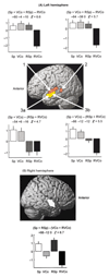
Significant voxels from the three contrasts used in the analysis, co-registered on to the left (A) and right (B) lateral MRI templates that are available in the image analysis software (SPM99b). The threshold was set at P < 0.00001 uncorrected, excluding clusters with <50 adjacent voxels. At this threshold, the activations were confined to the temporal lobes. Each contrast was centred around zero, and the ordinate of each plot is the mean size of the effect for each condition ± standard error of the mean, within the peak voxel. The coordinates of the peak voxel (x, y and z) in the stereotaxic space of SPM99b, and the _Z_-score, are shown at the head of each plot. (A) Left temporal lobe. The contrast {(Sp + VCo + RSp) – RVCo}, colour-coded in red, showed (1) the left superior temporal gyrus, both lateral and anterior to the primary auditory cortex, and (2) a separate region in the posterior superior temporal sulcus. The contrast {(Sp + VCo) – (RSp + RVCo)}, colour-coded in yellow, showed clear separation of intelligible from non-intelligible stimuli in the anterior superior temporal sulcus (3a). In the mid-superior temporal sulcus (3b), the response profile to the stimuli appeared to be transitional between the profiles in the posterior and anterior superior temporal sulcus. This ‘transition’ pattern may reflect a change in response to the rotated speech stimuli, but could also arise as a result of the smoothing applied to the data. (B) Right temporal lobe. The only significant voxels were revealed by the contrast {(Sp + RSp) – (VCo + RVCo}, colour-coded in white. They were located in the lateral superior temporal gyrus, anterior to the primary auditory cortex.
Fig. 3
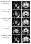
The five peak activations illustrated in Fig. 2A and B mapped on to sagittal (left column) and coronal (right column) slices of the T1-weighted MRI template. The upper arrow points at the sylvian sulcus, the lower arrow to the superior temporal sulcus. The contrasts which reveal these activations are shown for each pair of images. The x, y and z coordinates are in millimetres relative to the plane of the anterior commissure. STS = superior temporal sulcus; STG = superior temporal gyrus.
Comment in
- The new neuroanatomy of speech perception.
Binder J. Binder J. Brain. 2000 Dec;123 Pt 12:2371-2. doi: 10.1093/brain/123.12.2371. Brain. 2000. PMID: 11099441 No abstract available.
Similar articles
- The pathways for intelligible speech: multivariate and univariate perspectives.
Evans S, Kyong JS, Rosen S, Golestani N, Warren JE, McGettigan C, Mourão-Miranda J, Wise RJ, Scott SK. Evans S, et al. Cereb Cortex. 2014 Sep;24(9):2350-61. doi: 10.1093/cercor/bht083. Epub 2013 Apr 12. Cereb Cortex. 2014. PMID: 23585519 Free PMC article. - Separate neural subsystems within 'Wernicke's area'.
Wise RJ, Scott SK, Blank SC, Mummery CJ, Murphy K, Warburton EA. Wise RJ, et al. Brain. 2001 Jan;124(Pt 1):83-95. doi: 10.1093/brain/124.1.83. Brain. 2001. PMID: 11133789 Clinical Trial. - Hemispheric asymmetries in speech perception: sense, nonsense and modulations.
Rosen S, Wise RJ, Chadha S, Conway EJ, Scott SK. Rosen S, et al. PLoS One. 2011;6(9):e24672. doi: 10.1371/journal.pone.0024672. Epub 2011 Sep 30. PLoS One. 2011. PMID: 21980349 Free PMC article. - Segmental processing in the human auditory dorsal stream.
Zaehle T, Geiser E, Alter K, Jancke L, Meyer M. Zaehle T, et al. Brain Res. 2008 Jul 18;1220:179-90. doi: 10.1016/j.brainres.2007.11.013. Epub 2007 Nov 17. Brain Res. 2008. PMID: 18096139 Review. - [Auditory perception and language: functional imaging of speech sensitive auditory cortex].
Samson Y, Belin P, Thivard L, Boddaert N, Crozier S, Zilbovicius M. Samson Y, et al. Rev Neurol (Paris). 2001 Sep;157(8-9 Pt 1):837-46. Rev Neurol (Paris). 2001. PMID: 11677406 Review. French.
Cited by
- Speech comprehension aided by multiple modalities: behavioural and neural interactions.
McGettigan C, Faulkner A, Altarelli I, Obleser J, Baverstock H, Scott SK. McGettigan C, et al. Neuropsychologia. 2012 Apr;50(5):762-76. doi: 10.1016/j.neuropsychologia.2012.01.010. Epub 2012 Jan 17. Neuropsychologia. 2012. PMID: 22266262 Free PMC article. - A New Paradigm for Individual Subject Language Mapping: Movie-Watching fMRI.
Tie Y, Rigolo L, Ozdemir Ovalioglu A, Olubiyi O, Doolin KL, Mukundan S Jr, Golby AJ. Tie Y, et al. J Neuroimaging. 2015 Sep-Oct;25(5):710-20. doi: 10.1111/jon.12251. Epub 2015 May 12. J Neuroimaging. 2015. PMID: 25962953 Free PMC article. - Phoneme and word recognition in the auditory ventral stream.
DeWitt I, Rauschecker JP. DeWitt I, et al. Proc Natl Acad Sci U S A. 2012 Feb 21;109(8):E505-14. doi: 10.1073/pnas.1113427109. Epub 2012 Feb 1. Proc Natl Acad Sci U S A. 2012. PMID: 22308358 Free PMC article. - The functional neuroanatomy of language.
Hickok G. Hickok G. Phys Life Rev. 2009 Sep;6(3):121-43. doi: 10.1016/j.plrev.2009.06.001. Phys Life Rev. 2009. PMID: 20161054 Free PMC article. - Cracking the language code: neural mechanisms underlying speech parsing.
McNealy K, Mazziotta JC, Dapretto M. McNealy K, et al. J Neurosci. 2006 Jul 19;26(29):7629-39. doi: 10.1523/JNEUROSCI.5501-05.2006. J Neurosci. 2006. PMID: 16855090 Free PMC article.
References
- Belin P, Zatorre RJ, Lafaille P, Ahad P, Pike B. Voice-selective areas in human auditory cortex. Nature. 2000;403:309–12. - PubMed
- Binder JR, Frost JA, Hammeke TA, Rao SM, Cox RW. Function of the left planum temporale in auditory and linguistic processing. Brain. 1996;119:1239–47. - PubMed
- Binder JR, Frost MS. Functional MRI studies of language processing in the brain. Neurosci News. 1998;1:15–23.
- Blesser B. Speech perception under conditions of spectral transformation: I. Phonetic characteristics. J Speech Hear Res. 1972;15:5–41. - PubMed
Publication types
MeSH terms
LinkOut - more resources
Full Text Sources


