Genetic heterogeneity in Mycobacterium tuberculosis isolates reflected in IS6110 restriction fragment length polymorphism patterns as low-intensity bands - PubMed (original) (raw)
Genetic heterogeneity in Mycobacterium tuberculosis isolates reflected in IS6110 restriction fragment length polymorphism patterns as low-intensity bands
A S de Boer et al. J Clin Microbiol. 2000 Dec.
Abstract
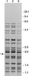
Mycobacterium tuberculosis isolates with identical IS6110 restriction fragment length polymorphism (RFLP) patterns are considered to originate from the same ancestral strain and thus to reflect ongoing transmission. In this study, we investigated 1,277 IS6110 RFLP patterns for the presence of multiple low-intensity bands (LIBs), which may indicate infections with multiple M. tuberculosis strains. We did not find any multiple LIBs, suggesting that multiple infections are rare in the Netherlands. However, we did observe a few LIBs in 94 patterns (7.4%) and examined the nature of this phenomenon. With single-colony cultures it was found that LIBs mostly represent mixed bacterial populations with slightly different RFLP patterns. Mixtures were expressed in RFLP patterns as LIBs when 10 to 30% of the DNA analyzed originated from a bacterial population with another RFLP pattern. Presumably, a part of the LIBs did not represent mixed bacterial populations, as in some clusters all strains exhibited LIBs in their RFLP patterns. The occurrence of LIBs was associated with increased age in patients. This may reflect either a gradual change of the bacterial population in the human body over time or IS6110-mediated genetic adaptation of M. tuberculosis to changes in the environmental conditions during the dormant state or reactivation thereafter.
Figures
FIG. 1
IS_6110_ RFLP patterns of isolates, showing LIBs, and SCCs, from three different patients (A to C). Lanes 1 show the banding patterns of the isolates, with LIBs indicated by arrows. Lanes 2 to 5 show the banding patterns of SCCs of these isolates. The numbers on the right indicate the sizes of standard DNA fragments in kilobase pairs.
FIG. 2
IS_6110_ RFLP patterns of different mixtures of the DNAs of two M. tuberculosis strains. Lanes 1 and 21 show the RFLP patterns of the pure DNAs of the two strains. Lanes 2 to 20 show the patterns of mixtures of the DNAs of these strains. The numbers in the second horizontal row indicate the ratios of the DNA mixtures. The numbers on the left indicate the sizes of standard DNA fragments in kilobase pairs.
FIG. 3
IS_6110_ RFLP patterns of different mixtures of the DNAs of two SCCs of an M. tuberculosis strain differing in a single IS_6110_ element. Lane 1 shows the pattern of pure DNA of one SCC. Lanes 2 to 21 depict patterns of mixtures of this DNA with an increasing amount of DNA of another SCC containing an additional IS_6110_ copy at a _Pvu_II restriction fragment of approximately 3.5 kb. The numbers in the second horizontal row indicate the ratios of the DNA mixtures. The numbers on the left indicate the sizes of standard DNA fragments in kilobase pairs.
FIG. 4
IS_6110_ RFLP patterns of three patient isolates of a cluster showing an LIB (indicated by the arrow) at the same _Pvu_II restriction fragment. The numbers on the right indicate the sizes of standard DNA fragments in kilobase pairs.
FIG. 5
Frequency distribution of 6,189 normal-intensity bands (NIB) and 69 LIBs per band position category (GelCompar band position and sizes of standard DNA fragments in kilobase pairs).
Comment in
- Genetic mutations occur gradually in in vivo populations of Mycobacterium tuberculosis bacteria.
de Boer AS, van Soolingen D, Borgdorff MW. de Boer AS, et al. J Clin Microbiol. 2001 Oct;39(10):3814. doi: 10.1128/JCM.39.10.3814.2001. J Clin Microbiol. 2001. PMID: 11599522 Free PMC article. No abstract available.
Similar articles
- Epidemiologic import of tuberculosis cases whose isolates have similar but not identical IS6110 restriction fragment length polymorphism patterns.
Cave MD, Yang ZH, Stefanova R, Fomukong N, Ijaz K, Bates J, Eisenach KD. Cave MD, et al. J Clin Microbiol. 2005 Mar;43(3):1228-33. doi: 10.1128/JCM.43.3.1228-1233.2005. J Clin Microbiol. 2005. PMID: 15750088 Free PMC article. - Stability of IS6110 restriction fragment length polymorphism patterns of Mycobacterium tuberculosis strains in actual chains of transmission.
Niemann S, Rüsch-Gerdes S, Richter E, Thielen H, Heykes-Uden H, Diel R. Niemann S, et al. J Clin Microbiol. 2000 Jul;38(7):2563-7. doi: 10.1128/JCM.38.7.2563-2567.2000. J Clin Microbiol. 2000. PMID: 10878044 Free PMC article. - Analysis of rate of change of IS6110 RFLP patterns of Mycobacterium tuberculosis based on serial patient isolates.
de Boer AS, Borgdorff MW, de Haas PE, Nagelkerke NJ, van Embden JD, van Soolingen D. de Boer AS, et al. J Infect Dis. 1999 Oct;180(4):1238-44. doi: 10.1086/314979. J Infect Dis. 1999. PMID: 10479153 - Chromosomal DNA fingerprinting analysis using the insertion sequence IS6110 and the repetitive element DR as strain-specific markers for epidemiological study of tuberculosis in French Polynesia.
Torrea G, Levee G, Grimont P, Martin C, Chanteau S, Gicquel B. Torrea G, et al. J Clin Microbiol. 1995 Jul;33(7):1899-904. doi: 10.1128/jcm.33.7.1899-1904.1995. J Clin Microbiol. 1995. PMID: 7665667 Free PMC article. - [New era in molecular epidemiology of tuberculosis in Japan].
Takashima T, Iwamoto T. Takashima T, et al. Kekkaku. 2006 Nov;81(11):693-707. Kekkaku. 2006. PMID: 17154049 Review. Japanese.
Cited by
- Multiple Mycobacterium tuberculosis strains in early cultures from patients in a high-incidence community setting.
Richardson M, Carroll NM, Engelke E, Van Der Spuy GD, Salker F, Munch Z, Gie RP, Warren RM, Beyers N, Van Helden PD. Richardson M, et al. J Clin Microbiol. 2002 Aug;40(8):2750-4. doi: 10.1128/JCM.40.8.2750-2754.2002. J Clin Microbiol. 2002. PMID: 12149324 Free PMC article. - Identifying mixed Mycobacterium tuberculosis infections from whole genome sequence data.
Sobkowiak B, Glynn JR, Houben RMGJ, Mallard K, Phelan JE, Guerra-Assunção JA, Banda L, Mzembe T, Viveiros M, McNerney R, Parkhill J, Crampin AC, Clark TG. Sobkowiak B, et al. BMC Genomics. 2018 Aug 14;19(1):613. doi: 10.1186/s12864-018-4988-z. BMC Genomics. 2018. PMID: 30107785 Free PMC article. - Proposal for standardization of optimized mycobacterial interspersed repetitive unit-variable-number tandem repeat typing of Mycobacterium tuberculosis.
Supply P, Allix C, Lesjean S, Cardoso-Oelemann M, Rüsch-Gerdes S, Willery E, Savine E, de Haas P, van Deutekom H, Roring S, Bifani P, Kurepina N, Kreiswirth B, Sola C, Rastogi N, Vatin V, Gutierrez MC, Fauville M, Niemann S, Skuce R, Kremer K, Locht C, van Soolingen D. Supply P, et al. J Clin Microbiol. 2006 Dec;44(12):4498-510. doi: 10.1128/JCM.01392-06. Epub 2006 Sep 27. J Clin Microbiol. 2006. PMID: 17005759 Free PMC article. - Evaluation of the inaccurate assignment of mixed infections by Mycobacterium tuberculosis as exogenous reinfection and analysis of the potential role of bacterial factors in reinfection.
Martín A, Herranz M, Navarro Y, Lasarte S, Ruiz Serrano MJ, Bouza E, García de Viedma D. Martín A, et al. J Clin Microbiol. 2011 Apr;49(4):1331-8. doi: 10.1128/JCM.02519-10. Epub 2011 Feb 23. J Clin Microbiol. 2011. PMID: 21346048 Free PMC article. - Exploring genotype concordance in epidemiologically linked cases of tuberculosis in New York City.
Robbins RS, Perri BR, Ahuja SD, Anger HA, Sullivan Meissner J, Shashkina E, Kreiswirth BN, Proops DC. Robbins RS, et al. Epidemiol Infect. 2017 Feb;145(3):503-514. doi: 10.1017/S0950268816002399. Epub 2016 Nov 21. Epidemiol Infect. 2017. PMID: 27866489 Free PMC article.
References
- Barnes P F, el-Hajj H, Preston-Martin S, Cave M D, Jones B E, Otaya M, Pogoda J, Eisenach K D. Transmission of tuberculosis among the urban homeless. JAMA. 1996;275:305–307. - PubMed
- Borgdorff M W, Nagelkerke N, van Soolingen D, de Haas P E W, Veen J, van Embden J D A. Analysis of tuberculosis transmission between nationalities in the Netherlands in the period 1993–1995 using DNA fingerprinting. Am J Epidemiol. 1998;147:187–195. - PubMed
- Butcher P D, Hutchinson N A, Doran T J, Dale J W. The application of molecular techniques to the diagnosis and epidemiology of mycobacterial diseases. Soc Appl Bacteriol Symp Ser. 1996;25:53S–71S. - PubMed
- Cole S T, Brosch R, Parkhill J, Garnier T, Churcher C, Harris D, Gordon S V, Eiglmeier K, Gas S, Barry III C E, Tekaia F, Badcock K, Basham D, Brown D, Chillingworth T, Connor R, Davies R, Devlin K, Feltwell T, Gentles S, Hamlin N, Holroyd S, Hornsby T, Jagels K, Krogh A, McLean J, Moule S, Murphy L, Oliver K, Osborne J, Quail M A, Rajandream M A, Rogers J, Rutter S, Seeger K, Skelton J, Squares R, Squares S, Sulston J E, Taylor K, Whitehead S, Barrell B G. Deciphering the biology of Mycobacterium tuberculosis from the complete genome sequence. Nature. 1998;393:537–544. - PubMed
Publication types
MeSH terms
Substances
LinkOut - more resources
Full Text Sources


