Perception and recognition memory in monkeys following lesions of area TE and perirhinal cortex - PubMed (original) (raw)
Perception and recognition memory in monkeys following lesions of area TE and perirhinal cortex
E A Buffalo et al. Learn Mem. 2000 Nov-Dec.
Abstract
Monkeys with lesions of perirhinal cortex (PR group) and monkeys with lesions of inferotemporal cortical area TE (TE group) were tested on a modified version of the delayed nonmatching to sample (DNMS) task that included very short delay intervals (0.5 sec) as well as longer delay intervals (1 min and 10 min). Lesions of the perirhinal cortex and lesions of area TE produced different patterns of impairment. The PR group learned the DNMS task as quickly as normal monkeys (N) when the delay between sample and choice was very short (0.5 sec). However, performance of the PR group, unlike that of the N group, fell to chance levels when the delay between sample and choice was lengthened to 10 min. In contrast to the PR group, the TE group was markedly impaired on the DNMS task even at the 0.5-sec delay, and three of four monkeys with TE lesions failed to acquire the task. The results provide support for the idea that perirhinal cortex is important not for perceptual processing, but for the formation and maintenance of long-term memory. Area TE is important for the perceptual processing of visual stimuli.
Figures
Figure 1
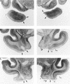
Photomicrographs of representative sections through the left and right temporal lobes of monkey PR 2, whose lesion most closely approximated the intended lesion. The sections are arranged from rostral (A) to caudal (E) (also see facing page), and the lesion is indicated by arrows at each level. rs, rhinal sulcus; sts, superior temporal sulcus; TE, area TE; A, amygdala; E, entorhinal cortex; H, hippocampal region.
Figure 2
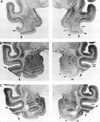
Photomicrographs of representative sections through the left and right temporal lobes of monkey TE 4, whose lesion most closely approximated the intended lesion. The sections are arranged from rostral (A) to caudal (E) (also see next page), and the lesion is indicated by arrows at each level. The asterisk indicates a processing artifact. rs, rhinal sulcus; sts, superior temporal sulcus; PR, perirhinal cortex; A, amygdala; E, entorhinal cortex; H, hippocampal region; PH, parahippocampal cortex.
Figure 3
(A) Initial learning of the automated visual delayed nonmatching to sample task (DNMS) at a delay of 0.5 sec by normal monkeys (N = 3), monkeys with lesions of the perirhinal cortex (PR = 4), and monkeys with lesions of area TE (TE = 4). Symbols show trials to criterion for individual monkeys. (B) Performance across delays for the normal monkeys (N = 3) and the monkeys with lesions of the perirhinal cortex (PR-4). Bars represent standard errors of the mean. (C) An expanded view of the performance of the N and PR groups at the 10-min delay. Symbols show the performance of individual monkeys.
Figure 4
The ventral surface of a macaque monkey brain showing the location of the perirhinal cortex (PR) and inferotemporal cortical area TE (TE). The PR forms a band of cortex along the ventromedial surface of the temporal lobe, lateral to the rhinal sulcus. Area TE is located immediately lateral to the PR and consists of a band of cortex lying primarily on the middle temporal gyrus. See Materials and Methods for a description of the boundaries of the PR and area TE.
Similar articles
- Dissociation between the effects of damage to perirhinal cortex and area TE.
Buffalo EA, Ramus SJ, Clark RE, Teng E, Squire LR, Zola SM. Buffalo EA, et al. Learn Mem. 1999 Nov-Dec;6(6):572-99. doi: 10.1101/lm.6.6.572. Learn Mem. 1999. PMID: 10641763 Free PMC article. - The hippocampal/parahippocampal regions and recognition memory: insights from visual paired comparison versus object-delayed nonmatching in monkeys.
Nemanic S, Alvarado MC, Bachevalier J. Nemanic S, et al. J Neurosci. 2004 Feb 25;24(8):2013-26. doi: 10.1523/JNEUROSCI.3763-03.2004. J Neurosci. 2004. PMID: 14985444 Free PMC article. - Functional double dissociation between two inferior temporal cortical areas: perirhinal cortex versus middle temporal gyrus.
Buckley MJ, Gaffan D, Murray EA. Buckley MJ, et al. J Neurophysiol. 1997 Feb;77(2):587-98. doi: 10.1152/jn.1997.77.2.587. J Neurophysiol. 1997. PMID: 9065832 - Development and plasticity of the neural circuitry underlying visual recognition memory.
Webster MJ, Ungerleider LG, Bachevalier J. Webster MJ, et al. Can J Physiol Pharmacol. 1995 Sep;73(9):1364-71. doi: 10.1139/y95-191. Can J Physiol Pharmacol. 1995. PMID: 8748986 Review. - The parahippocampal region and object identification.
Murray EA, Bussey TJ, Hampton RR, Saksida LM. Murray EA, et al. Ann N Y Acad Sci. 2000 Jun;911:166-74. doi: 10.1111/j.1749-6632.2000.tb06725.x. Ann N Y Acad Sci. 2000. PMID: 10911873 Review.
Cited by
- cAMP responsive element-binding protein phosphorylation is necessary for perirhinal long-term potentiation and recognition memory.
Warburton EC, Glover CP, Massey PV, Wan H, Johnson B, Bienemann A, Deuschle U, Kew JN, Aggleton JP, Bashir ZI, Uney J, Brown MW. Warburton EC, et al. J Neurosci. 2005 Jul 6;25(27):6296-303. doi: 10.1523/JNEUROSCI.0506-05.2005. J Neurosci. 2005. PMID: 16000619 Free PMC article. - Long-Term Cocaine Self-administration Produces Structural Brain Changes That Correlate With Altered Cognition.
Jedema HP, Song X, Aizenstein HJ, Bonner AR, Stein EA, Yang Y, Bradberry CW. Jedema HP, et al. Biol Psychiatry. 2021 Feb 15;89(4):376-385. doi: 10.1016/j.biopsych.2020.08.008. Epub 2020 Aug 18. Biol Psychiatry. 2021. PMID: 33012519 Free PMC article. - The primate working memory networks.
Constantinidis C, Procyk E. Constantinidis C, et al. Cogn Affect Behav Neurosci. 2004 Dec;4(4):444-65. doi: 10.3758/cabn.4.4.444. Cogn Affect Behav Neurosci. 2004. PMID: 15849890 Free PMC article. Review. - Impairment in delayed nonmatching to sample following lesions of dorsal prefrontal cortex.
Moore TL, Schettler SP, Killiany RJ, Rosene DL, Moss MB. Moore TL, et al. Behav Neurosci. 2012 Dec;126(6):772-80. doi: 10.1037/a0030493. Epub 2012 Oct 22. Behav Neurosci. 2012. PMID: 23088539 Free PMC article. - Why does brain damage impair memory? A connectionist model of object recognition memory in perirhinal cortex.
Cowell RA, Bussey TJ, Saksida LM. Cowell RA, et al. J Neurosci. 2006 Nov 22;26(47):12186-97. doi: 10.1523/JNEUROSCI.2818-06.2006. J Neurosci. 2006. PMID: 17122043 Free PMC article.
References
- Alvarez-Royo P, Zola-Morgan S, Squire LR. Impairment of long-term memory and sparing of short-term memory in monkeys with medial temporal lobe lesions: A response to Ringo. Behav Brain Res. 1992;52:1–5. - PubMed
- Brodmann K. Leipzig: Barth; 1909.
- Buckley MJ, Gaffan D. Impairment of visual object-discrimination learning after perirhinal cortex ablation. Behav Neurosci. 1997;111:467–475. - PubMed
- ————— Learning and transfer of object-reward associations and the role of the perirhinal cortex. Behav Neurosci. 1998a;112:15–23. - PubMed
Publication types
MeSH terms
Grants and funding
- R37 MH024600/MH/NIMH NIH HHS/United States
- MH58933/MH/NIMH NIH HHS/United States
- R01 MH024600/MH/NIMH NIH HHS/United States
- T32 AG000216/AG/NIA NIH HHS/United States
- MH24600/MH/NIMH NIH HHS/United States
- 2T32AG00216/AG/NIA NIH HHS/United States
LinkOut - more resources
Full Text Sources
Medical
Research Materials


