Chronic antidepressant treatment increases neurogenesis in adult rat hippocampus - PubMed (original) (raw)
Chronic antidepressant treatment increases neurogenesis in adult rat hippocampus
J E Malberg et al. J Neurosci. 2000.
Abstract
Recent studies suggest that stress-induced atrophy and loss of hippocampal neurons may contribute to the pathophysiology of depression. The aim of this study was to investigate the effect of antidepressants on hippocampal neurogenesis in the adult rat, using the thymidine analog bromodeoxyuridine (BrdU) as a marker for dividing cells. Our studies demonstrate that chronic antidepressant treatment significantly increases the number of BrdU-labeled cells in the dentate gyrus and hilus of the hippocampus. Administration of several different classes of antidepressant, but not non-antidepressant, agents was found to increase BrdU-labeled cell number, indicating that this is a common and selective action of antidepressants. In addition, upregulation of the number of BrdU-labeled cells is observed after chronic, but not acute, treatment, consistent with the time course for the therapeutic action of antidepressants. Additional studies demonstrated that antidepressant treatment increases the proliferation of hippocampal cells and that these new cells mature and become neurons, as determined by triple labeling for BrdU and neuronal- or glial-specific markers. These findings raise the possibility that increased cell proliferation and increased neuronal number may be a mechanism by which antidepressant treatment overcomes the stress-induced atrophy and loss of hippocampal neurons and may contribute to the therapeutic actions of antidepressant treatment.
Figures
Fig. 1.
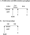
Experimental paradigms. _A,_Proliferation and differentiation paradigms. Animals were administered antidepressants for 1–28 d. Four days after the last antidepressant treatment, animals were given BrdU and killed (S) 24 hr after BrdU administration. In one experiment designed to measure cell proliferation, animals were killed 2 hr after BrdU injection. To determine cell differentiation or phenotype, rats were killed 28 d after BrdU injection. B, Survival paradigm. To examine the influence of antidepressant treatment on the survival of BrdU-labeled cells, BrdU was given to drug-naïve animals before initiating antidepressant treatment (14 d). Rats were then killed 28 d after BrdU injection (14 d after the last antidepressant treatment).
Fig. 2.
The number of BrdU-positive cells in the adult hippocampus is increased after chronic antidepressant treatment. Rats received injections of BrdU 4 d after the last ECS (10 d) or drug (14–21 d) treatment and were killed 24 hr after the last BrdU injection. Shown are representative photomicrographs (10× magnification) from vehicle (A), tranylcypromine (B), ECS (C), or fluoxetine (D). The majority of the BrdU-labeled cells are located in the subgranular zone (SGZ, indicated by_arrow_ in A) of the hippocampus, the region between the granule cell layer (GCL) and hilus (H).
Fig. 3.
Chronic antidepressant treatment increases BrdU labeling in the adult hippocampus. Rats received BrdU injections 4 d after the last ECS or drug treatment, as described in Figure 2. The results are the mean ± SEM number of BrdU-positive cells in hippocampus (n = 8 per group). _ECS,_Electroconvulsive shock; TCP, tranylcypromine. *p < 0.05 significantly different from vehicle control (F(3,28) = 7.05;p < 0.05).
Fig. 4.
Chronic, but not acute, fluoxetine administration increases BrdU labeling in the adult hippocampus. Rats were administered fluoxetine for 1, 5, 14, or 28 d and were then given BrdU for analysis of cell proliferation, as described in Figure1. The results are the mean ± SEM number of BrdU-positive cells in hippocampus at each time point (n = 8 per group). *p < 0.05 significantly different from vehicle control (F(4,35) = 4.35;p < 0.05).
Fig. 5.
Representative photomicrographs of proliferating and mature BrdU-labeled cells. A–C, Photomicrographs of labeled cells 2 hr after BrdU injection (1000×). At this time point proliferating cells are localized to the SGZ and often appear in clusters. The cell clusters in A and _B_contain multiple progenitor cells. Mitotic figures (B,C, indicated by arrow) were also evident in sections from every animal. D, _E,_Photomicrographs of labeled cells 4 weeks after BrdU injection (1000×). At this time point mature cells are found throughout the granule cell layer and appear ovoid or round, similar to surrounding granule cells. The characteristics of the new and mature cells from control and antidepressant-treated sections were not different. Only the number of proliferating cells was significantly increased by antidepressant treatment.
Fig. 6.
The number of BrdU-positive cells is increased 4 weeks after chronic antidepressant treatment. Rats received BrdU injections 4 d after the last ECS (10 d) or fluoxetine (14 d) treatment and were killed 4 weeks later. The results are the mean ± SEM number of BrdU-positive cells (n = 7 per group). *p < 0.05 significantly different from vehicle control (F(2,18) = 5.8;p < 0.01).
Fig. 7.
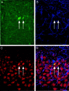
Triple labeling confirms that BrdU-positive cells mature into neurons. Rats received BrdU injections 4 d after the last ECS treatment and were killed 4 weeks later. A representative confocal laser-scanning image (66×) of a section from a fluoxetine-treated rat that has been triple-labeled with BrdU (A, green; BrdU-positive cells indicated by_arrows_), GFAP (B, blue), and NeuN (C, red). The merged image (D) demonstrates cells that are double-labeled in the GCL for BrdU and NeuN but not GFAP.
Similar articles
- Brain-derived neurotrophic factor and antidepressant drugs have different but coordinated effects on neuronal turnover, proliferation, and survival in the adult dentate gyrus.
Sairanen M, Lucas G, Ernfors P, Castrén M, Castrén E. Sairanen M, et al. J Neurosci. 2005 Feb 2;25(5):1089-94. doi: 10.1523/JNEUROSCI.3741-04.2005. J Neurosci. 2005. PMID: 15689544 Free PMC article. - Chronic treatment with AMPA receptor potentiator Org 26576 increases neuronal cell proliferation and survival in adult rodent hippocampus.
Su XW, Li XY, Banasr M, Koo JW, Shahid M, Henry B, Duman RS. Su XW, et al. Psychopharmacology (Berl). 2009 Oct;206(2):215-22. doi: 10.1007/s00213-009-1598-0. Epub 2009 Jul 15. Psychopharmacology (Berl). 2009. PMID: 19603152 - Chronic SSRI Treatment, but Not Norepinephrine Reuptake Inhibitor Treatment, Increases Neurogenesis in Juvenile Rats.
Hovorka M, Ewing D, Middlemas DS. Hovorka M, et al. Int J Mol Sci. 2022 Jun 22;23(13):6919. doi: 10.3390/ijms23136919. Int J Mol Sci. 2022. PMID: 35805924 Free PMC article. - Depression and adult neurogenesis: Positive effects of the antidepressant fluoxetine and of physical exercise.
Micheli L, Ceccarelli M, D'Andrea G, Tirone F. Micheli L, et al. Brain Res Bull. 2018 Oct;143:181-193. doi: 10.1016/j.brainresbull.2018.09.002. Epub 2018 Sep 17. Brain Res Bull. 2018. PMID: 30236533 Review. - Implications of adult hippocampal neurogenesis in antidepressant action.
Malberg JE. Malberg JE. J Psychiatry Neurosci. 2004 May;29(3):196-205. J Psychiatry Neurosci. 2004. PMID: 15173896 Free PMC article. Review.
Cited by
- The impact of voluntary wheel-running exercise on hippocampal neurogenesis and behaviours in response to nicotine cessation in rats.
Zaniewska M, Brygider S, Majcher-Maślanka I, Gawliński D, Głowacka U, Glińska S, Balcerzak Ł. Zaniewska M, et al. Psychopharmacology (Berl). 2024 Dec;241(12):2585-2607. doi: 10.1007/s00213-024-06705-7. Epub 2024 Oct 27. Psychopharmacology (Berl). 2024. PMID: 39463206 Free PMC article. - Phosphatidic acid is involved in regulation of autophagy in neurons in vitro and in vivo.
Schiller M, Wilson GC, Keitsch S, Soddemann M, Wilker B, Edwards MJ, Scherbaum N, Gulbins E. Schiller M, et al. Pflugers Arch. 2024 Dec;476(12):1881-1894. doi: 10.1007/s00424-024-03026-8. Epub 2024 Oct 8. Pflugers Arch. 2024. PMID: 39375214 Free PMC article. - BMP Antagonist Gremlin 2 Regulates Hippocampal Neurogenesis and Is Associated with Seizure Susceptibility and Anxiety.
Frazer NB, Kaas GA, Firmin CG, Gamazon ER, Hatzopoulos AK. Frazer NB, et al. eNeuro. 2024 Oct 17;11(10):ENEURO.0213-23.2024. doi: 10.1523/ENEURO.0213-23.2024. Print 2024 Oct. eNeuro. 2024. PMID: 39349059 Free PMC article. - Quantitative cytoarchitectural phenotyping of deparaffinized human brain tissues.
Meo DD, Sorelli M, Ramazzotti J, Cheli F, Bradley S, Perego L, Lorenzon B, Mazzamuto G, Emmi A, Porzionato A, Caro R, Garbelli R, Biancheri D, Pelorosso C, Conti V, Guerrini R, Pavone FS, Costantini I. Meo DD, et al. bioRxiv [Preprint]. 2024 Sep 14:2024.09.10.612232. doi: 10.1101/2024.09.10.612232. bioRxiv. 2024. PMID: 39314456 Free PMC article. Preprint. - Fast-acting antidepressant-like effects of ketamine in aged male rats.
Hernández-Hernández E, Ledesma-Corvi S, Jornet-Plaza J, García-Fuster MJ. Hernández-Hernández E, et al. Pharmacol Rep. 2024 Oct;76(5):991-1000. doi: 10.1007/s43440-024-00636-y. Epub 2024 Aug 19. Pharmacol Rep. 2024. PMID: 39158787 Free PMC article.
References
- Backhouse B, Barochovsky O, Malik C, Patel A, Lewis P. Effect of haloperidol on cell proliferation in the early postnatal rat brain. Neuropathol Appl Neurobiol. 1982;8:109–116. - PubMed
- Cameron HA, Woolley CS, McEwen BS, Gould E. Differentiation of newly born neurons and glia in the dentate gyrus of the adult rat. Neuroscience. 1993;56:337–344. - PubMed
- Dawirs RR, Hildebrandt K, Teuchert-Noodt G. Adult treatment with haloperidol increases dentate granule cell proliferation in the gerbil hippocampus. J Neural Transm. 1998;105:317–327. - PubMed
- Duman RS. Novel therapeutic approaches beyond the serotonin receptor. Biol Psychiatry. 1998;44:324–335. - PubMed
Publication types
MeSH terms
Substances
Grants and funding
- P01 MH025642/MH/NIMH NIH HHS/United States
- 2 PO1 MH25642/MH/NIMH NIH HHS/United States
- MH45481/MH/NIMH NIH HHS/United States
- R37 MH045481/MH/NIMH NIH HHS/United States
- R01 MH045481/MH/NIMH NIH HHS/United States
- MH53199/MH/NIMH NIH HHS/United States
LinkOut - more resources
Full Text Sources
Other Literature Sources
Medical


