Augmented senile plaque load in aged female beta-amyloid precursor protein-transgenic mice - PubMed (original) (raw)
Augmented senile plaque load in aged female beta-amyloid precursor protein-transgenic mice
M J Callahan et al. Am J Pathol. 2001 Mar.
Abstract
Transgenic mice (Tg2576) overexpressing human beta-amyloid precursor protein with the Swedish mutation (APP695SWE) develop Alzheimer's disease-like amyloid beta protein (Abeta) deposits by 8 to 10 months of age. These mice show elevated levels of Abeta40 and Abeta42, as well as an age-related increase in diffuse and compact senile plaques in the brain. Senile plaque load was quantitated in the hippocampus and neocortex of 8- to 19-month-old male and female Tg2576 mice. In all mice, plaque burden increased markedly after the age of 12 months. At 15 and 19 months of age, senile plaque load was significantly greater in females than in males; in 91 mice studied at 15 months of age, the area occupied by plaques in female Tg2576 mice was nearly three times that of males. By enzyme-linked immunosorbent assay, female mice also had more Abeta40 and Abeta42 in the brain than did males, although this difference was less pronounced than the difference in histological plaque load. These data show that senescent female Tg2576 mice deposit more amyloid in the brain than do male mice, and may provide an animal model in which the influence of sex differences on cerebral amyloid pathology can be evaluated.
Figures
Figure 1.
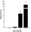
Tg2576 mice have an age-related increase in the percent area of the hippocampus and neocortex occupied by Aβ deposits as quantitated using a point-counting technique. Data evaluated by analysis of variance followed by a post hoc Newman-Keuls test. ***, P < 0.001, compared to all other age groups.
Figure 2.
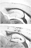
Campbell-Switzer silver AD-stained sagittal tissue sections under low (×4 objective) magnification from male (a) and female (b) Tg2576 mice representing mean percent plaque areas of 2.31% and 6.11%, respectively, at 15 months of age. The Aβ deposits are stained black or dark brown with this method. The silver stain normally turns myelinated pathways a golden brown color; these are easily distinguished from Aβ deposits under the microscope. Scale bar, 200 μm.
Figure 3.
At 15 months of age, female Tg2576 mice have a threefold greater percent area of the hippocampus and neocortex occupied by senile plaques. Data evaluated by analysis of variance followed by a post hoc Newman-Keuls test. ***, P < 0.001 compared to male mice.
Figure 4.
In 15-month-old Tg2576 mice, soluble and insoluble Aβ levels, measured by ELISA, were increased in females compared to males. This increase was significant for Aβ40 in both the soluble and insoluble extracts. Data evaluated by analysis of variance followed by post hoc Newman-Keuls test. *, P < 0.05; **, P < 0.01 compared to male mice.
Comment in
- Alzheimer's disease in man and transgenic mice: females at higher risk.
Turner RS. Turner RS. Am J Pathol. 2001 Mar;158(3):797-801. doi: 10.1016/S0002-9440(10)64026-6. Am J Pathol. 2001. PMID: 11238027 Free PMC article. Review. No abstract available.
Similar articles
- Quantitative Comparison of Dense-Core Amyloid Plaque Accumulation in Amyloid-β Protein Precursor Transgenic Mice.
Liu P, Reichl JH, Rao ER, McNellis BM, Huang ES, Hemmy LS, Forster CL, Kuskowski MA, Borchelt DR, Vassar R, Ashe KH, Zahs KR. Liu P, et al. J Alzheimers Dis. 2017;56(2):743-761. doi: 10.3233/JAD-161027. J Alzheimers Dis. 2017. PMID: 28059792 Free PMC article. - Gender differences in the amount and deposition of amyloidbeta in APPswe and PS1 double transgenic mice.
Wang J, Tanila H, Puoliväli J, Kadish I, van Groen T. Wang J, et al. Neurobiol Dis. 2003 Dec;14(3):318-27. doi: 10.1016/j.nbd.2003.08.009. Neurobiol Dis. 2003. PMID: 14678749 - beta-amyloid deposits in transgenic mice expressing human beta-amyloid precursor protein have the same characteristics as those in Alzheimer's disease.
Terai K, Iwai A, Kawabata S, Tasaki Y, Watanabe T, Miyata K, Yamaguchi T. Terai K, et al. Neuroscience. 2001;104(2):299-310. doi: 10.1016/s0306-4522(01)00095-1. Neuroscience. 2001. PMID: 11377835 - Alzheimer's disease in man and transgenic mice: females at higher risk.
Turner RS. Turner RS. Am J Pathol. 2001 Mar;158(3):797-801. doi: 10.1016/S0002-9440(10)64026-6. Am J Pathol. 2001. PMID: 11238027 Free PMC article. Review. No abstract available. - [The lesions of Alzheimer's disease: which therapeutic perspectives?].
Duyckaerts C, Perruchini C, Lebouvier T, Potier MC. Duyckaerts C, et al. Bull Acad Natl Med. 2008 Feb;192(2):303-18; discussion 318-21. Bull Acad Natl Med. 2008. PMID: 18819685 Review. French.
Cited by
- Anxiety and Alzheimer's disease: Behavioral analysis and neural basis in rodent models of Alzheimer's-related neuropathology.
Pentkowski NS, Rogge-Obando KK, Donaldson TN, Bouquin SJ, Clark BJ. Pentkowski NS, et al. Neurosci Biobehav Rev. 2021 Aug;127:647-658. doi: 10.1016/j.neubiorev.2021.05.005. Epub 2021 May 9. Neurosci Biobehav Rev. 2021. PMID: 33979573 Free PMC article. Review. - Sex-related dimorphism in dentate gyrus atrophy and behavioral phenotypes in an inducible tTa:APPsi transgenic model of Alzheimer's disease.
Melnikova T, Park D, Becker L, Lee D, Cho E, Sayyida N, Tian J, Bandeen-Roche K, Borchelt DR, Savonenko AV. Melnikova T, et al. Neurobiol Dis. 2016 Dec;96:171-185. doi: 10.1016/j.nbd.2016.08.009. Epub 2016 Aug 26. Neurobiol Dis. 2016. PMID: 27569580 Free PMC article. - The role of Beta-adrenergic receptor blockers in Alzheimer's disease: potential genetic and cellular signaling mechanisms.
Lương Kv, Nguyen LT. Lương Kv, et al. Am J Alzheimers Dis Other Demen. 2013 Aug;28(5):427-39. doi: 10.1177/1533317513488924. Epub 2013 May 20. Am J Alzheimers Dis Other Demen. 2013. PMID: 23689075 Free PMC article. Review. - Sex-dependent alterations in social behaviour and cortical synaptic activity coincide at different ages in a model of Alzheimer's disease.
Bories C, Guitton MJ, Julien C, Tremblay C, Vandal M, Msaid M, De Koninck Y, Calon F. Bories C, et al. PLoS One. 2012;7(9):e46111. doi: 10.1371/journal.pone.0046111. Epub 2012 Sep 24. PLoS One. 2012. PMID: 23029404 Free PMC article. - Inducible proteopathies.
Walker LC, Levine H 3rd, Mattson MP, Jucker M. Walker LC, et al. Trends Neurosci. 2006 Aug;29(8):438-43. doi: 10.1016/j.tins.2006.06.010. Epub 2006 Jun 27. Trends Neurosci. 2006. PMID: 16806508 Free PMC article. Review.
References
- Selkoe DJ: Biology of β-amyloid precursor protein and the mechanism of Alzheimer disease. ed 2 Terry RD Katzman R Bick KL Sisodia SS eds. Alzheimer Disease, 1999, :pp 293-310 Lippincott Williams and Wilkins, Philadelphia
- Mullan M, Crawford F, Axelman K, Houlden H, Lilius L, Winblad B, Lannfelt L: A pathogenic mutation for probable Alzheimer’s disease in the APP gene at the N-terminus of beta-amyloid. Nat Genet 1992, 1:345-347 - PubMed
- Hardy J: Amyloid, the presenilins and Alzheimer’s disease. Trends Neurosci 1997, 20:154-159 - PubMed
- Price DL, Sisodia SS: Mutant genes in familial Alzheimer’s disease and transgenic models. Ann Rev Neurosci 1998, 21:479-505 - PubMed
- Molsa PK, Marttila RJ, Rinne UK: Epidemiology of dementia in a Finnish population. Acta Neurol Scand 1982, 65:541-552 - PubMed
Publication types
MeSH terms
Substances
LinkOut - more resources
Full Text Sources
Other Literature Sources
Medical
Molecular Biology Databases


