Spontaneous corticospinal axonal plasticity and functional recovery after adult central nervous system injury - PubMed (original) (raw)
Spontaneous corticospinal axonal plasticity and functional recovery after adult central nervous system injury
N Weidner et al. Proc Natl Acad Sci U S A. 2001.
Abstract
Although it is believed that little recovery occurs after adult mammalian spinal cord injury, in fact significant spontaneous functional improvement commonly occurs after spinal cord injury in humans. To investigate potential mechanisms underlying spontaneous recovery, lesions of defined components of the corticospinal motor pathway were made in adult rats in the rostral cervical spinal cord or caudal medulla. Following complete lesions of the dorsal corticospinal motor pathway, which contains more than 95% of all corticospinal axons, spontaneous sprouting from the ventral corticospinal tract occurred onto medial motoneuron pools in the cervical spinal cord; this sprouting was paralleled by functional recovery. Combined lesions of both dorsal and ventral corticospinal tract components eliminated sprouting and functional recovery. In addition, functional recovery was also abolished if dorsal corticospinal tract lesions were followed 5 weeks later by ventral corticospinal tract lesions. We found extensive spontaneous structural plasticity as a mechanism correlating with functional recovery in motor systems in the adult central nervous system. Experimental enhancement of spontaneous plasticity may be useful to promote further recovery after adult central nervous system injury.
Figures
Figure 1
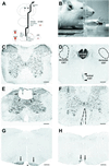
Lesion models and functional testing. (A) Lesion model. Animals received bilateral lesions of the CST in the MED just rostral to the pyramidal decussation, to the dCST at C3, to the vCST at cervical level C2, or to the d/vCST at C3 and C2. The CST was anterogradely labeled by BDA injected into sensorimotor cortex. (B) Behavioral testing: skilled forelimb task. Illustrated is an animal reaching through an aperture to retrieve a food pellet (arrow). (C–H) Morphology of lesions. Nissl stain (C) and BDA-labeled (D) coronal sections at C3 in an intact animal. Most BDA-labeled CST axons are located in the dorsal CST. The dorsolateral CST is dispersed throughout the dorsolateral columns (dashed circles). The ventral CST projects in the ventromedial portion of the cord (arrowheads). Rare BDA-labeled axons comprising the lateral CST are scattered throughout lateral aspects of the lateral columns and are not detectable at this magnification. (E) Representative Nissl-stained section through the center of a dCST lesion at C3 (arrows). The lesion affects the dorsal CST and much of the dorsal sensory columns without disrupting the surrounding spinal cord. (F) Representative Nissl-stained section through the center of a vCST lesion. The lesion is confined to the ventromedial columns bilaterally. (G) Coronal section located 200 μm rostral to a MED CST lesion showing the intact ventral BDA-labeled CST rostral to the lesion (arrows). Asterisk indicates basilar artery. (H) Coronal section from the epicenter of a bilateral medullary CST lesion. The lesion is confined to the CST bilaterally, leaving surrounding structures intact. The lateral portion of the BDA-labeled CST is completely disrupted, whereas the medial portion (arrows) is incompletely lesioned to avoid damage to the basilar artery. The wire knife could not be apposed as closely to the basilar artery on the left side as on the right side because of a restricted visual field, sparing slightly more CST axons on the left (scale bars,C–F = 283 μm; G and_H_ = 458 μm).
Figure 2
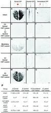
Extent of CST lesions. CST axons were anterogradely labeled by bilateral BDA injections into sensorimotor cortex. Coronal sections were examined at C4, caudal to all lesions. (A,D, G, J, and_M_) Dorsal CST. (B, E,H, K, and N) Ventral CST. (C, F, I,L, and O) Dorsolateral CST. In intact animals (A–C), the tightly bundled dorsal CST is evident, together with the less dense ventral CST aligned along the median fissure. The dorsolateral tract is loosely organized, and the lateral CST is diffusely interspersed within lateral regions of the white matter (not shown). In animals with combined dorsal and ventral CST lesions (d/vCST; D–F), virtually all dorsal and ventral CST axons are eliminated, and numbers of dorsolateral CST axons are unchanged (see also P). Subjects with MED CST lesions (G–I) show a loss of 86% of dorsal CST axons, 94% of ventral CST axons, 79% of dorsolateral CST axons, and 88% of lateral CST axons. Animals with dCST lesions (J–L) exhibit a loss of all dorsal CST axons. In addition, although dorsal CST lesions were restricted to the midline dorsal white matter of the cord, numbers of ventral CST axons were reduced by 36%, and numbers of lateral CST axons were reduced by 76%, possibly because of collateralization of CST axons at the medullary level (49). Animals with ventral CST lesions (vCST; M–O) showed a 90% reduction in ventral CST axons but no losses in any other portions of the CST projection. (P) Quantification of axon numbers in each component of the CST after specific lesions (* ANOVA, P < 0.001; scale bars, A–O = 73 μm).
Figure 3
Behavior of pellet retrieval. Data are presented for each group as percentage of successful pellet retrievals on the last day of testing 2, 3, and 4 weeks postoperatively. Preoperatively, groups did not differ significantly from one another (P = 0.45; data not shown). Significant postoperative group differences were present; asterisks indicate significant post hoc differences compared with intact animals (post hoc Fisher's). Animals with lesions of ventral plus dorsal components of the CST exhibited long-lasting, stable deficits in skilled forelimb use. Animals with isolated lesions of either the dorsal CST or ventral CST showed significant deficits compared with intact animals 2 weeks postlesion, which improved by 4 weeks postlesion. Thus, recovery occurred over several weeks after isolated lesions of the dorsal or ventral CST.
Figure 4
Behavior. Detailed analysis of forelimb movements revealed the nature of errors in pellet retrieval 4 weeks postoperatively. Animals with long-lasting deficits in pellet retrieval (d/vCST, MED) exhibited particular dysfunction of proximal forelimb use. Animals with d/vCST lesions showed a 9-fold increase in failed advances and a 6.4-fold increase in missed aims compared with intact animals. In contrast, animals with dCST lesions had only a small increase in failed advances (1.4-fold), missed aims (2-fold), and pellet drops (1.9-fold) and failed to exhibit long-term deficiencies in pellet retrieval rate (see Fig. 3).
Figure 5
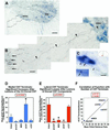
Spontaneous compensatory CST sprouting after injury. Contacts of BDA-labeled CST axons (brown-black) with ChAT-immunolabeled motoneurons (blue) were quantified in the medial motoneuron column (mmc) and lateral motoneuron columns (lmc) at C4. (A) Ventral horn and ventral white matter are shown, with ChAT-labeled motoneurons in the medial and lateral motor columns. Animal with dorsal CST lesion. (B) Higher magnification of the boxed area in_A_. A BDA-labeled axon (arrowheads) branches off the ventral CST (arrows) and projects to the medial motoneuron column, Rexed's lamina IX. (C) A BDA-labeled CST axon (arrowheads) projects from the ventral white matter into the medial motoneuron column and contacts a ChAT-labeled motoneuron, exhibiting a bouton-like swelling at the site of this contact (arrow). This point of putative contact is shown at higher magnification in the_Inset_. The total number of bouton-like contacts was quantified through the full thickness of ten coronal sections at C4 in every animal. (D and E) Total number of BDA-labeled CST axons contacting ChAT-labeled motoneurons in lamina IX at C4. (D) BDA-labeled CST axon contacts in the medial motoneuron column of lamina IX are significantly increased only in dCST-lesioned animals compared with intact animals (ANOVA,P < 0.01). (E) All lesions disrupting the dorsal CST (d/vCST, MED, dCST) yield a significant reduction of BDA-labeled CST terminations in the lateral motoneuron column of lamina IX (ANOVA, P < 0.001). (F) The total number of CST terminations on medial + lateral motor neurons correlates significantly with functional performance on the pellet retrieval task (_r_2 = 0.89; P = 0.03; scale bars: A = 91 μm,B = 13.4 μm, C = 11.1).
Similar articles
- Competition with Primary Sensory Afferents Drives Remodeling of Corticospinal Axons in Mature Spinal Motor Circuits.
Jiang YQ, Zaaimi B, Martin JH. Jiang YQ, et al. J Neurosci. 2016 Jan 6;36(1):193-203. doi: 10.1523/JNEUROSCI.3441-15.2016. J Neurosci. 2016. PMID: 26740661 Free PMC article. - Corticospinal circuit plasticity in motor rehabilitation from spinal cord injury.
Serradj N, Agger SF, Hollis ER 2nd. Serradj N, et al. Neurosci Lett. 2017 Jun 23;652:94-104. doi: 10.1016/j.neulet.2016.12.003. Epub 2016 Dec 6. Neurosci Lett. 2017. PMID: 27939980 Review. - A Single Bolus of Docosahexaenoic Acid Promotes Neuroplastic Changes in the Innervation of Spinal Cord Interneurons and Motor Neurons and Improves Functional Recovery after Spinal Cord Injury.
Liu ZH, Yip PK, Adams L, Davies M, Lee JW, Michael GJ, Priestley JV, Michael-Titus AT. Liu ZH, et al. J Neurosci. 2015 Sep 16;35(37):12733-52. doi: 10.1523/JNEUROSCI.0605-15.2015. J Neurosci. 2015. PMID: 26377463 Free PMC article. - Re-Establishment of Cortical Motor Output Maps and Spontaneous Functional Recovery via Spared Dorsolaterally Projecting Corticospinal Neurons after Dorsal Column Spinal Cord Injury in Adult Mice.
Hilton BJ, Anenberg E, Harrison TC, Boyd JD, Murphy TH, Tetzlaff W. Hilton BJ, et al. J Neurosci. 2016 Apr 6;36(14):4080-92. doi: 10.1523/JNEUROSCI.3386-15.2016. J Neurosci. 2016. PMID: 27053214 Free PMC article. - Corticospinal Motor Circuit Plasticity After Spinal Cord Injury: Harnessing Neuroplasticity to Improve Functional Outcomes.
Kazim SF, Bowers CA, Cole CD, Varela S, Karimov Z, Martinez E, Ogulnick JV, Schmidt MH. Kazim SF, et al. Mol Neurobiol. 2021 Nov;58(11):5494-5516. doi: 10.1007/s12035-021-02484-w. Epub 2021 Aug 3. Mol Neurobiol. 2021. PMID: 34341881 Review.
Cited by
- Spinal interneurons and forelimb plasticity after incomplete cervical spinal cord injury in adult rats.
Gonzalez-Rothi EJ, Rombola AM, Rousseau CA, Mercier LM, Fitzpatrick GM, Reier PJ, Fuller DD, Lane MA. Gonzalez-Rothi EJ, et al. J Neurotrauma. 2015 Jun 15;32(12):893-907. doi: 10.1089/neu.2014.3718. Epub 2015 May 5. J Neurotrauma. 2015. PMID: 25625912 Free PMC article. - Intensity sensitive modulation effect of theta burst form of median nerve stimulation on the monosynaptic spinal reflex.
Yeh KL, Fong PY, Huang YZ. Yeh KL, et al. Neural Plast. 2015;2015:704849. doi: 10.1155/2015/704849. Epub 2015 Mar 4. Neural Plast. 2015. PMID: 25821603 Free PMC article. - Endogenous neurogenesis replaces oligodendrocytes and astrocytes after primate spinal cord injury.
Yang H, Lu P, McKay HM, Bernot T, Keirstead H, Steward O, Gage FH, Edgerton VR, Tuszynski MH. Yang H, et al. J Neurosci. 2006 Feb 22;26(8):2157-66. doi: 10.1523/JNEUROSCI.4070-05.2005. J Neurosci. 2006. PMID: 16495442 Free PMC article. - Effects of bromelain on motor responses following intra-medial forebrain bundle 6-OHDA injection in rat model of parkinsonism.
Adu TS, Mabandla MV. Adu TS, et al. Metab Brain Dis. 2019 Dec;34(6):1557-1564. doi: 10.1007/s11011-019-00462-9. Epub 2019 Jul 22. Metab Brain Dis. 2019. PMID: 31332728
References
- Bracken M B, Shepard M J, Collins W F, Jr, Holford T R, Baskin D S, Eisenberg H M, Flamm E, Leo-Summers L, Maroon J C, Marshall L F, et al. J Neurosurg. 1992;76:23–31. - PubMed
- Frankel H K. In: Outcomes in Neurological and Surgical Disorders. Swash M, editor. Cambridge: Cambridge Univ. Press; 1998. pp. 181–194.
- Ferrucci L, Bandinelli S, Guralnik J M, Lamponi M, Bertini C, Falchini M, Baroni A. Stroke. 1993;24:200–205. - PubMed
- Naeser M A, Palumbo C L, Prete M N, Fitzpatrick P M, Mimura M, Samaraweera R, Albert M L. Brain Lang. 1998;62:1–28. - PubMed
- Sbordone R J, Liter J C, Pettler-Jennings P. Brain Inj. 1995;9:285–299. - PubMed
Publication types
MeSH terms
LinkOut - more resources
Full Text Sources
Other Literature Sources
Medical


